-
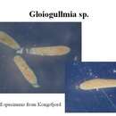
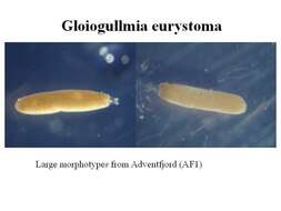
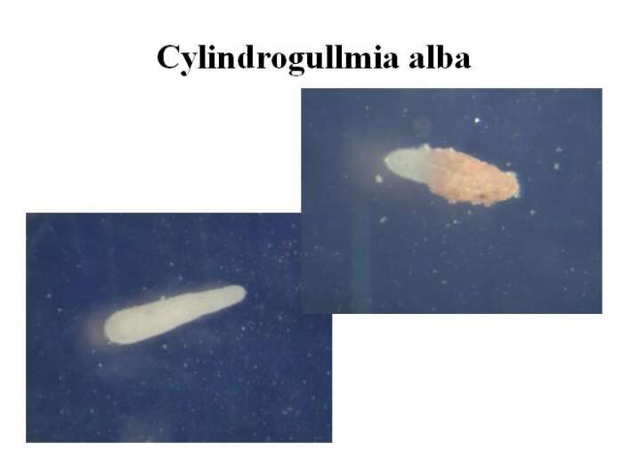
Specimens were isolated from surface sediments samples collected in Kongsfjorden, Isfjorden and Adventfjorden during the cruise of r/v « Oceania » between 22 July and 2 August 2004. The sediment samples were sieved at 500 um and 125 um sized meshes, and living specimens were picked under dissecting microscope on board. The specimens were photographed, measured and fixed for further DNA extraction. Source: http://www.iopan.gda.pl/projects/biodaff/EMBS-06.html
-
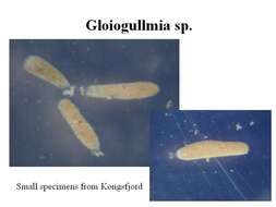
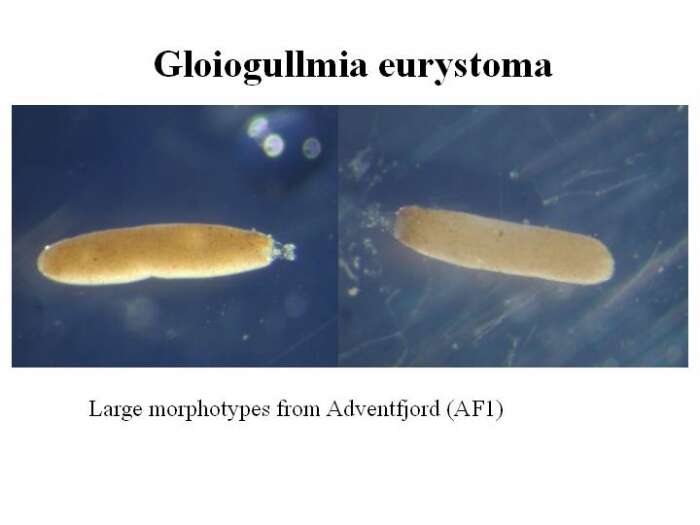
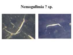
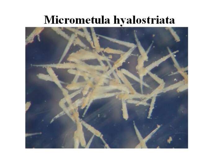
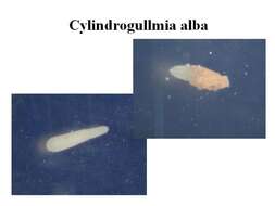
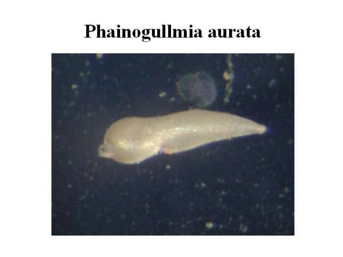
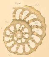
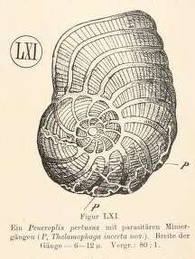
Rhumbler L. (1911 (cover 1909)). Die Foraminiferen (Talamophoren) der Plankton-Expedition. Zugleich Entwurf eines natürlichen Systems der Foraminiferen auf Grund selektionistischer und mechanisch-physiologischer Faktoren. Ergebnisse der Plankton-Expedition der Humboldt-Stiftung. vol. 3 L.c.: 1-331., available online at (http://www.biodiversitylibrary.org/item/18706#page/7/) page(s): p. 229 tf. 61, pl. 12 fig. 12
-
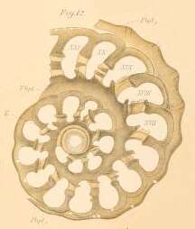
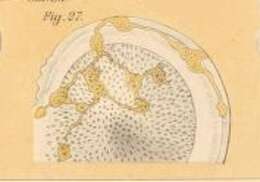
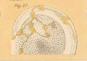
Rhumbler L. (1911 (cover 1909)). Die Foraminiferen (Talamophoren) der Plankton-Expedition. Zugleich Entwurf eines natürlichen Systems der Foraminiferen auf Grund selektionistischer und mechanisch-physiologischer Faktoren. Ergebnisse der Plankton-Expedition der Humboldt-Stiftung. vol. 3 L.c.: 1-331., available online at (http://www.biodiversitylibrary.org/item/18706#page/7/) page(s): pp. 228-231 pl. 39 fig. 27
-
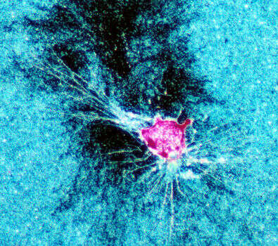
False-color image of a foraminiferan (pink) rending and consuming a bacterial biofilm (blue). The dark area is the region cleared by the foram in approximately 12 hours. Species not identified. Image courtesy of Joan Bernhard, WHOI. A version of this image appeared in Bernhard, J., and Bowser, S.S. (1992) Mar. Ecol. Prog. Ser. 83:263-272.
-
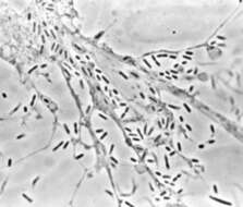
A brightfield image of a portion of a reticulopod and several rod-shaped bacteria. The oral zone of the foraminiferan is at upper left. Image courtesy of Samuel S. Bowser, Wadsworth Center.
-
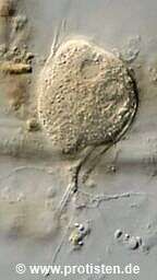
Lieberkuehnia from the Severn Estuary as promised.
-
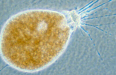
This image clearly shows the prominent "collar" around the oral zone for which the species is named. Image courtesy of Samuel S. Bowser, Wadsworth Center.
-
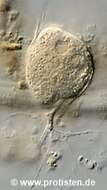
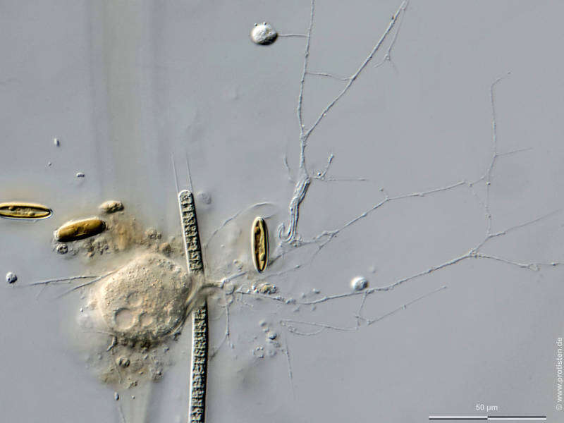
This species has an organic-walled test, which is thin, flexible and somewhat transparent. It is visible as the hazy "halo" around the cell body. Image courtesy of Samuel S. Bowser, Wadsworth Center.
-
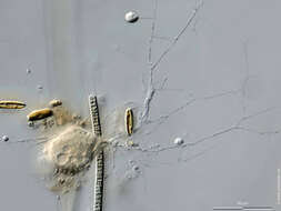
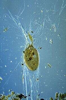
This individual is surrounded by the empty frustules of diatoms it has consumed. Image courtesy of Jeffrey L. Travis, University at Albany.
-
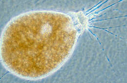
Marine thecate foraminiferan with reticulopods extended. Isolated from culture provided by Jeff L. Travis. Microscopy by L.W.Parfrey
-
-
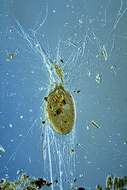
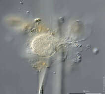
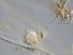
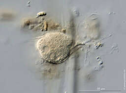
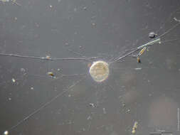
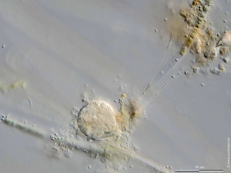
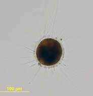
Scale bars indicate 100 µm (1), 50 µm (3–5).Five images.Please click on < or > on the image edges or on the dots at the bottom edge of the images to browse through the slides!Place name: Tropical freshwater aquarium Latitude: 54.3018013 Longitude: 10.07120132Microscope Zeiss Axioplan, camera Olympus OM-D M5 MKII.© Wolfgang Bettighofer,images under Creative Commons License V 3.0 (CC BY-NC-SA).For permission to use of (high resolution) images please contact
postmaster@protisten.de.For further information about the image, please click here:
Link to protisten.de page
-
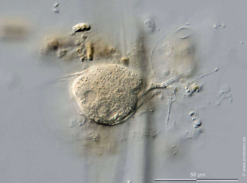
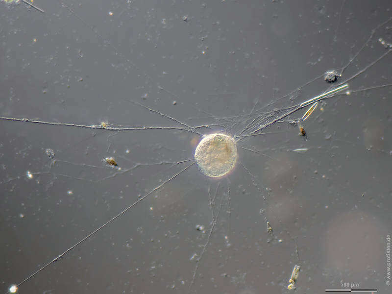
Scale bars indicate 100 µm (1), 50 µm (3–5).Five images.Please click on < or > on the image edges or on the dots at the bottom edge of the images to browse through the slides!Place name: Tropical freshwater aquarium Latitude: 54.3018013 Longitude: 10.07120132Microscope Zeiss Axioplan, camera Olympus OM-D M5 MKII.© Wolfgang Bettighofer,images under Creative Commons License V 3.0 (CC BY-NC-SA).For permission to use of (high resolution) images please contact
postmaster@protisten.de.For further information about the image, please click here:
Link to protisten.de page