-
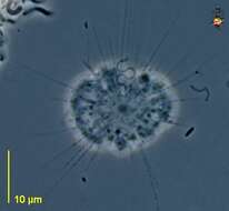
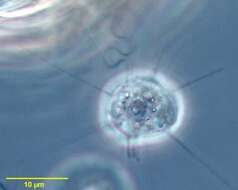
Ciliophrys (silly-off-rees), a pedinellid stramenopile, with a single flagellum. The cell may exist as a non-swimming form with radiating arms, and with the flagellum inactive or beating in a languid figure of 8 motion. The arms can be resorbed, and the cell can then swim with the flagellum pulling the cell forward. This is the heliozoon form, with a nucleus in the centre of the cell. Phase contrast.
-
-
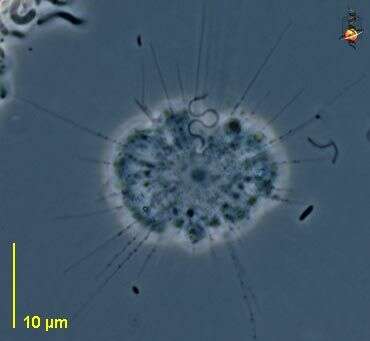
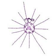
Ciliophrys (silly-off-riss) infusionum Cienkowski, 1876. Helioflagellate, in the heliozoan stage the cells are about 4 - 9 microns across, and have a central nucleus and one flagellum held in a figure of eight. The cells are spherical with delicate pseudopodia extending radially from the body and bearing extrusomes. The cells may change from the heliozoan stage with pseudopodia and a slow beating flagellum to a swimming flagellate without pseudopodia and with the flagellum beating rapidly. In swimming cells, the nucleus is located apically. Observed to consume suspended bacteria. When feeding, bacteria adhere to the pseudopodia and then are drawn to the body. The cells eat diatoms up to 18 microns long. Sometimes common.
-
Ciliophrys infusionum Cienkowski, 1876. Ciliophrys, in the heliozoan stage the cells are about 4 - 9 microns across, and have a central nucleus and one flagellum held in a figure of eight. The cells are spherical with delicate pseudopodia extending radially from the body and bearing extrusomes. The cells may change from the heliozoan stage with pseudopodia and a slow beating flagellum to a swimming flagellate without pseudopodia and with the flagellum beating rapidly. In swimming cells, the nucleus is located apically. Observed to consume suspended bacteria. When feeding, bacteria adhere to the pseudopodia and then are drawn to the body. The cells eat diatoms up to 18 microns long.
-
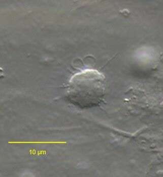
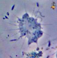
Portrait of Ciliophrys infusionum (Cienkowski,1876), one of the colourless pedinellid flagellates (also referred to as a helioflagellate). When at rest on the substrate tentacles(axopodia supported by one triplet of microtubules) of with fine granules are distributed over the entire surface and the single flagellum is held in a tight S configuration hardly moving. When swimming, tentacles withdraw and the cell surface appears smooth, the flagellum beating more rapidly in sine wave fashion. From standing freshwater near Boise, Idaho. Phase contrast.
-
Portrait of Ciliophrys infusionum (Cienkowski,1876), one of the colourless pedinellid flagellates (also referred to as a helioflagellates). When at rest on the substrate, tentacles (axopodia supported by one triplet of microtubules) with fine granules are distributed over the entire cell surface and the single flagellum is held in a tight S configuration hardly moving. When swimming, tentacles withdraw and the cell surface appears smooth, the flagellum beating more rapidly in sine wave fashion. From a commercial marine aquarium in Boise, Idaho. DIC.
-
Place name: Pond Birkensee near Rödelsee (Lower Franconia, Germany) Latitude: 49.71819841 Longitude: 10.27807474Microscope Zeiss Axioplan, camera Canon DSLR.© Wolfgang Bettighofer,images under Creative Commons License V 3.0 (CC BY-NC-SA).For permission to use of (high resolution) images please contact
postmaster@protisten.de.For further information about the image, please click here:
Link to protisten.de page
-
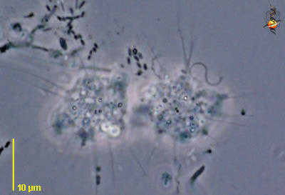
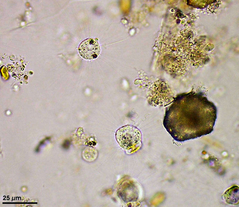
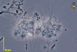
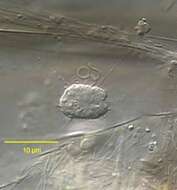
Sampling date 10/2007. Scale bar indicates 25 µm.Cells of Ciliophrys infusionum being in heliozoic state usually have one flagellum and numerous axopodia. The specimen just below the center of the image is starting with binary fission, the flagellum has already been duplicated.Place name: Hiddensee Bodden (Germany) Latitude: 54.582633 Longitude: 13.115051Microscope Zeiss Universal, camera Olympus C7070WZ.© Wolfgang Bettighofer,images under Creative Commons License V 3.0 (CC BY-NC-SA).For permission to use of (high resolution) images please contact
postmaster@protisten.de.For further information about the image, please click here:
Link to protisten.de page
-
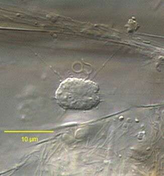
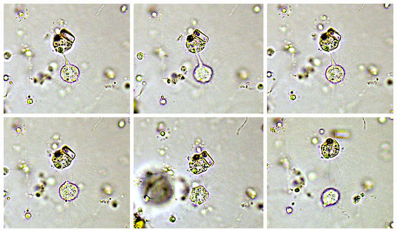
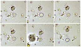
Series of images from the end of the binary fission, the beginning of which can be seen in the previous image.Place name: Hiddensee Bodden (Germany) Latitude: 54.582633 Longitude: 13.115051Microscope Zeiss Universal, camera Olympus C7070WZ.© Wolfgang Bettighofer,images under Creative Commons License V 3.0 (CC BY-NC-SA).For permission to use of (high resolution) images please contact
postmaster@protisten.de.For further information about the image, please click here:
Link to protisten.de page
-
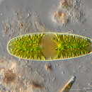
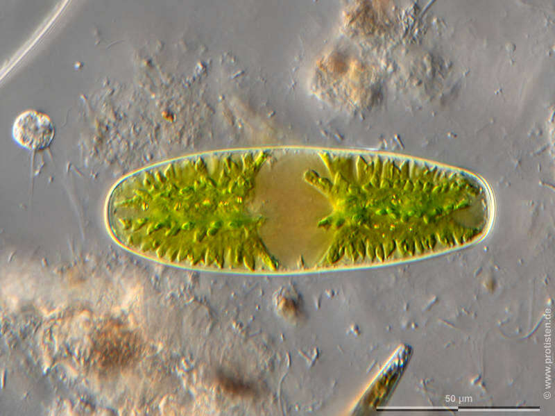
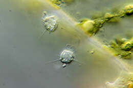
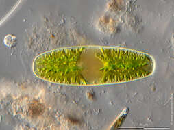
Sampling date 06/2023. Scale bar indicates 50 µm.Ciliophrys infusionum on the upper left, the streptophyte alga Netrium digitus is in the center oft the image.Place name: Wetland Lauchseemoor, Fieberbrunn (Tyrol, Austria) Latitude: 47.46954439 Longitude: 12.53826499Microscope Zeiss Axioplan, camera Olympus OM-D M5 MKII. DOF image.© Wolfgang Bettighofer,images under Creative Commons License V 3.0 (CC BY-NC-SA).For permission to use of (high resolution) images please contact
postmaster@protisten.de.For further information about the image, please click here:
Link to protisten.de page