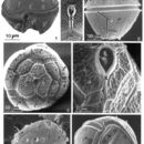
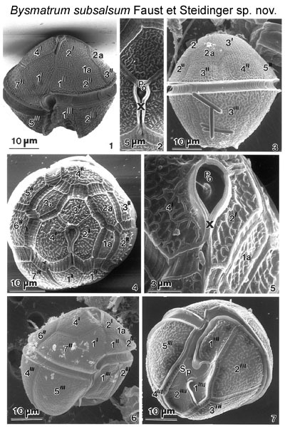
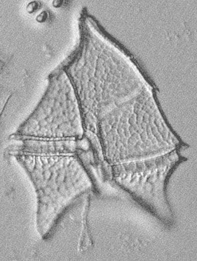
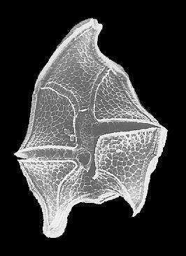
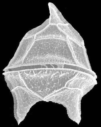
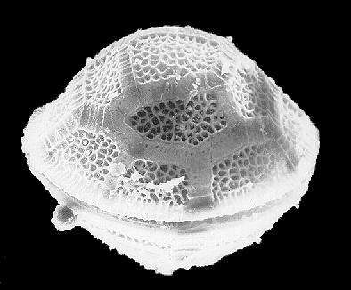
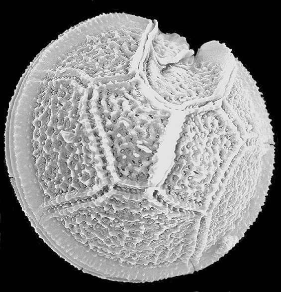
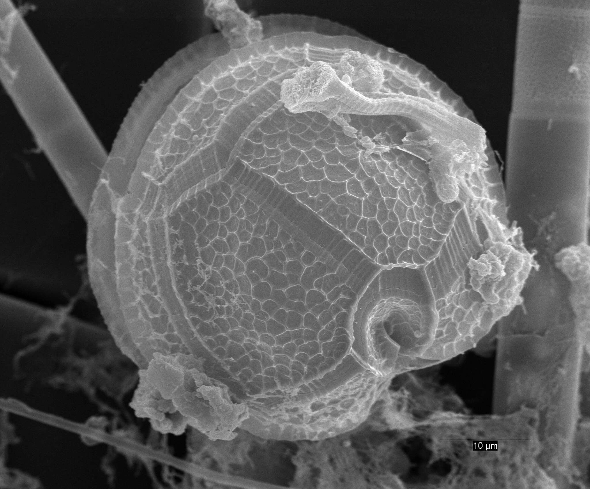
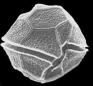
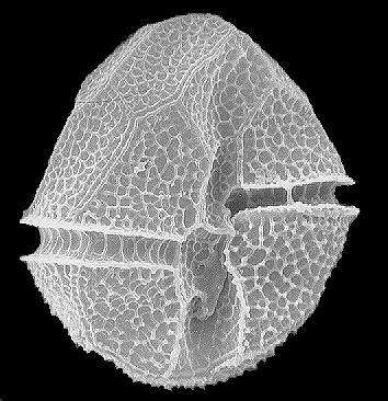
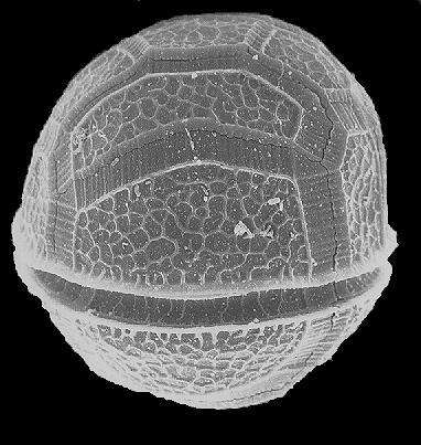
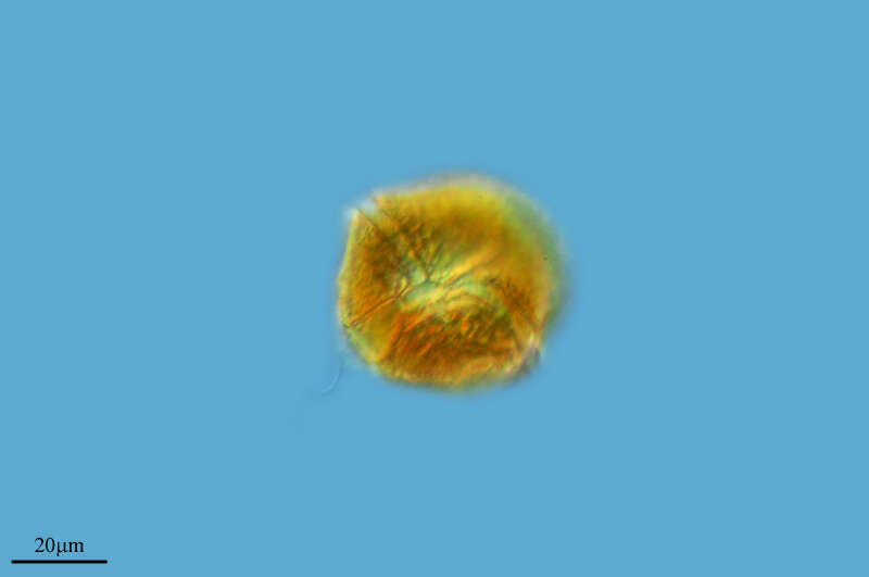
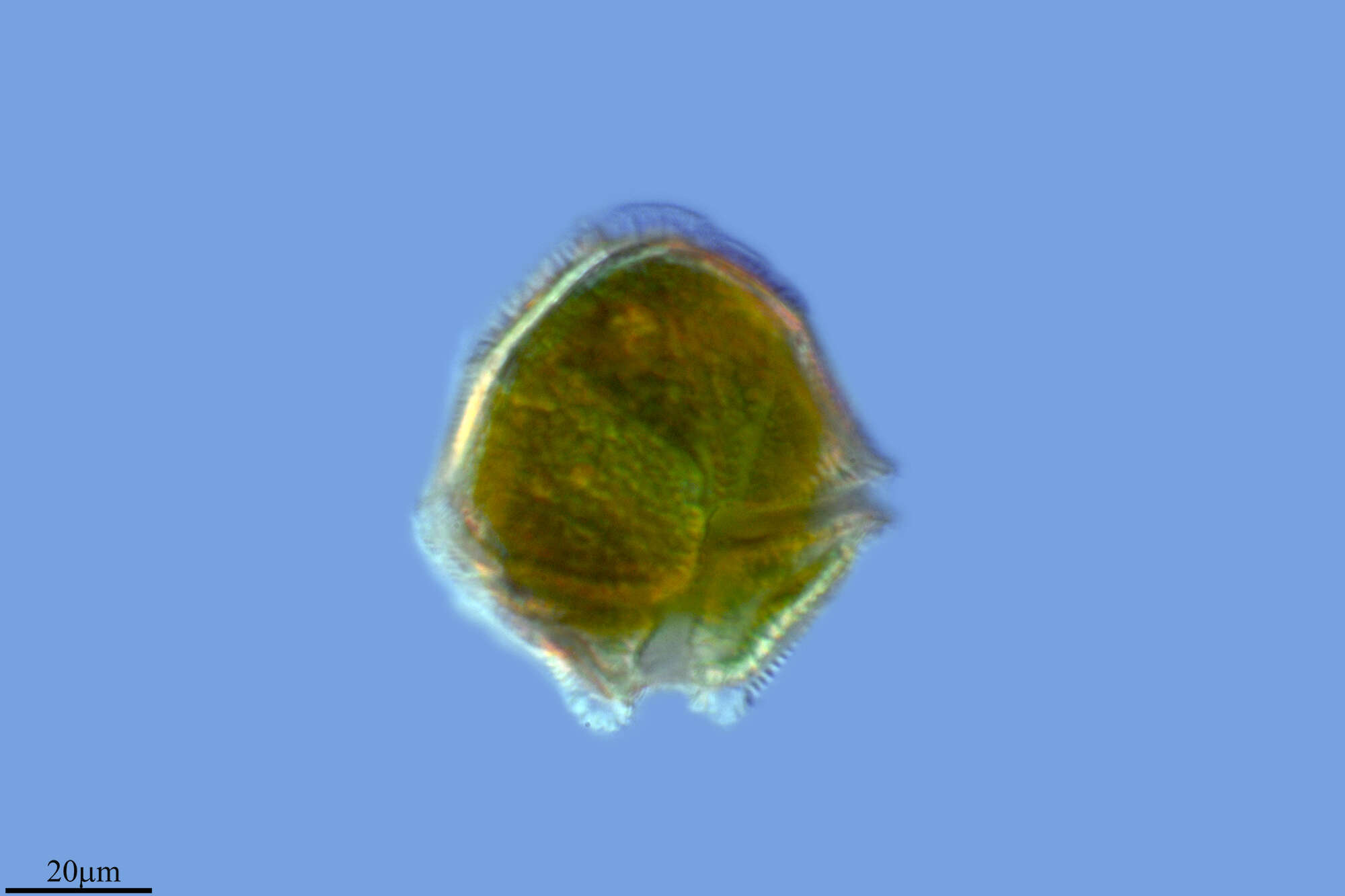
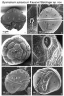
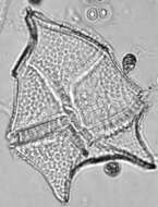
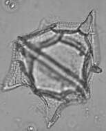
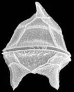
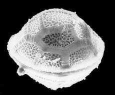
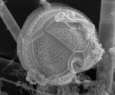
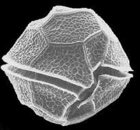
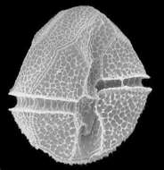
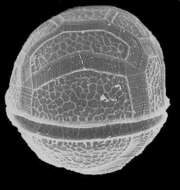
Figs 1-7. Lateral view of a cell with reticulated thecal surface, a conical epitheca, wide and deep, displaced cingulum, and a trapezoid hypotheca. The apical pore complex is situated ventrally. The apical plate 1' is asymmetric and pentagonal. The hypotheca is ventrally indented forming two lobes separating plates 2'" and 5'". The cingulum is displaced and finely striated with small pores aligned along the cingular lists. Fig. 2.The apical pore complex is a recessed chamber with a centrally located raised dome surrounded by a collar; it includes the apical pore plate (PO) and canal plate (X). Fig.3. Lateral view of a cell: a conical epitheca, wide and deep cingulum, and trapezoid hypotheca. Fig.4. Architecture of the epitheca including the position of the apical pore complex. Intercalary plates 2a and 3a are separated by plate 3'. The intercalary bands are striated.
EMu: Holotype SEM negative # 23040; SEM stub # ?; Field # 78-87; Accession # 407159; Catalog #1730; Figure # 1.
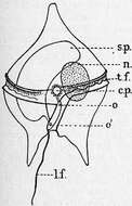
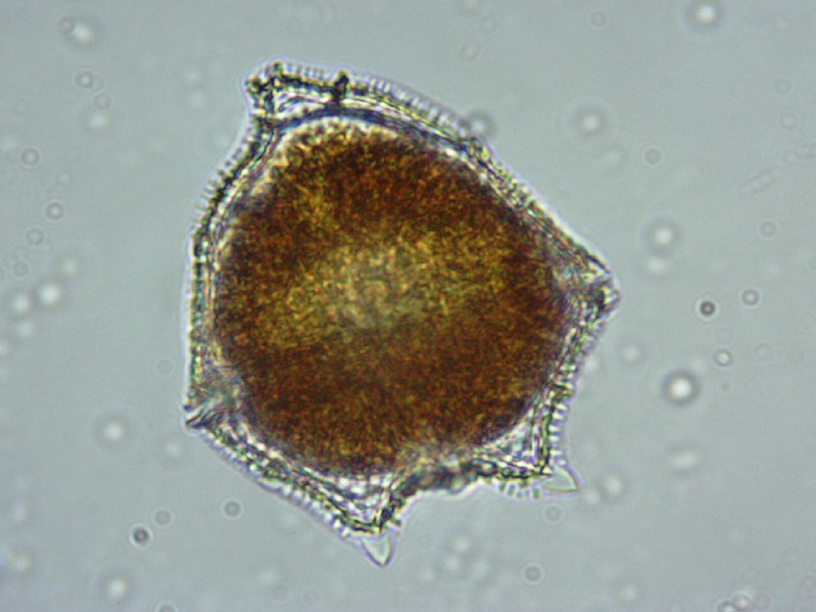
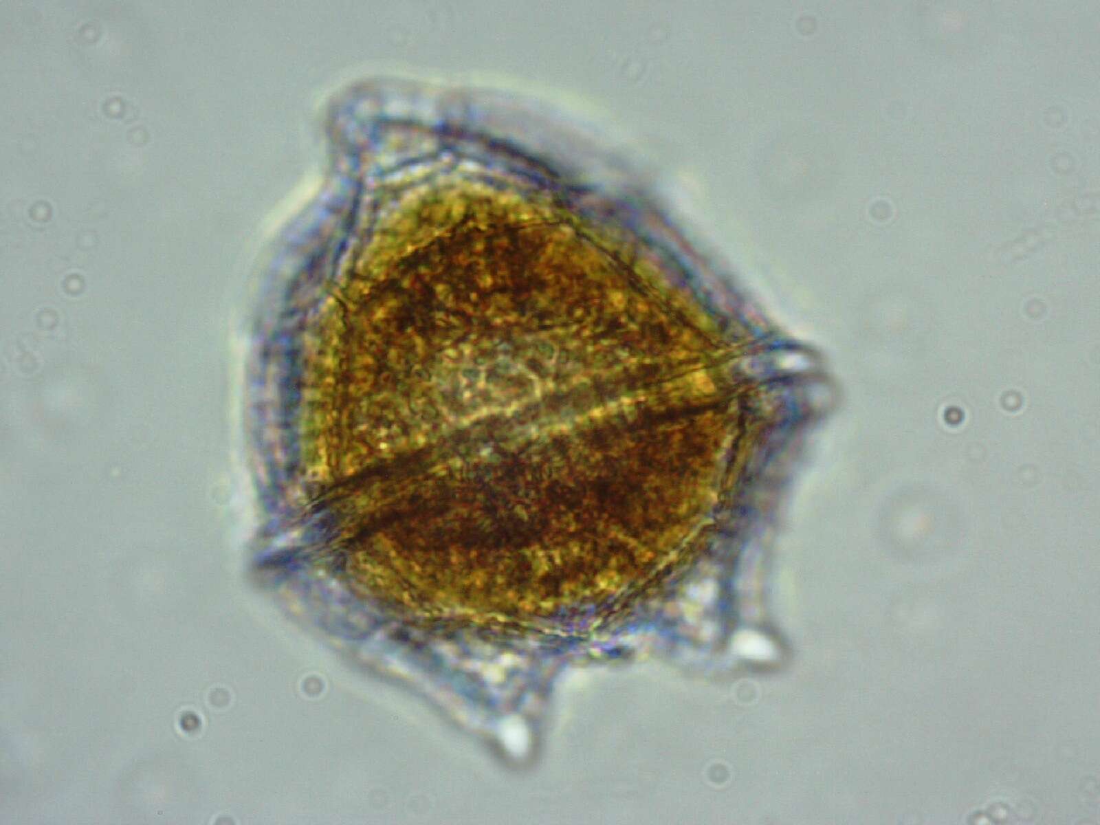
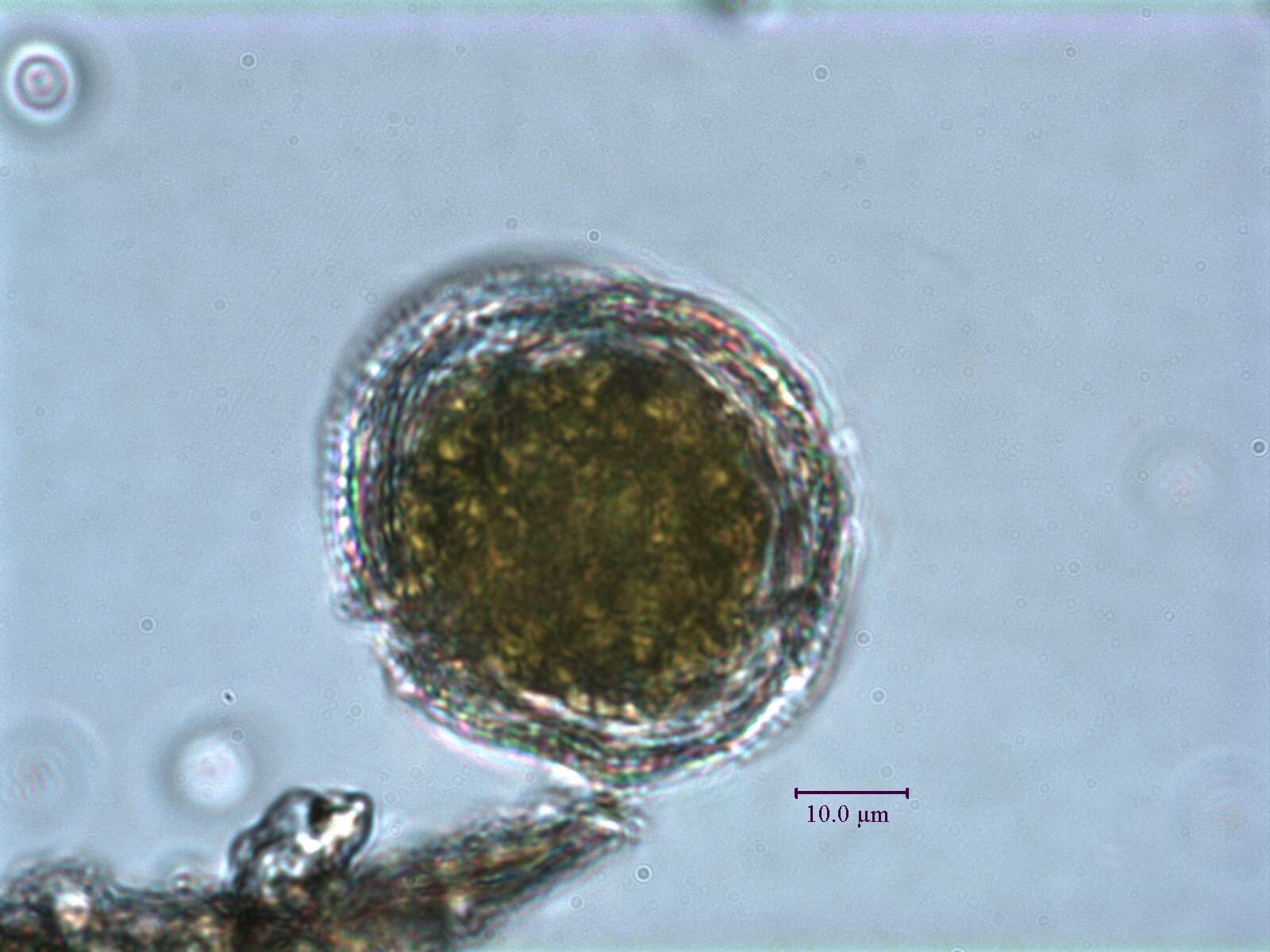
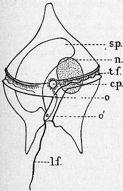
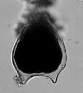
Description: English: Peridinium divergens showing longitudinal and transverse grooves in which lie the respective flagella l.f., t.f.; s.p., large “sack pusule” discharging through a tube by pore o’; c.p., “collective pusule discharging at o, and surrounded by a ring of formative” or “daughter pusules”; n, nucleus. After F. Schutt in Engler and Prantl’s Pflanzenfamilien, by permission of Wm Engelmann. Date: 1911. Source: Encyclopædia Britannica, 1911. Author: Encyclopædia Britannica, Volume 8, Slice 5., available freely at Project Gutenberg.