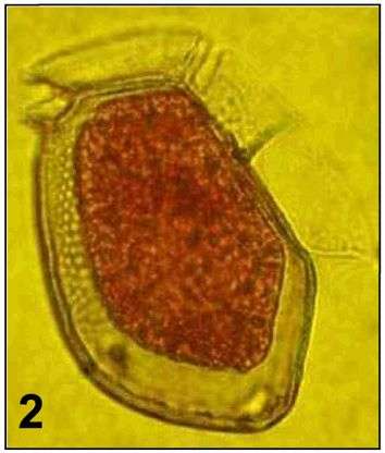
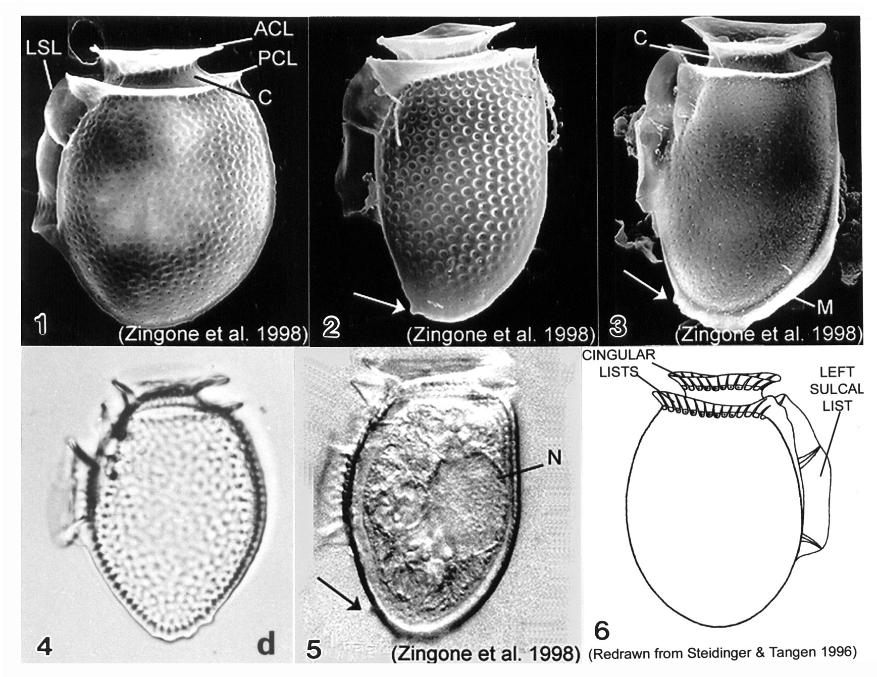
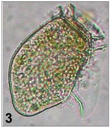
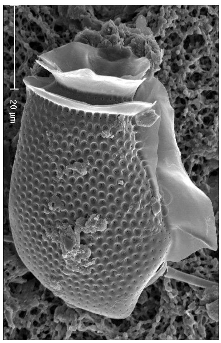
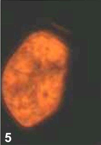
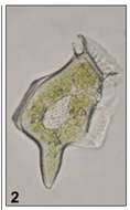
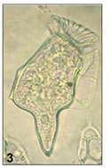
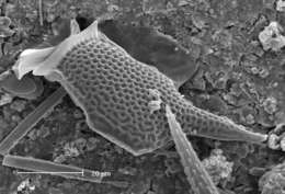
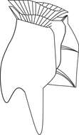
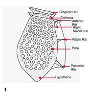
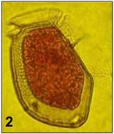
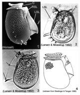
Plate 11. Dinophysis acuminata. Figs. 1-5. SEM: lateral view. Fig. 1. Cell oval and rotund; thecal surface with shallow depressions and scattered pores. Left sulcal list (LSL) extends beyond midpoint of cell. Well-developed cingular lists: anterior cingular list (ACL); posterior cingular list (PCL). C=cingulum. Fig. 2. Long and narrow cell with prominent surface areolae, each with a pore. Antapex tapered and ventrally off-center. Small posterior protrusion present (arrow). Fig. 3. Long and narrow cell. Thecal surface smooth with small scattered pores. Megacytic zone (M) void of pores. Posterior protrusions on antapex (arrow). Figs. 4-5. LM: lateral view. Fig. 4. Surface areolae and tapered antapex (from Larsen & Moestrup 1992: fig. 1d). Fig. 5. Large dorsal nucleus (N). Small, blunt projections on tapered antapex (arrow). Fig. 6. Line drawing.
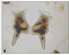
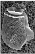
Plate 12. Dinophysis acuta. Fig. 1. SEM: lateral view. Cell oblong and robust; theca heavily areolated. Well developed cingular lists (CL) and left sulcal list (LSL). Pointed antapex. Figs. 2-3. LM: lateral view (from Larsen & Moestrup 1992: fig.s 2a,d; scale bars=20 _). Fig. 2. Large areolae, each with a pore (arrows). Fig. 3. Widest point below mid-section (dashed line) aligned with third sulcal rib (arrow). Fig. 4. Line drawing.
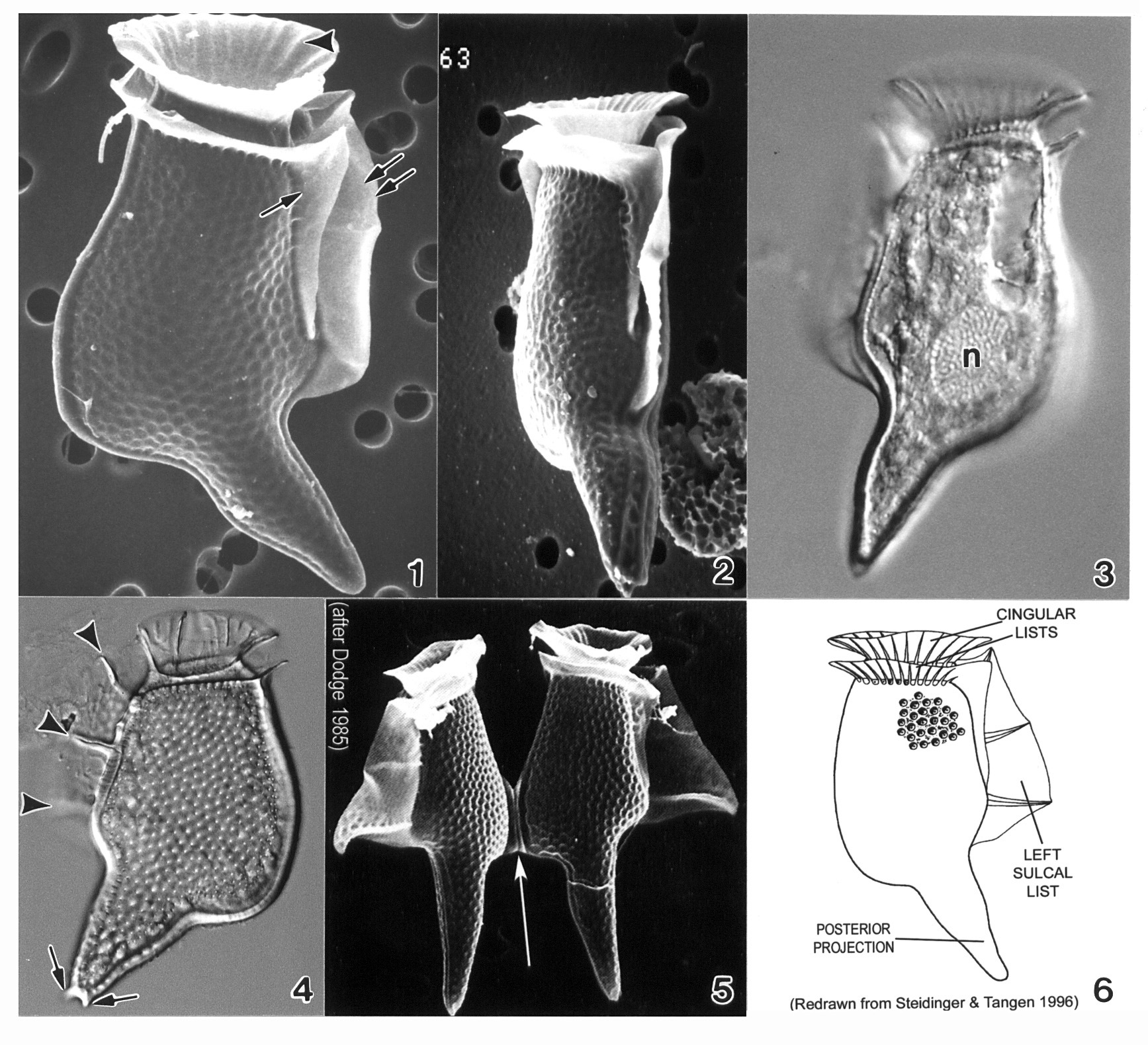
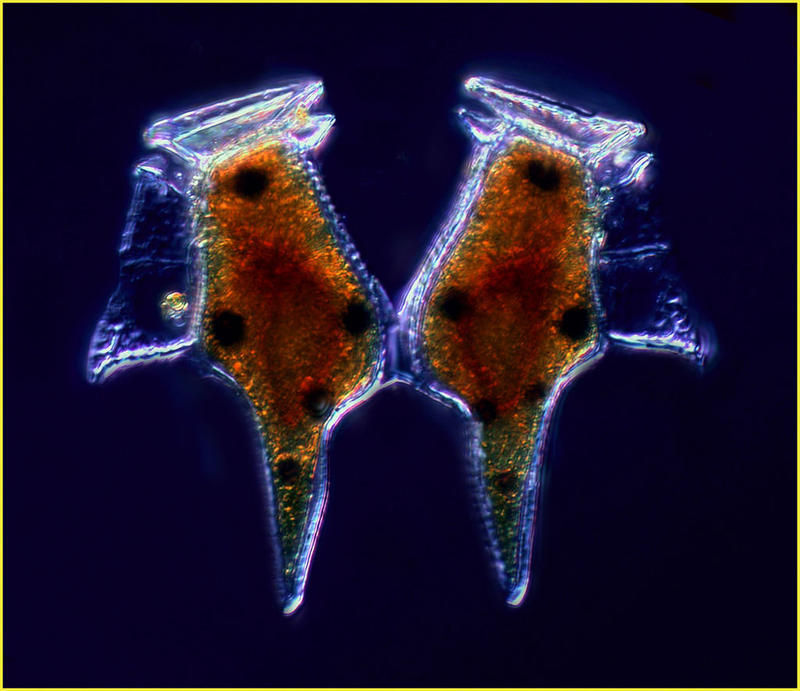
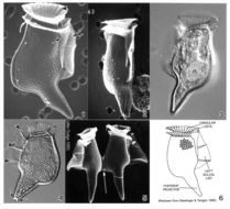
Plate 13. Dinophysis caudata. Figs. 1-2. SEM. Fig. 1. Large, long and distinctive cell with extended ventral hypothecal process. Cingulum narrow; lists supported by ribs (arrowhead). Strong left sulcal list (double arrows). Right sulcal list present (single arrow). Fig. 2. Ventral view: cell compressed laterally. Figs. 3-4. LM. Fig. 3. Large posterior nucleus (n). Fig. 4. Left sulcal list with three supporting ribs (arrowheads); posterior projection with small knob-like spines (arrows). Surface areolae evident. Fig. 5. SEM. Paired cells joined at dorsal expansion (arrow). Fig. 6. Line drawing.
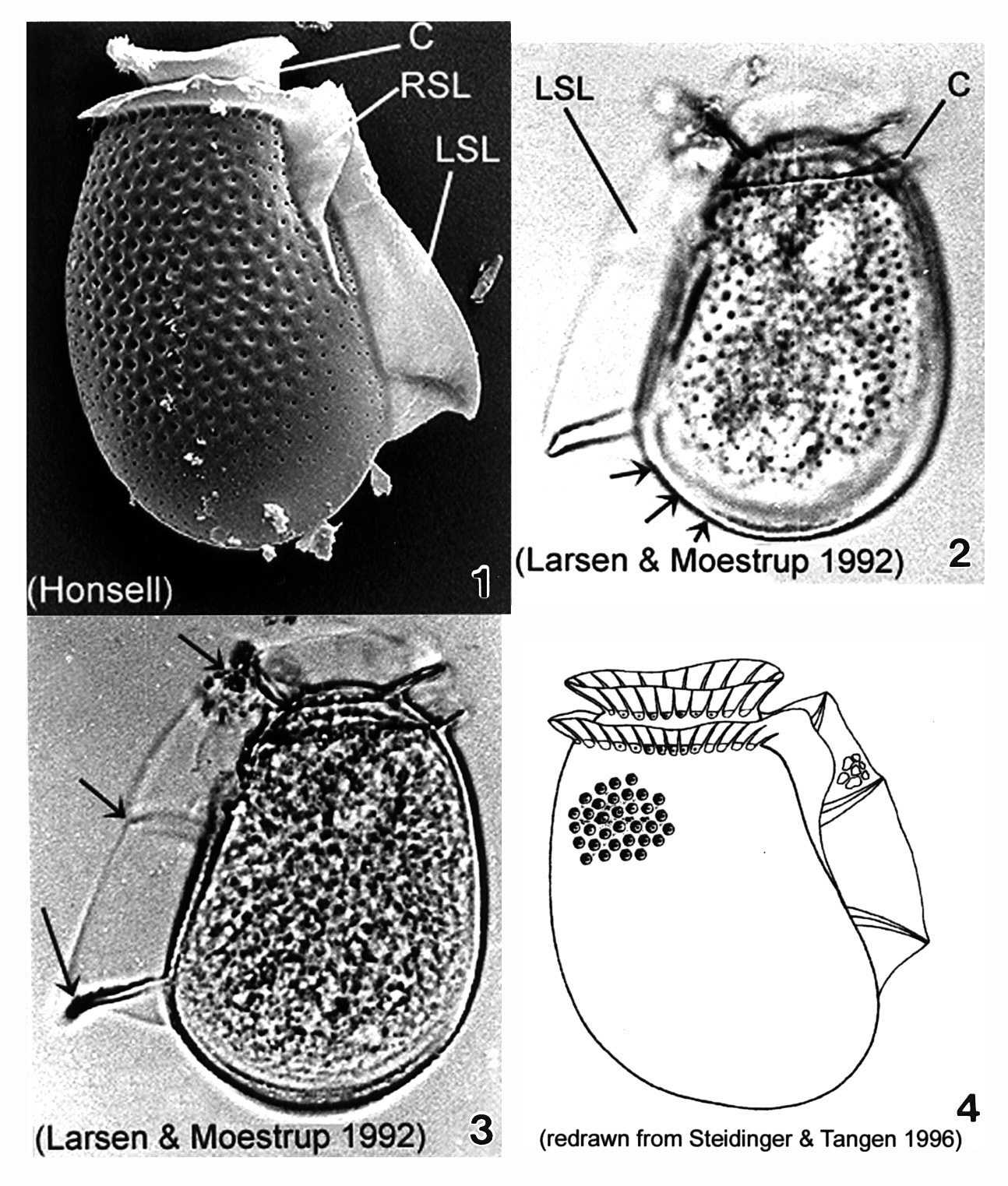
Plate 14. Dinophysis fortii. Fig. 1. SEM: lateral view. Left sulcul list (LSL) long and well-developed. Right sulcal list (RSL) present. Cingulum (C) obscures low and small epitheca. Thecal surface covered with areolae. Figs. 2-3. LM: lateral view. Fig. 2. Cell subovate with a wide round posterior bottom (dorsal bulge)(arrows). Fig. 3. LSL supported by three strong ribs (arrows). Smoothly convex dorsal margin. Fig. 4. Line drawing.
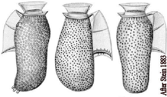
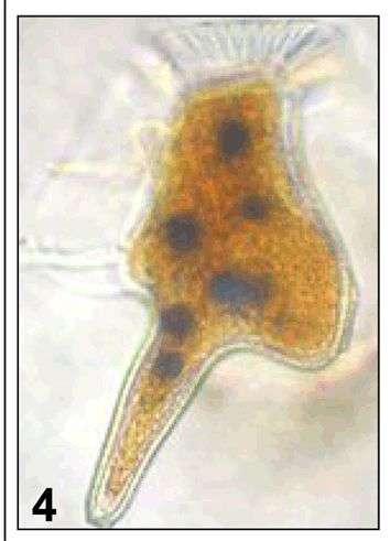
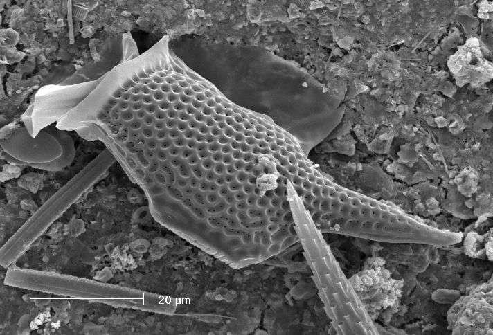
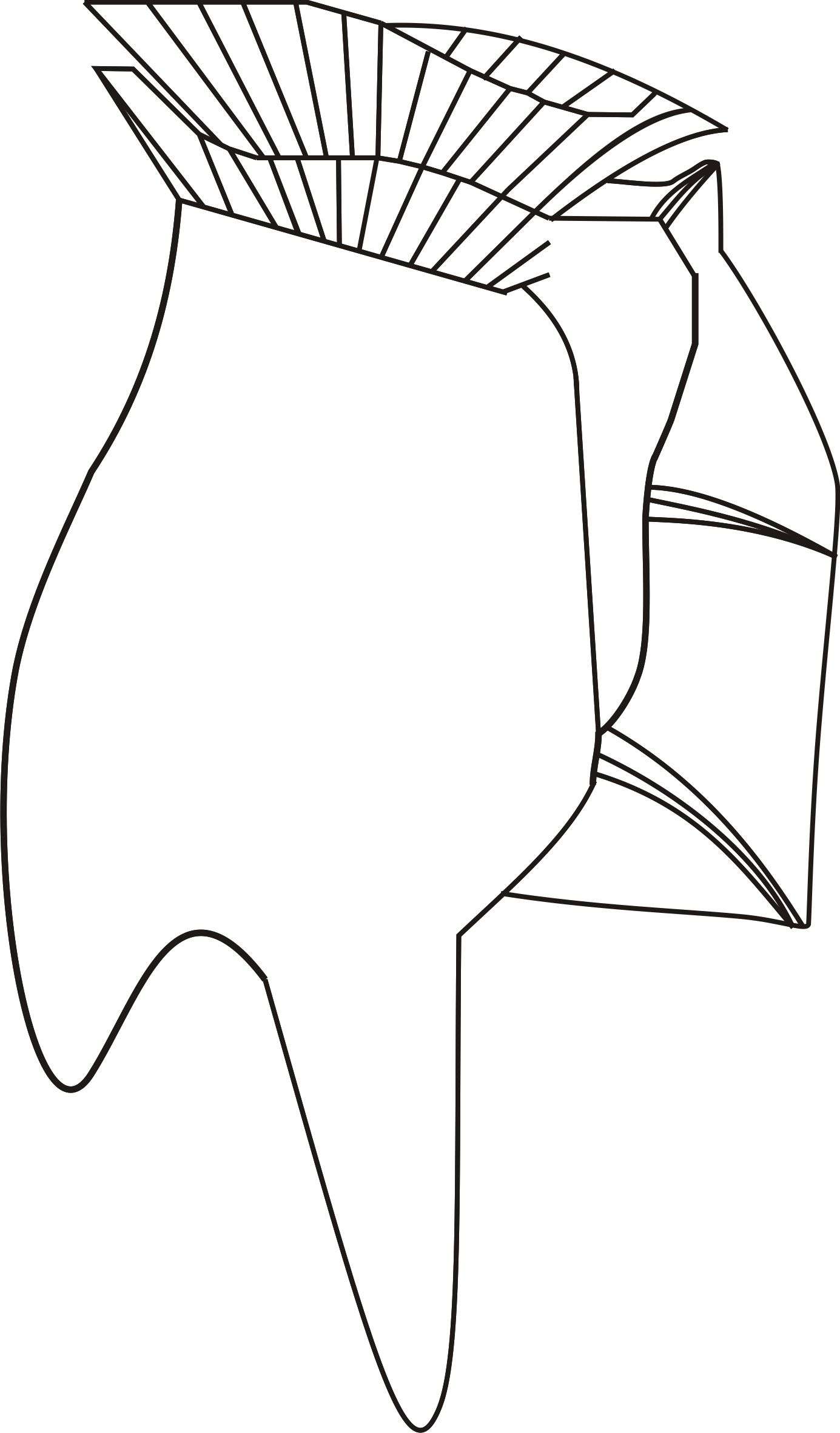
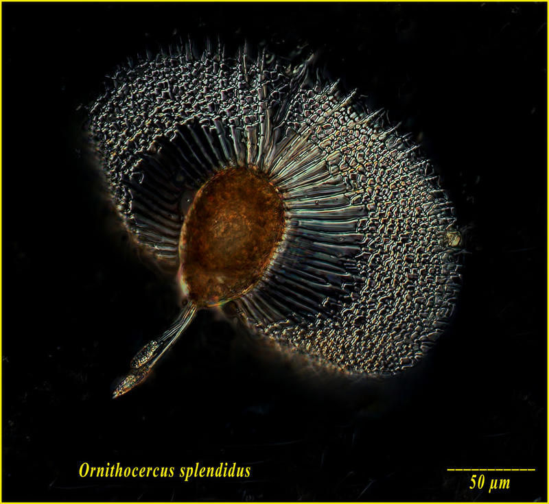
He thought it splendid. Figures from the original description of Ornitheroceras splendidus by Franz Schütt in 1893. The paper is available on the Classic Taxonomic Monographs page.