-
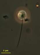
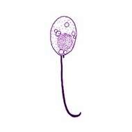
Small gliding flagellate with one long trailing flagellum and one short one. Phase contrast microscopy.
-
Kiitoksia kaloista Tong et al., 1997. Small spherical gliding cells with a near spherical body measuring 2- 4 microns in diameter. Two flagella insert about a third of the way up the cell and are directed backwards during gliding. One flagellum is about twice the cell length. The other is about half the cell length or shorter. The short flagellum is difficult to see using light microscopy and could possibly be overlooked. Gliding is smooth, with the cell and long flagellum applied to the substratum. Cells sometimes waggle slightly from side to side.
-
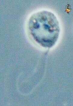
Kiitoksia (kee-tox-ee-a) is a very small gliding flagellate with a rounded cell body and a single long flagellum which trails behind the gliding cell. From marine habitats. Two species - Kiitoksia ystava and K. kaloista (which in Finnish means, - Thank you my old friend, and - Thank you for the fish. Phase contrast.
-
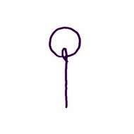
Kiitoksia ystava V+rs, 1992. Cell circular or slightly reniform, 2-3 microns A single flagellum, about twice cell length, inserts into a small dent to one side of the posterior surface. A fine hair-like portion is sometimes seen at the distal end of the flagellum. Cells glide rapidly and jerkily, with the cell nodding up and down from the surface. Seen occasionally in cultures, associated with detrital flocs.
-
-
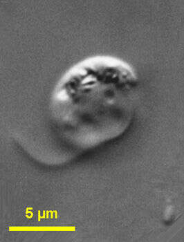
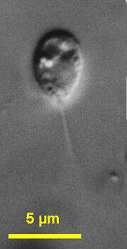
Metopion (met-ope-ee-on) fluens Larsen and Patterson, 1990. Cell outline is ovate. Cells are 4 to 9 microns long, laterally compressed and with a small rostrum anterior to the flagellar insertion. Small bodies are seen in the protrusion or at the proximal anterior part of the cell. Two flagella of unequal size emerge from a ventral groove located in the left side of the cell. The long flagellum is about 1.5 times cell length, is thickened and is not tapered at the tip, and the short flagellum may be difficult to see. There are small granules in the posterior part of the cell. The nucleus is situated near the groove. The cells move by gliding. Rarely observed.
-
Metopion fluens Larsen and Patterson, 1990. The cell outline is ovate. Cells are 4 to 9 microns long, laterally compressed and with a small rostrum anterior to the flagellar insertion. Small bodies are seen in the protrusion or at the proximal anterior part of the cell. Two flagella of unequal size emerge from a ventral groove located in the left side of the cell. The long flagellum is about 1.5 times cell length, is thickened and is not tapered at the tip, and the short flagellum may be difficult to see. There are small granules in the posterior part of the cell. The nucleus is situated near the groove. The cells move by gliding.
-
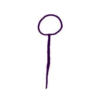
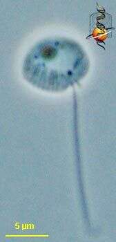
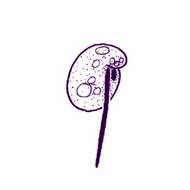
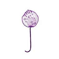
Metromonas (met-row-moan-ass) is a predator. Usually attached to the substrate by the curving posterior end of the longer flagellum (see, there is also a short one in most cells). They nod backwards and forwards. In cultures they usually appear in large numbers after the small bacterivorous flagellates - evidently preying upon them. Prey are ingested after encountering the margins of the cell. Unhappy cells tend to migrate by gliding along the substrate, with the flagellum no longer coiled but trailing behind the cells. Phase contrast
-
Metromonas (met-row-moan-ass) is a predator. Usually attached to the substrate by the curving posterior end of the longer flagellum (see, there is also a short one in most cells). They nod backwards and forwards. In cultures they usually appear in large numbers after the small bacterivorous flagellates - evidently preying upon them. Prey are ingested after encountering the margins of the cell. There are thin cylindrical structures near the margin - which we may assume to be associated with the capture of food. Unhappy cells tend to migrate by gliding along the substrate, with the flagellum no longer coiled but trailing behind the cells. Phase contrast
-
Metromonas (met-row-moan-ass) is a predator. Usually attached to the substrate by the curving posterior end of the longer flagellum (see, there is also a short one in most cells). They nod backwards and forwards. In cultures they usually appear in large numbers after the small bacterivorous flagellates - evidently preying upon them. Prey are ingested after encountering the margins of the cell. Unhappy cells tend to migrate by gliding along the substrate, with the flagellum no longer coiled but trailing behind the cells. Phase contrast
-
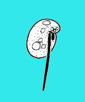
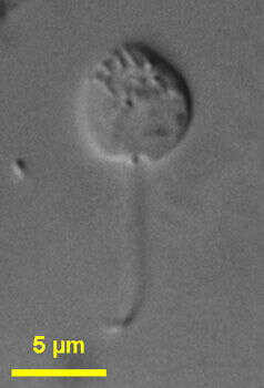
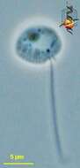
Metromonas (met-row-moan-ass) grandis Larsen and Patterson, 1990. Cell outline is leaf shaped or slightly roundish. Cells are 5 to 11 microns long (mostly 7 to 10 microns), 4 to 10 microns wide, about 2 microns deep and dorso-ventrally flattened. One side of the cell appears folded. The cells have two flagella, a long flagellum is 1.2 to 2.5 times the length of the cell and trails behind the cell when gliding. There is a short inactive flagellum, less than 2 microns long, which inserts to the right of the major flagellum and is always present. The cells attach to the substratum with the longer flagellum and move with a nodding action - like a pendulum. The nucleus is near the flagellar insertion. Relatively common.
-
Metromonas grandis Larsen and Patterson, 1990. Cell outline is leaf shaped or slightly roundish. Cells are 5 to 11 microns long (mostly 7 to 10 microns), 4 to 10 microns wide, about 2 microns deep and dorso-ventrally flattened. One side of the cell appears folded. The cells have two flagella, a long flagellum is 1.2 to 2.5 times the length of the cell and trails behind the cell when gliding. There is a short inactive flagellum, less than 2 microns long, which inserts to the right of the major flagellum and is always present. The cells attach to the substratum with the longer flagellum and move with a nodding action - like a pendulum. The nucleus is near the flagellar insertion.
-
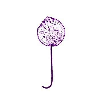
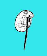
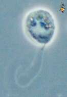
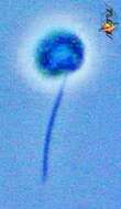
Metromonas (met-row-moan-ass) simplex (Griessmann, 1913) Larsen and Patterson, 1990. Cells are obovate, 3 to 8 microns long (mostly 4 to 7 microns), 2 to 6 microns wide and dorso-ventrally flattened, and have smooth pellicle. The abflagellar margin of the cell is thicker than the (posterior) margin. Two flagella of very unequal length arise from the posterior part of the cell. The major flagellum is always present, is about 1.5 to 3.0 times the length of the cell and may be attached to the substrate. The short inactive flagellum is about 1 microns long and inserts to the right of the major flagellum. It may be difficult to see. The cells normally attach to the substratum and swing from side to side like a pendulum and the cells may also glide with the cell body in front of the flagellum. More common than M. grandis.
-
Metromonas simplex (Griessmann, 1913) Larsen and Patterson, 1990. Cells are obovate, 3 to 8 microns long (mostly 4 to 7 microns), 2 to 6 microns wide and dorso-ventrally flattened, and have smooth pellicle. The margin of the cell that is away from the flagella is thicker than the (posterior) margin. Two flagella of very unequal length arise from the posterior part of the cell. The major flagellum is always present, is about 1.5 to 3.0 times the length of the cell and may be attached to the substrate. The short inactive flagellum is about 1 microns long and inserts to the right of the major flagellum. It may be difficult to see. The cells normally attach to the substratum and swing from side to side like a pendulum and the cells may also glide with the cell body in front of the flagellum.
-
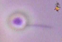
This is a long exposure phase contrast micrograph of Metromonas simplex in situ. This is one of my all time favourites as it so effectively captures the movements of this distinctive predator. Attaching by the crook of the long flagellum, the cell swings actively in an arc - increasing the probability of encountering another protist - its food.