-
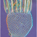
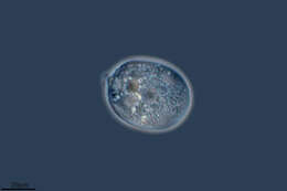
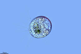
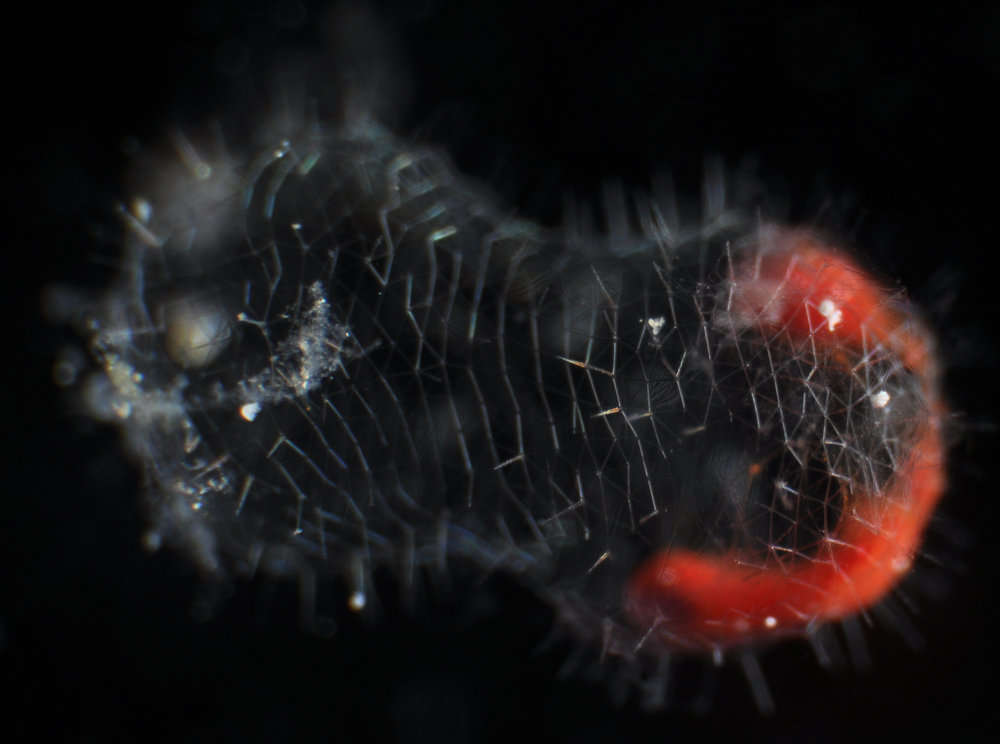
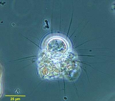
Protocystis found wearing a crown of diatoms. Sample from the Amundsen Sea.
-
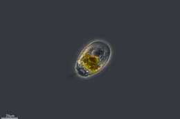
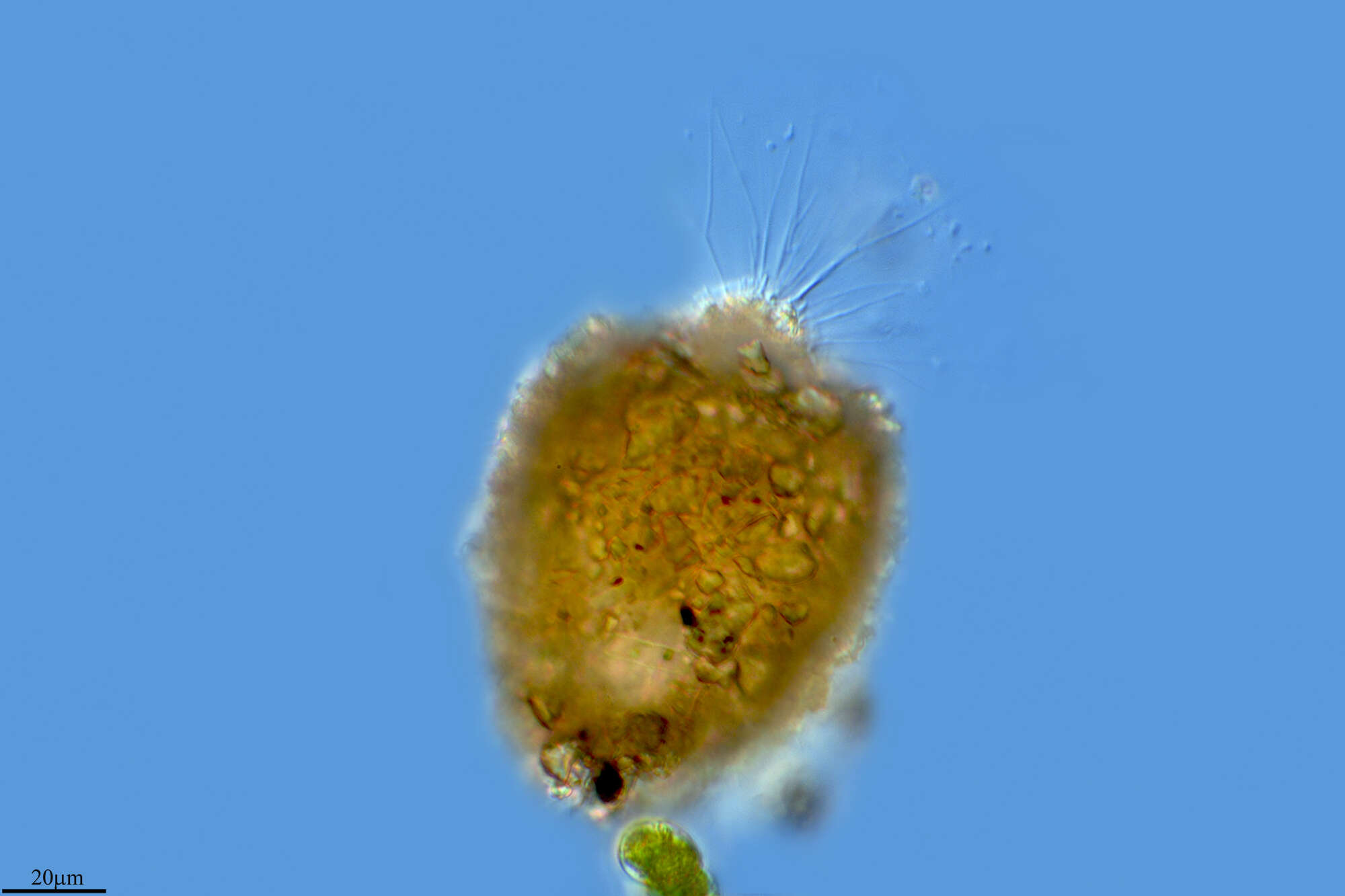
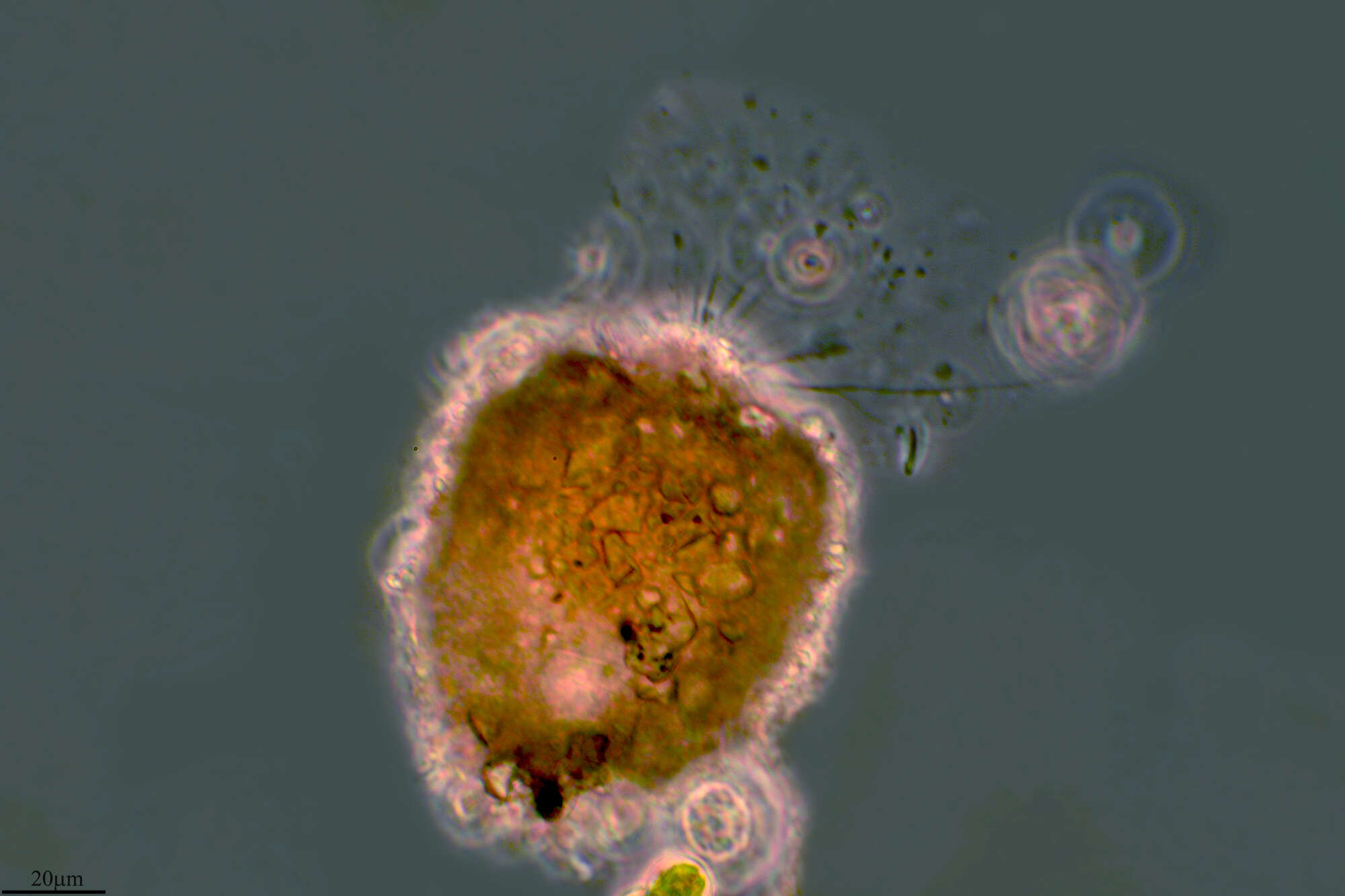
A specimen from Villefranche in Feb 2012 showing what appears to be a feeding tube. A short video is on the Aquaparadox video page.
-
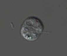
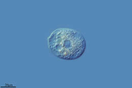
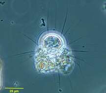
A radiolarian, Protocystis xiphodon, showing a what looks like a feeding tube.
-
All Biocode files are based on field identifications to the best of the researcher’s ability at the time.
-
Summary.mw-parser-output table.commons-file-information-table,.mw-parser-output.fileinfotpl-type-information{border:1px solid #a2a9b1;background-color:#f8f9fa;padding:5px;font-size:95%;border-spacing:2px;box-sizing:border-box;margin:0;width:100%}.mw-parser-output table.commons-file-information-table>tbody>tr,.mw-parser-output.fileinfotpl-type-information>tbody>tr{vertical-align:top}.mw-parser-output table.commons-file-information-table>tbody>tr>td,.mw-parser-output table.commons-file-information-table>tbody>tr>th,.mw-parser-output.fileinfotpl-type-information>tbody>tr>td,.mw-parser-output.fileinfotpl-type-information>tbody>tr>th{padding:4px}.mw-parser-output.fileinfo-paramfield{background:#ccf;text-align:right;padding-right:0.4em;width:15%;font-weight:bold}.mw-parser-output.commons-file-information-table+table.commons-file-information-table,.mw-parser-output.commons-file-information-table+div.commons-file-information-table>table{border-top:0;padding-top:0;margin-top:-8px}@media only screen and (max-width:719px){.mw-parser-output table.commons-file-information-table,.mw-parser-output.commons-file-information-table.fileinfotpl-type-information{border-spacing:0;padding:0;word-break:break-word;width:100%!important}.mw-parser-output.commons-file-information-table>tbody,.mw-parser-output.fileinfotpl-type-information>tbody{display:block}.mw-parser-output.commons-file-information-table>tbody>tr>td,.mw-parser-output.commons-file-information-table>tbody>tr>th,.mw-parser-output.fileinfotpl-type-information>tbody>tr>td,.mw-parser-output.fileinfotpl-type-information>tbody>tr>th{padding:0.2em 0.4em;text-align:left;text-align:start}.mw-parser-output.commons-file-information-table>tbody>tr,.mw-parser-output.fileinfotpl-type-information>tbody>tr{display:flex;flex-direction:column}.mw-parser-output.commons-file-information-table+table.commons-file-information-table,.mw-parser-output.commons-file-information-table+div.commons-file-information-table>table{margin-top:-1px}.mw-parser-output.fileinfo-paramfield{box-sizing:border-box;flex:1 0 100%;width:100%}} Description: English: Rhogostoma minus (dorsal view) with visible cleft-like aperture and extended pseudopodium. Date: 3 April 2023. Source: Own work. Author:
PicoProtist.
-
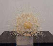
Description: Glasmodell von
Aulosphaera elegantissima von Leopold und Rudolf Blaschka um 1885 aus der Zoologischen Sammlung Tübingen in der Ausstellung "Wie Schönes Wissen schafft", 19. April bis 1. September 2013 im Museum der Universität Tübingen (MUT). Date: May 2013. Source:
Wie Schönes Wissen schafft im MUT. Author: NearEMPTiness.
-
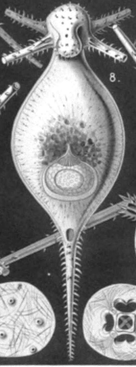
Summary.mw-parser-output table.commons-file-information-table,.mw-parser-output.fileinfotpl-type-information{border:1px solid #a2a9b1;background-color:#f8f9fa;padding:5px;font-size:95%;border-spacing:2px;box-sizing:border-box;margin:0;width:100%}.mw-parser-output table.commons-file-information-table>tbody>tr,.mw-parser-output.fileinfotpl-type-information>tbody>tr{vertical-align:top}.mw-parser-output table.commons-file-information-table>tbody>tr>td,.mw-parser-output table.commons-file-information-table>tbody>tr>th,.mw-parser-output.fileinfotpl-type-information>tbody>tr>td,.mw-parser-output.fileinfotpl-type-information>tbody>tr>th{padding:4px}.mw-parser-output.fileinfo-paramfield{background:#ccf;text-align:right;padding-right:0.4em;width:15%;font-weight:bold}.mw-parser-output.commons-file-information-table+table.commons-file-information-table,.mw-parser-output.commons-file-information-table+div.commons-file-information-table>table{border-top:0;padding-top:0;margin-top:-8px}@media only screen and (max-width:719px){.mw-parser-output table.commons-file-information-table,.mw-parser-output.commons-file-information-table.fileinfotpl-type-information{border-spacing:0;padding:0;word-break:break-word;width:100%!important}.mw-parser-output.commons-file-information-table>tbody,.mw-parser-output.fileinfotpl-type-information>tbody{display:block}.mw-parser-output.commons-file-information-table>tbody>tr>td,.mw-parser-output.commons-file-information-table>tbody>tr>th,.mw-parser-output.fileinfotpl-type-information>tbody>tr>td,.mw-parser-output.fileinfotpl-type-information>tbody>tr>th{padding:0.2em 0.4em;text-align:left;text-align:start}.mw-parser-output.commons-file-information-table>tbody>tr,.mw-parser-output.fileinfotpl-type-information>tbody>tr{display:flex;flex-direction:column}.mw-parser-output.commons-file-information-table+table.commons-file-information-table,.mw-parser-output.commons-file-information-table+div.commons-file-information-table>table{margin-top:-1px}.mw-parser-output.fileinfo-paramfield{box-sizing:border-box;flex:1 0 100%;width:100%}} Description: English: Illustration from Report on the Radiolaria collected by H.M.S. Challenger during the years 1873-1876. Part III. Fig. 8. Tuscaridium lithornithium, n. sp., × 20 View from the ventral side. Central capsule and calymma as in fig. 2. Fig. 8a. Peristome from the ventral side. Fig. 8b. Peristome from the right side. Date: 1887. Source:
https://archive.org/details/reportonradiolar00haecrich. Author: Ernst Haeckel (1834-1919); engravings by Adolf Giltsch (1852-1911).
-
Description: Ebria tripartita (Schumann) Lemmermann 1899 (Dictyocha tripartita Schumann 1867); Ebridea English: North-West
Black Sea, surface water Русский: Северо-Запад
Чёрного моря, поверхностные воды. Date: 19 March 2008. Source: Own work. Author:
Minami Himemiya.
-
Lumbreras, La Rioja, Spain
-
Rancho de la Herradura, Andalusia, Spain
-
Lumbreras, La Rioja, Espaa
-
Soba, Cantabria, Spain
-
Soba, Cantabria, Spain
-
Talveila, Castille and Leon, Spain
-
Talveila, Castille and Leon, Spain
-
Puras de Villafranca, Castille and Leon, Spain
-
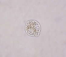
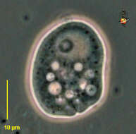
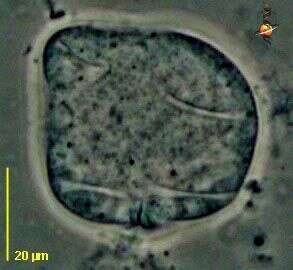
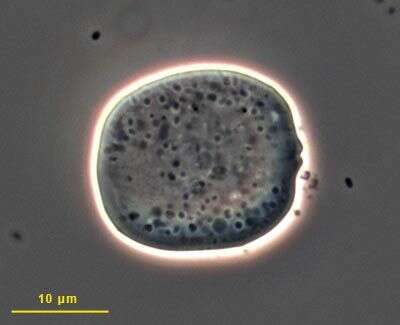
Lecythium is a shelled amoeba, with an organic shell. This phase contrast image is of the pseudopodia as they spread across the microscope slide. From a freshwater pond in Idaho.
-
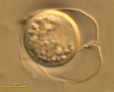
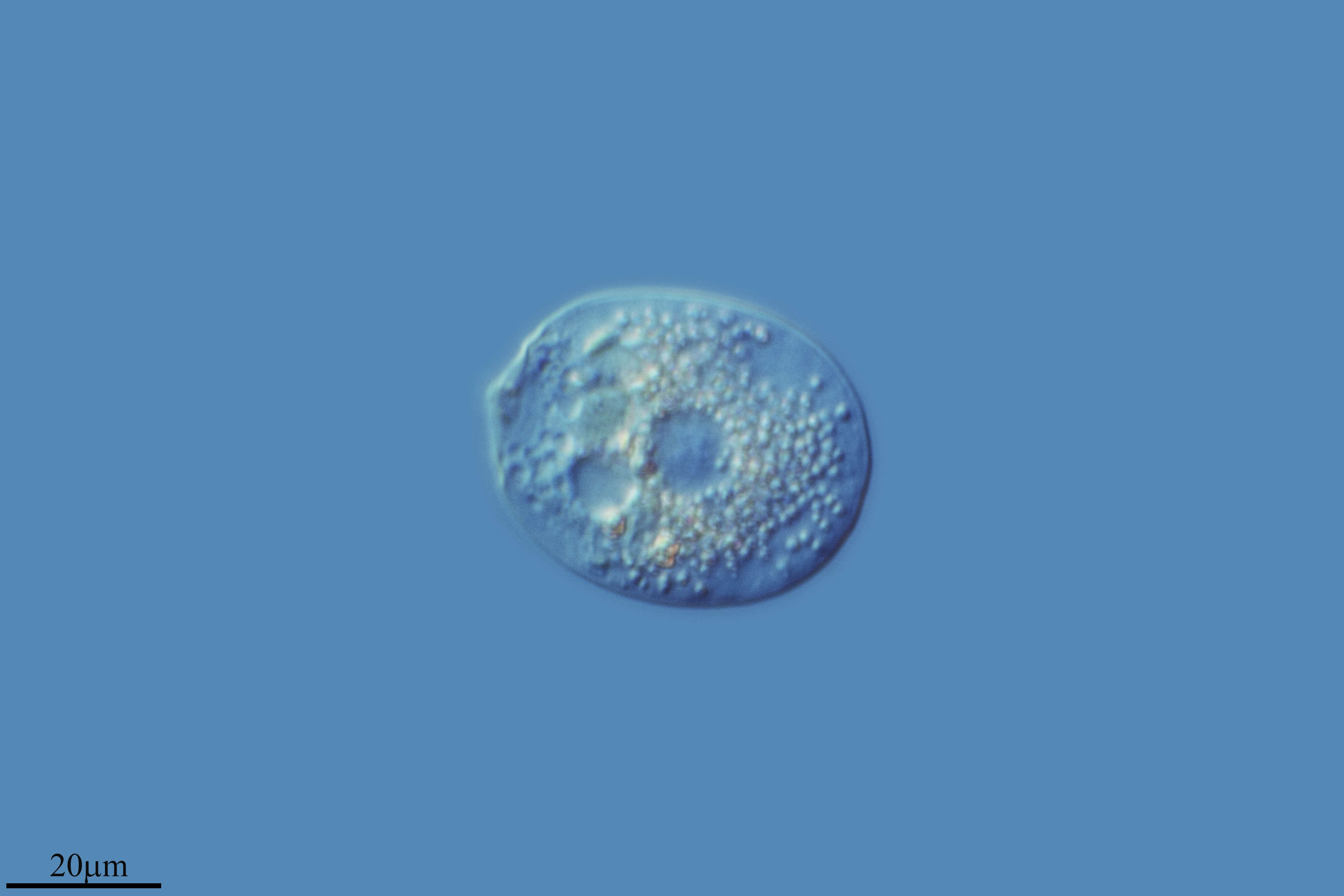
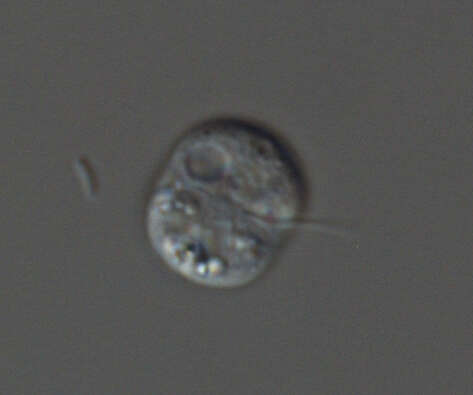
Rhogostoma (row-gaw-stow-ma) is a rarely reported amoeba with a flexible surrounding test and with a small opening through which pseudopodia can emerge. The opening is at the bottom of the cell and looks as if it has two lips. Large nucleus with nucleolus at the other side of the cell. Eats bacteria. Phase contrast.
-
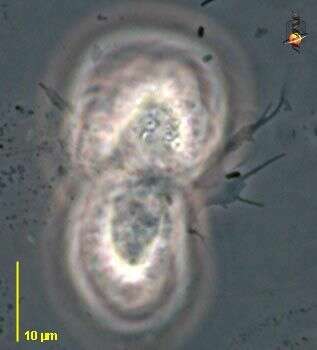
Rhogostoma (row-gaw-stow-ma) is a rarely reported amoeba with a flexible surrounding test and with a small opening through which pseudopodia can emerge. Two cells with common cytoplasm, showing the thin nature of the pseudopodia. Phase contrast.
-
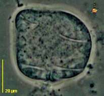
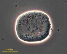
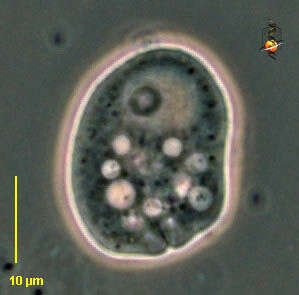
Rhogostoma (row-gaw-stow-ma) is a rarely reported amoeba with a flexible surrounding test and with a small opening through which pseudopodia can emerge. The opening is at the bottom of the cell and looks as if it has two lips. Folds in the test are evident. Phase contrast.
-
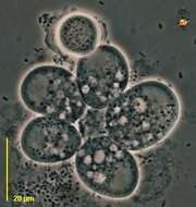
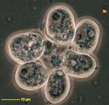
Rhogostoma (row-gaw-stow-ma) is a rarely reported amoeba with a flexible surrounding test and with a small opening through which pseudopodia can emerge. Cluster of cells with common cytoplasm, at the centre of the cluster. With one cyst. Phase contrast.
-
Rhogostoma (row-gaw-stow-ma) is a rarely reported amoeba with a flexible surrounding test and with a small opening through which pseudopodia can emerge. Cluster of cells with common cytoplasm, at the centre of the cluster. Phase contrast.
-
-