-
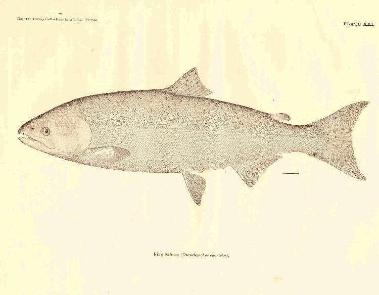
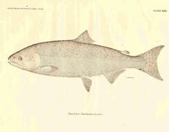
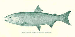
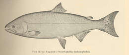
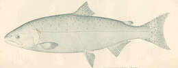
King Salmon (Oncorhynchus chouicha).
-
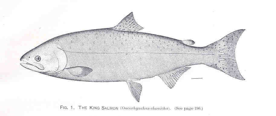
King Salmon (Oncorhynchus chouicha).
-
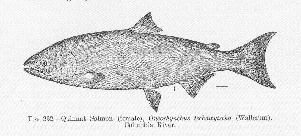
Quinnat Salmon (female), Oncorhynchus tschawytscha (Walbaum). Columbia River.
-
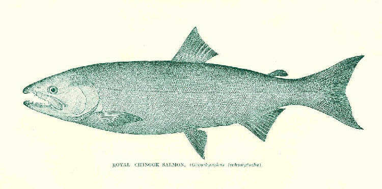
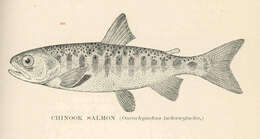
Royal Chinook Salmon. (Oncorhynchus tschawytscha)
-
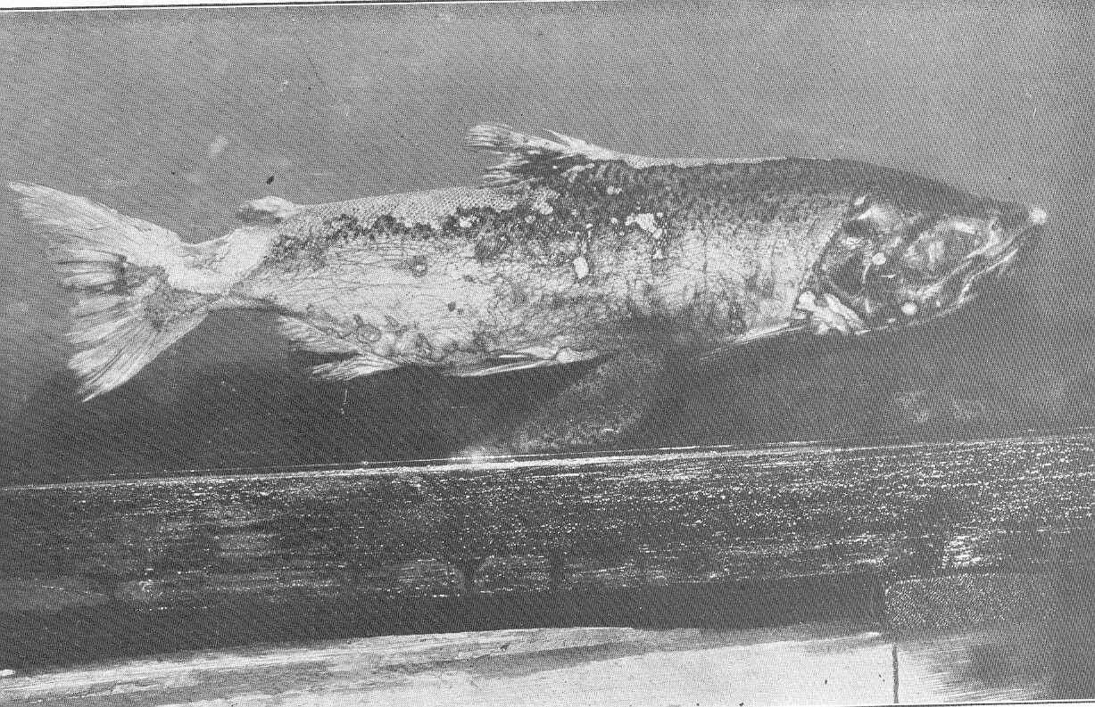
Young Male Quinnat Salmon, Oncorhynchus tschawytscha, dying after spawning. Sacramento River.
-
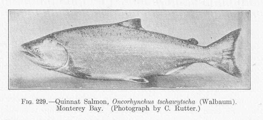
Quinnat Salmon, Oncorhynchus tschawytscha (Walbaum). Monterey Bay.
-
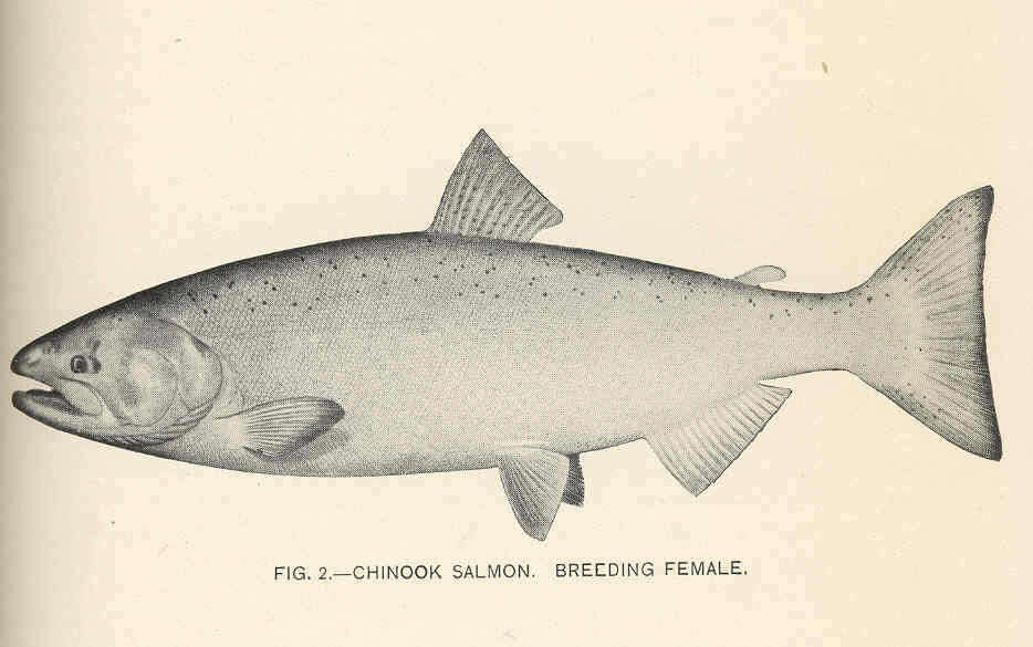
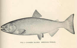
Chinook Salmon, Breeding Female.
-
Chinook Salmon, Artificially Raised, Marked by Removal of Fin Material.
-
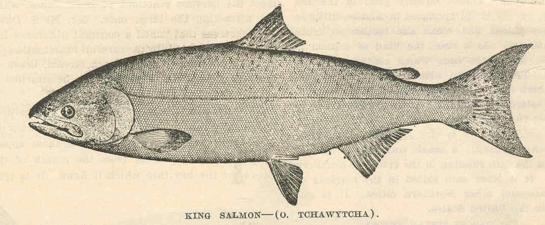
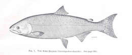
King Salmjon--(O. Tchawytcha).
-
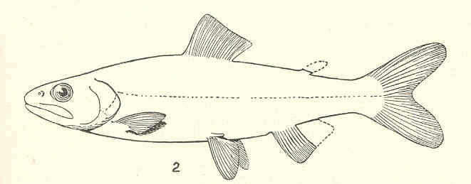
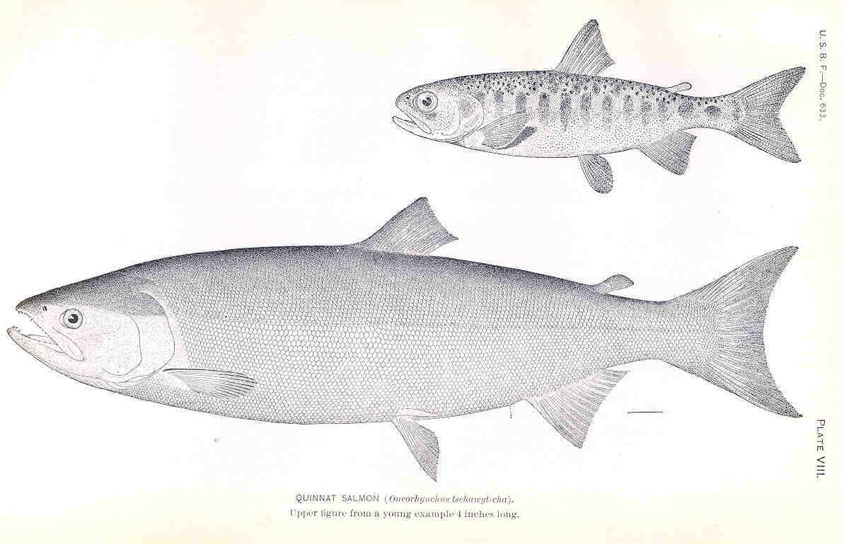
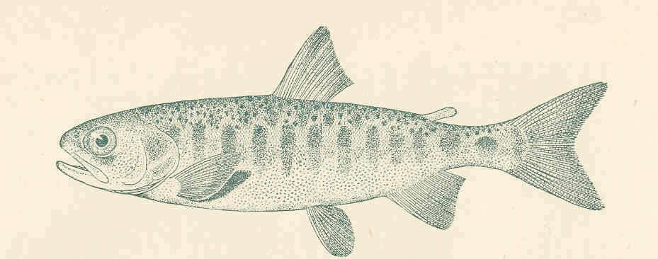
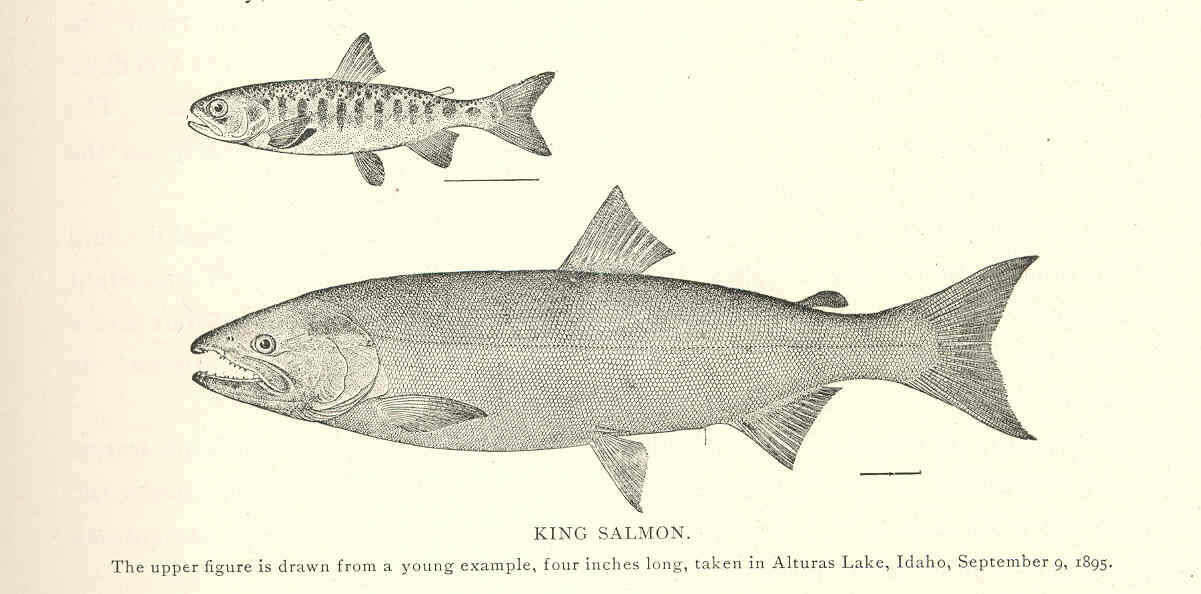
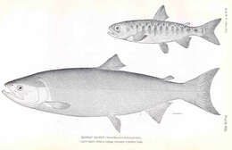
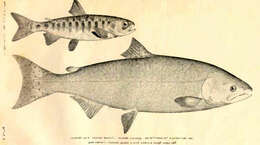
Quinnat Salmon (Oncorhynchus tschawyt-cha). Upper figure from a young example 4 inches long
-
Oncorhynchus Tschawytscha. Quinnat Salmon; Chinook Salmon; King Salmon.. The upper figure is drawn from a young example, 4 inches long.
-
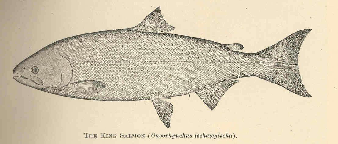
King Salmon (Oncorhynchus tschawytscha).
-
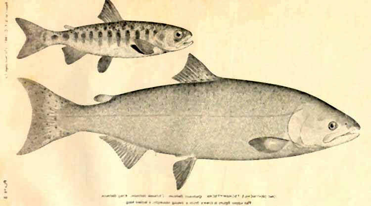
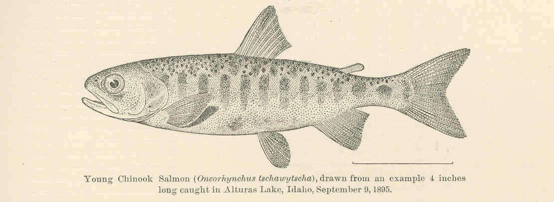
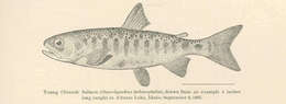
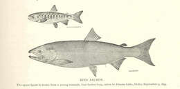
Young Chinook Salmon (Oncorhynchus tschawytscha), drawn form an example 4 inches long caught in Alturas Lake, Idaho, September 9, 1895.
-
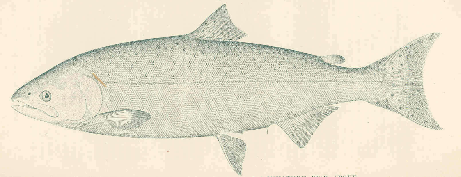
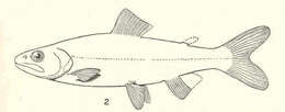
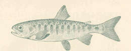
Chinook Salmon (Oncorhynchus tschawytscha) : Immature fish.
-
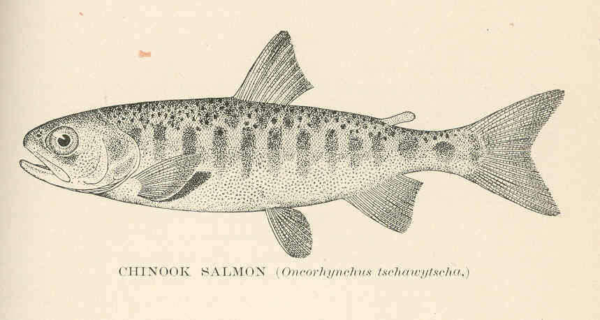
Chinook Salmon (Oncorhynchus tschawytscha).
-
Chinook Salmon (Oncorhynchus tschawytscyha).
-
King Salmon (Oncorhynchus tshasytscha Walbaum). Introduced. The upper figure is drawn from a young example, four inches long, taken in Alturas Lake, Idaho, September 9, 1895.
-
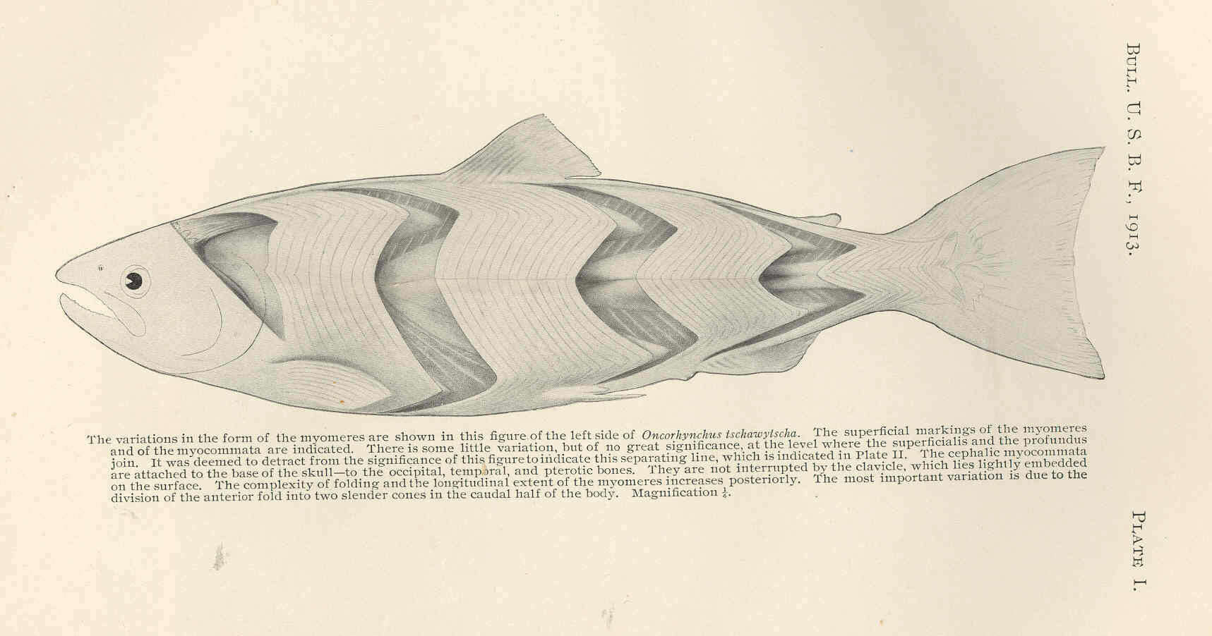
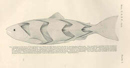
King Salmon: Variotions in the form of the myomeres are shown in this figure of the left side on Oncorhynchus tschawytscha. The superficial markings of the myomeres and of the myocommata are indicated. There is some little variation, but of no great significance, at the level where the superficialis and the profundus join. It was deemed to detract from the significance of this figure to indicate this separating line.... The cephalic myocommata are attached to the base of the skull--to the occipital, temporal and pterotic bones. They are not interrupted by the clavicle, which lies lightly embedded on the surface. The complexity of folding and the lingitudinal extent of the myomeres increases posteriorly. The most important variation is due to the division of the anterior fold into two slender cones in the caudal half of the body.