King Salmon: Variotions in the form of the myomeres are shown in this figure of the left side on Oncorhynchus tschawytscha. The superficial markings of the myomeres and of the myocommata are indicated. There is some little variation, but of no great signi
Description:
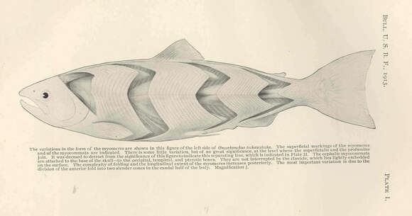
King Salmon: Variotions in the form of the myomeres are shown in this figure of the left side on Oncorhynchus tschawytscha. The superficial markings of the myomeres and of the myocommata are indicated. There is some little variation, but of no great significance, at the level where the superficialis and the profundus join. It was deemed to detract from the significance of this figure to indicate this separating line.... The cephalic myocommata are attached to the base of the skull--to the occipital, temporal and pterotic bones. They are not interrupted by the clavicle, which lies lightly embedded on the surface. The complexity of folding and the lingitudinal extent of the myomeres increases posteriorly. The most important variation is due to the division of the anterior fold into two slender cones in the caudal half of the body.
Included On The Following Pages:
- Life (creatures)
- Cellular (cellular organisms)
- Eukaryota (eukaryotes)
- Opisthokonta (opisthokonts)
- Metazoa (Animal)
- Bilateria
- Deuterostomia (deuterostomes)
- Chordata (Chordates)
- Vertebrata (vertebrates)
- Gnathostomata (jawed fish)
- Osteichthyes
- Actinopterygii (ray-finned fishes)
- Neopterygii
- Teleostei
- Euteleostei
- Protacanthopterygii
- Salmoniformes
- Salmonidae (trouts and salmons)
- Oncorhynchus (Salmon)
- Oncorhynchus tshawytscha (Chinook Salmon)
This image is not featured in any collections.
Source Information
- license
- cc-publicdomain
- publisher
- Freshwater and Marine Image Bank, University of Washington Libraries Digital Collections
- provider
- Freshwater and Marine Image Bank
- original
- original media file
- visit source
- partner site
- Freshwater and Marine Image Bank U Washington
- ID