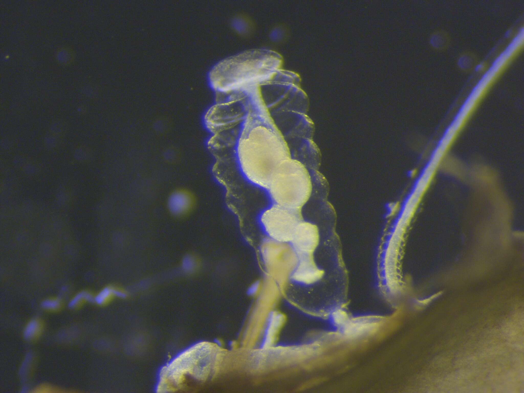
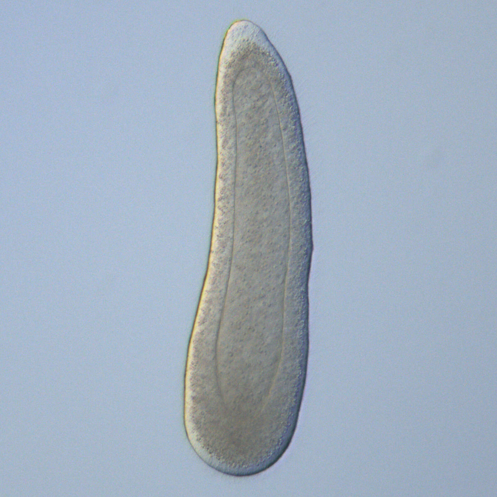
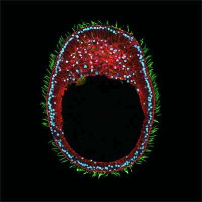
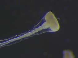
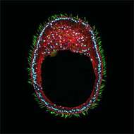
Description: English: DIC (Normalski) observation of Clytia hemisphaerica 2day old planula larvae (2 days after fertilisation). The embryo consists of two layers, notably epidermis (ectoderm) and gastrodermis (endoderm). Date: 22 April 2021, 20:27:31. Source: Own work. Author: Tsuyoshi Momose.
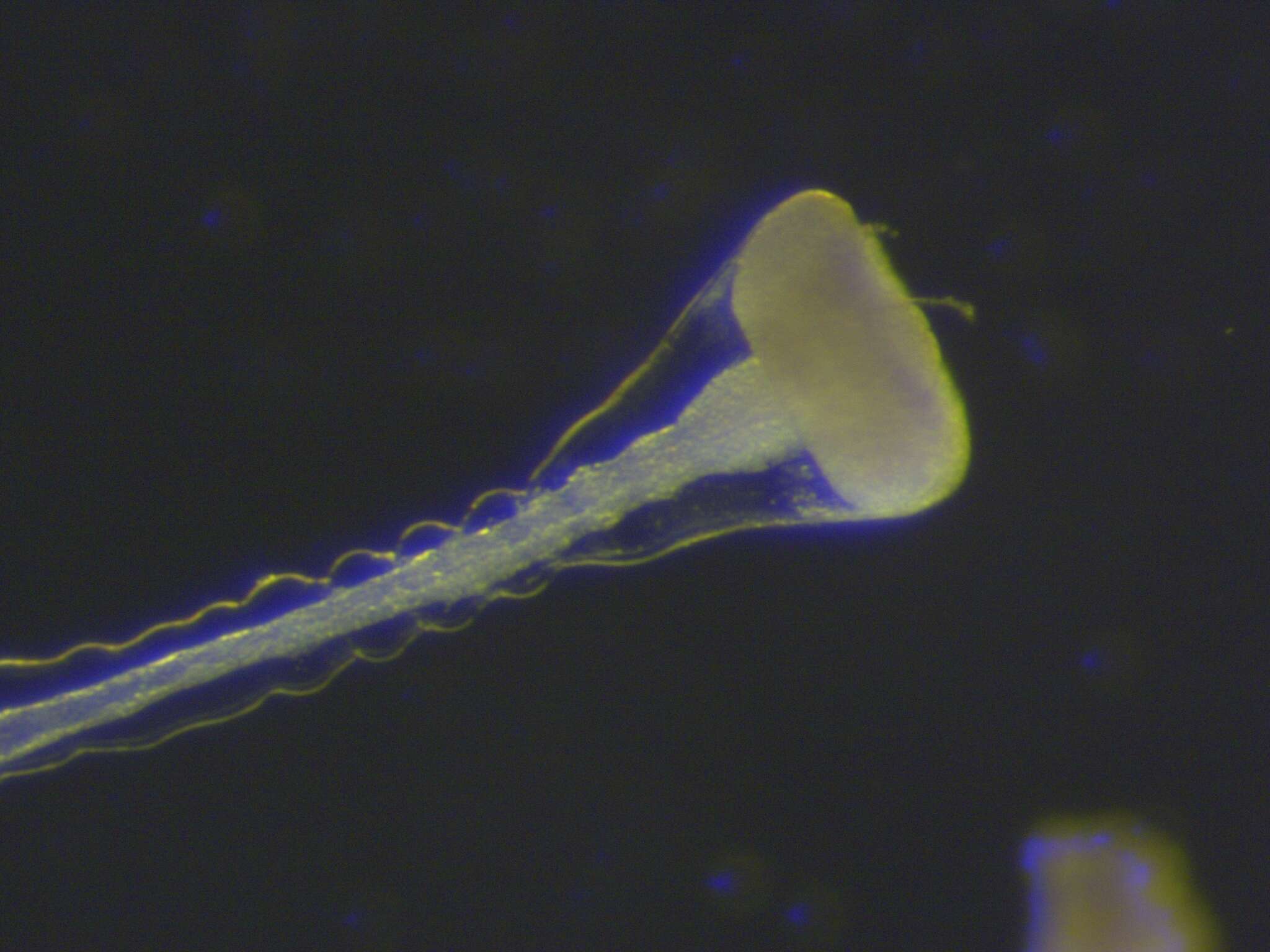
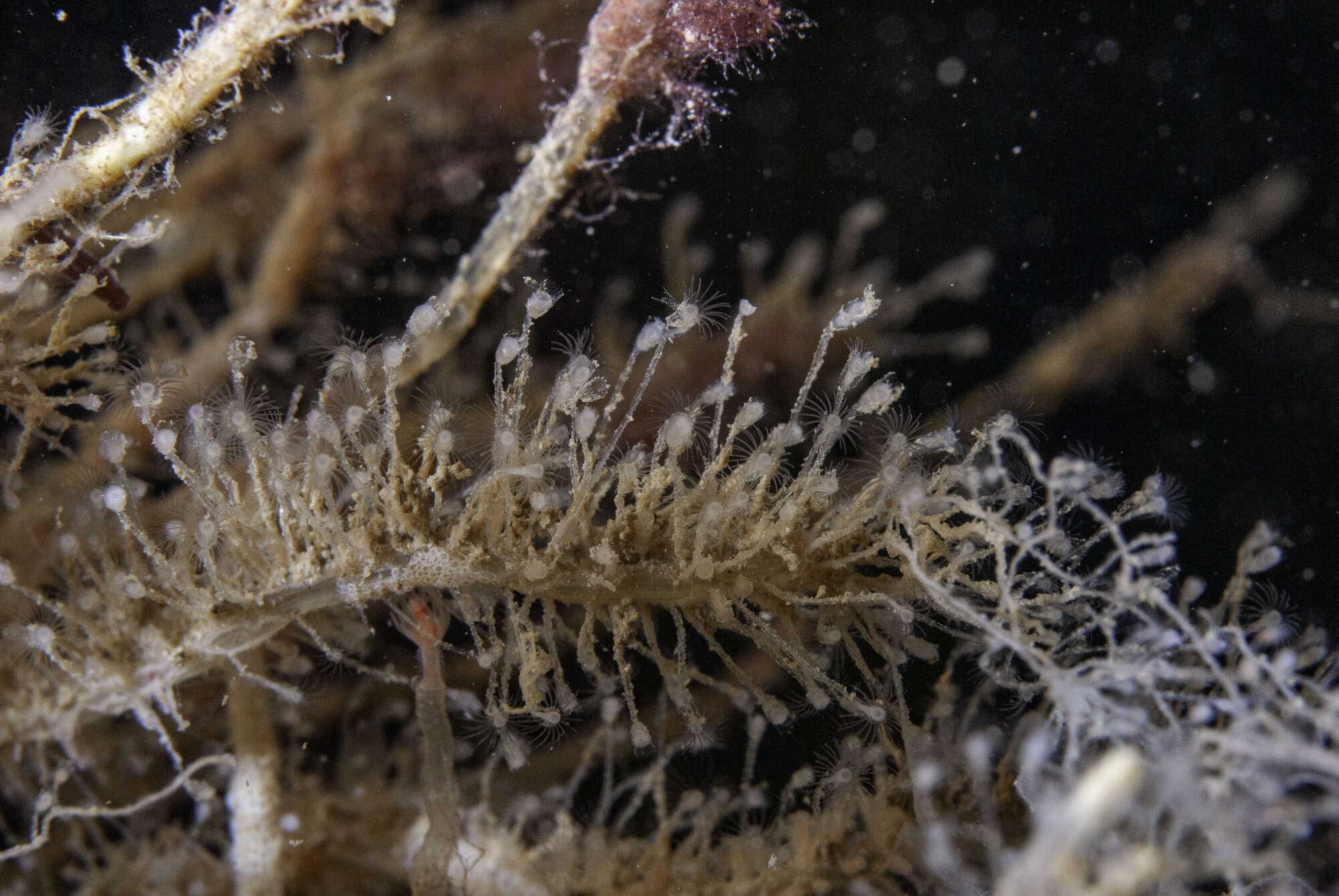
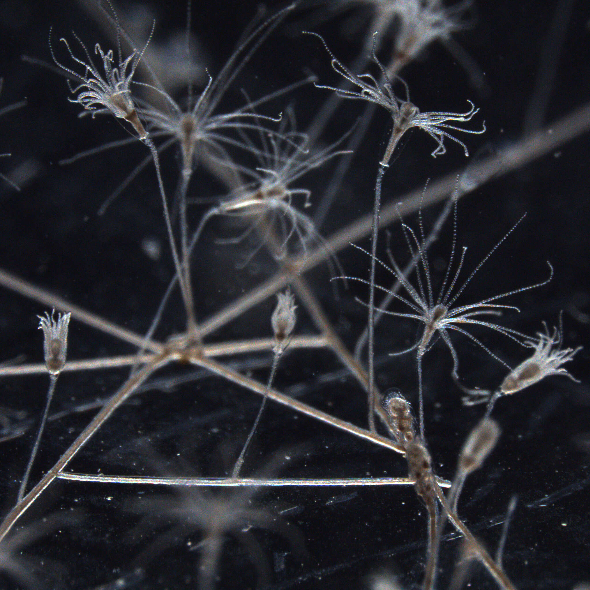
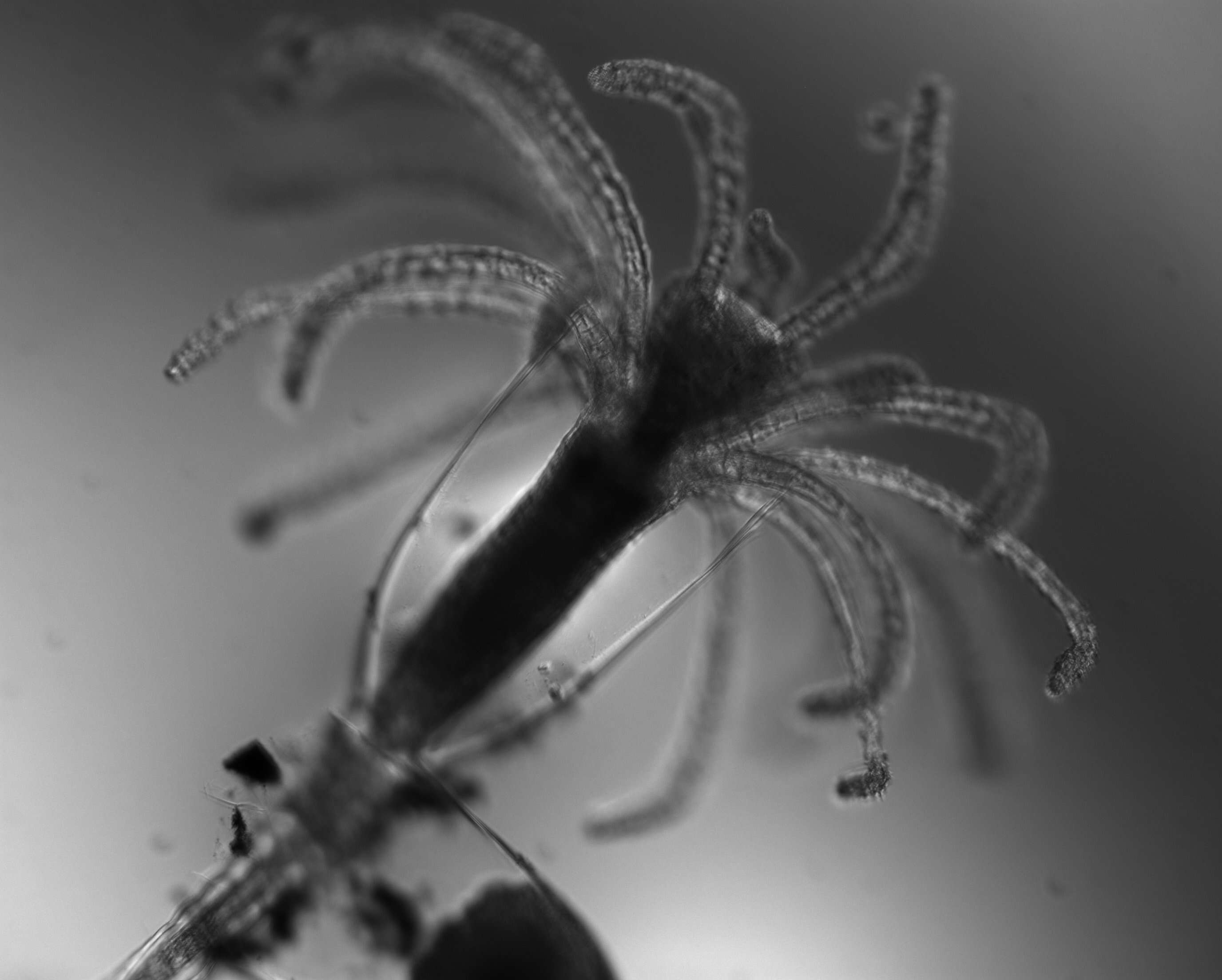
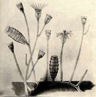
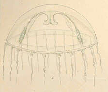
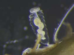
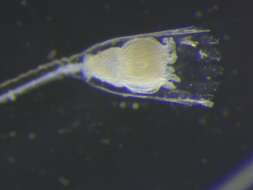
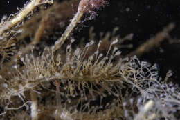
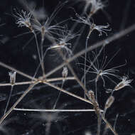
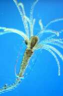
Description: English: Clytia hemisphaerica polyp grows asexually (vegitatively) by extending stolons on the solid surface, and makes many clonal polyps. The colony will form specialised type of polyp called gonozooid (visible boom right of the picture), if it is fed enough. The gonozooid forms medusa buds on it. Juvenile medusa (jellyfish) then detach and grow to mature jellyfish in a few weeks. Date: 23 April 2021, 19:35:31. Source: Own work. Author: Tsuyoshi Momose.
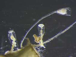
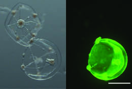
Description: English: Wildtype (Z23 strain, bottom) and GFP1/GFP2 mutant jellyfish (top) 1 day after being spawned from polyp colonies. Bright field image to to left. Fluorescent observation to the right. Mutant jellyfish does not exhibit any endogenous green fluorescence. See https://doi.org/10.1038/s41598-018-30188-0. Date: 30 April 2021. Source: Own work. Author: Tsuyoshi Momose.
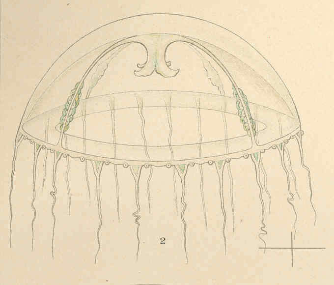
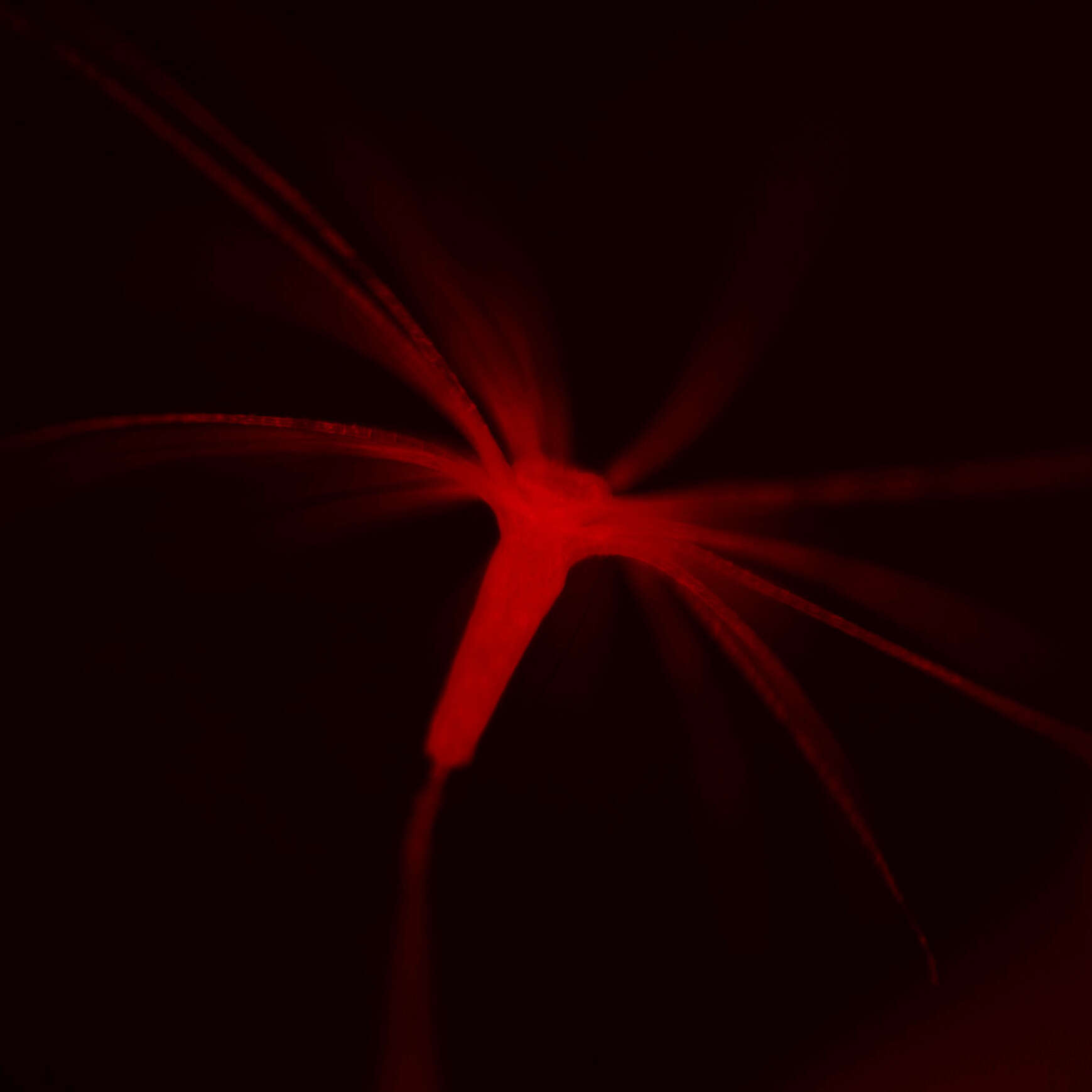
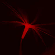
Description: English: A jellyfish Clytia hemisphaerica is a developmental and evolutionary biology model animal. Having fully transparent body, it is also an ideal model animal for live imaging of cell behaviour and neuronal activity. For this objective, we developed a transgenesis method using a transposon vector Tol2, which efficiently and stably introduce exogenous DNA into Clytia chromosome. Once ncorporated to the host genome, the transgenes can be transmitted to the offsprings allowing genetics studies. The image is red fluorescent protein mCherry signal ubiquitously expressed in the polyp stage of Clytia (F1 generation). Date: 24 November 2021. Source: Own work. Author: Tsuyoshi Momose.


