-
All Biocode files are based on field identifications to the best of the researcher’s ability at the time.
-
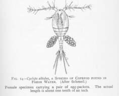
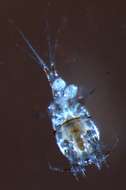
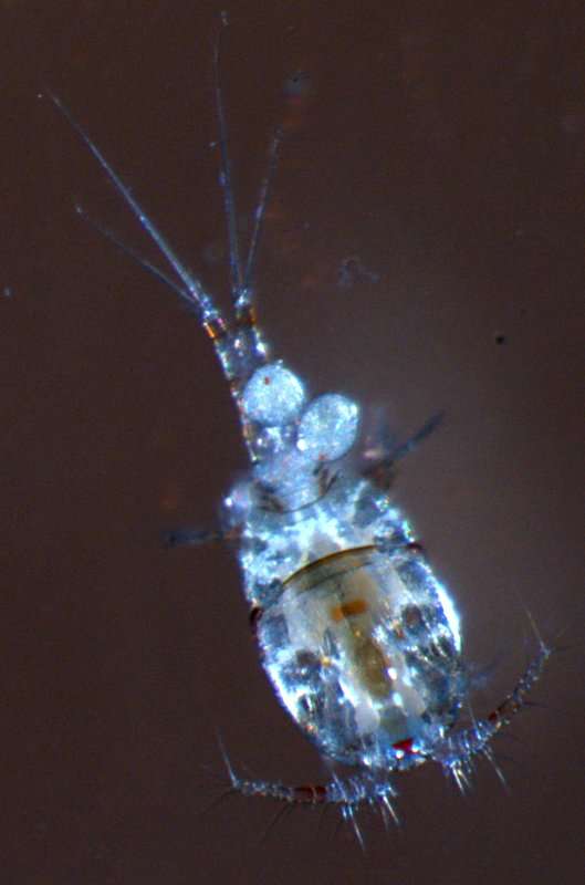
Cyclops albidus, a species of Copepod founr in fresh water. Female specimen carrying a pair of egg-packets. Actual length is about one tenth of an inch
-
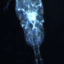
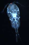
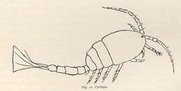
Cyclops.
-
Tomislav Karanovic, Mark J. Grygier, Wonchoel Lee
Zookeys
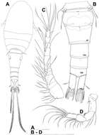
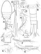
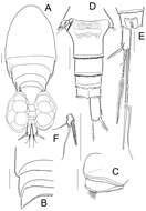
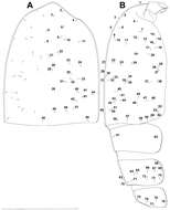
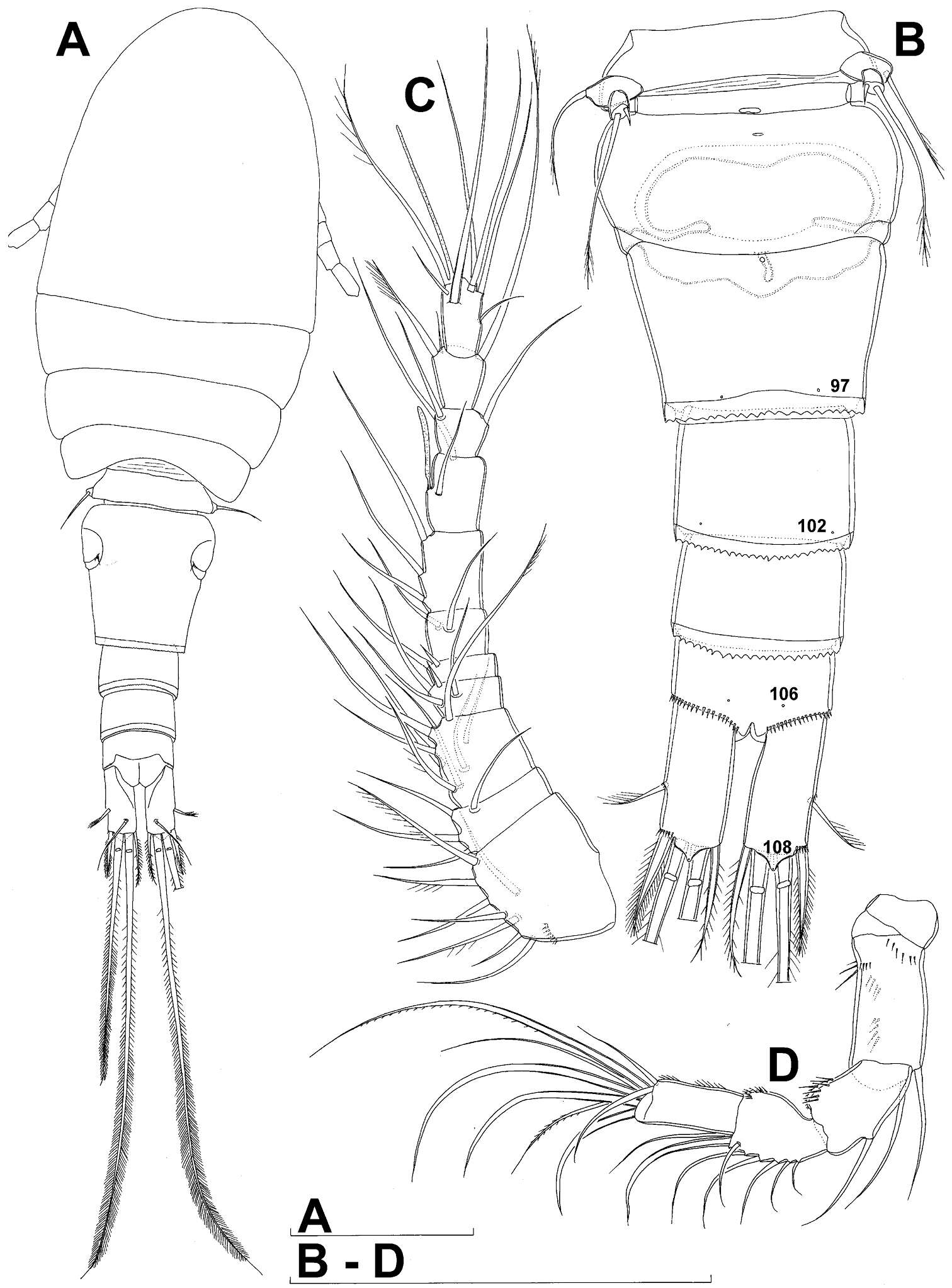
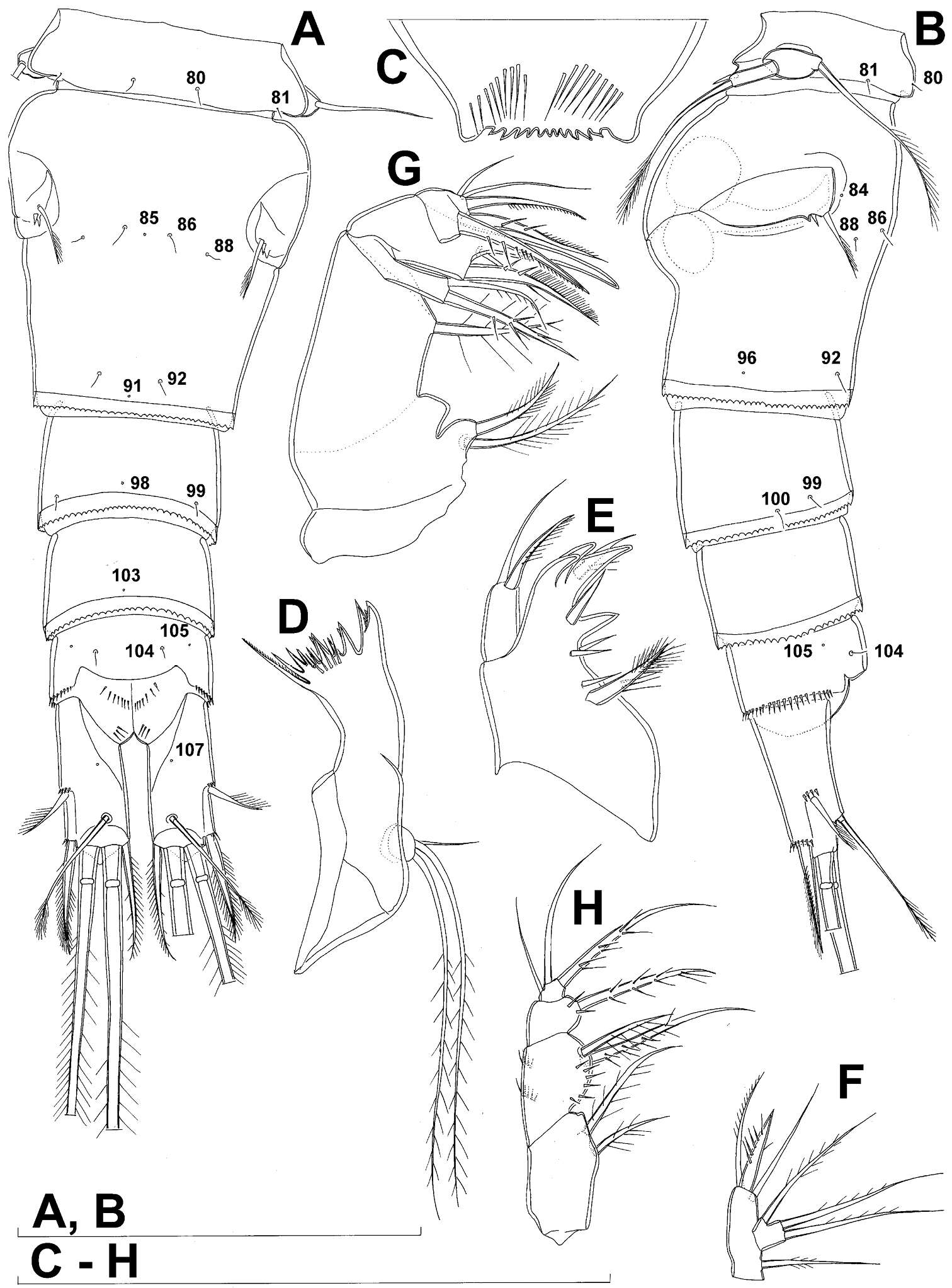
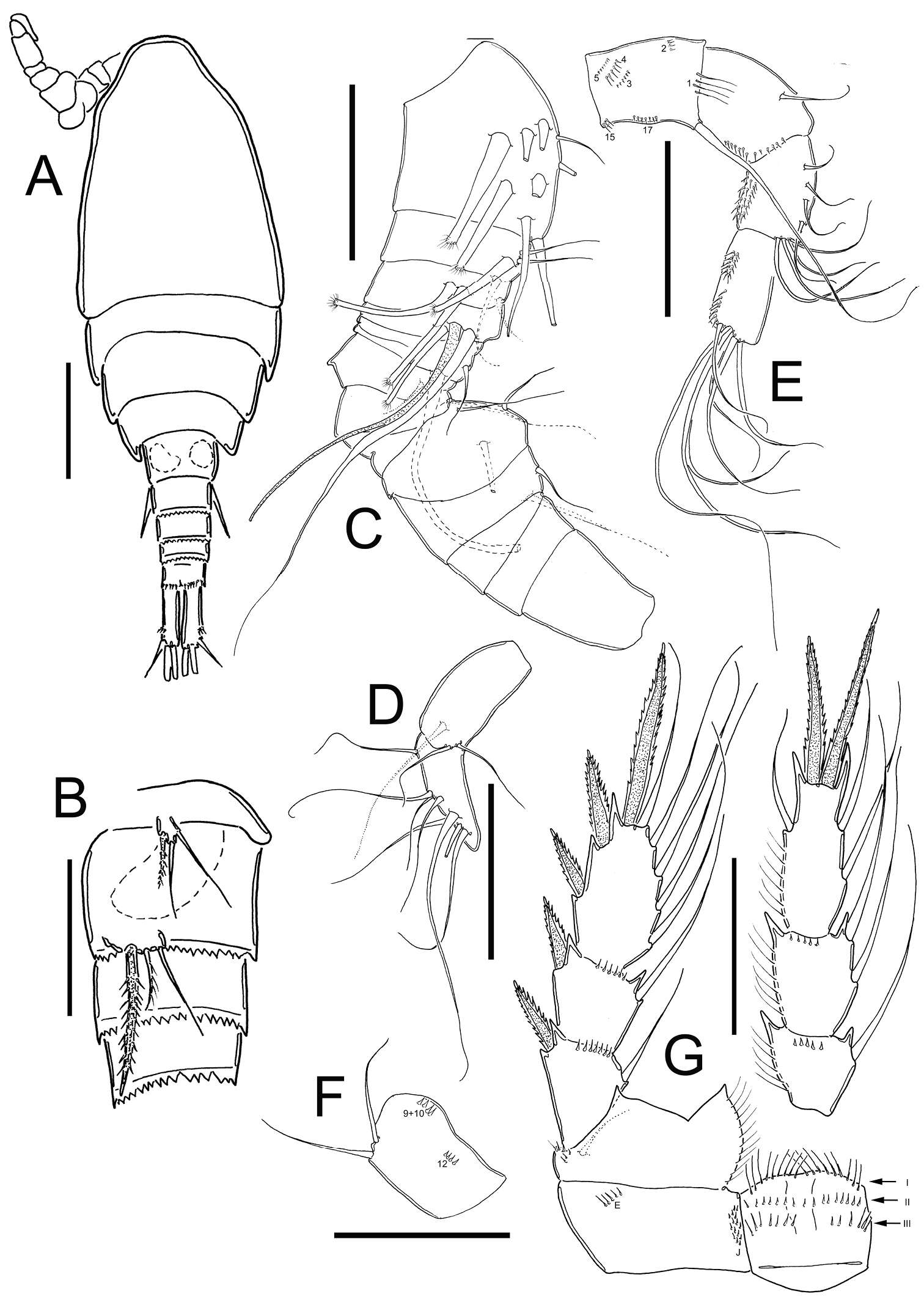
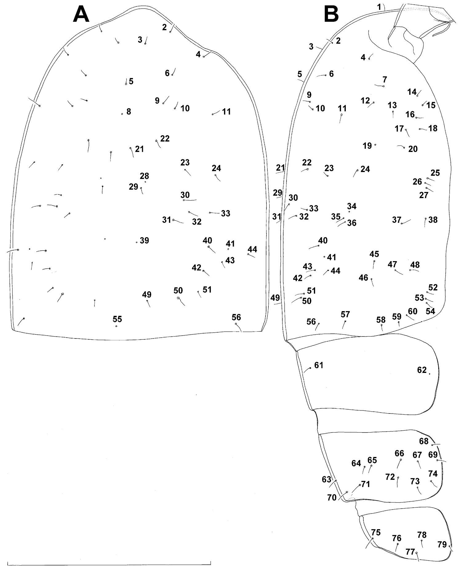
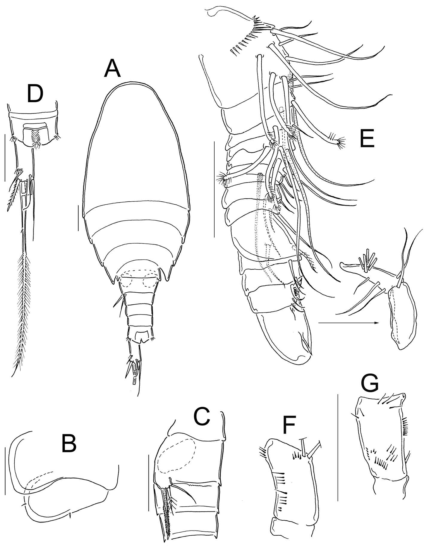
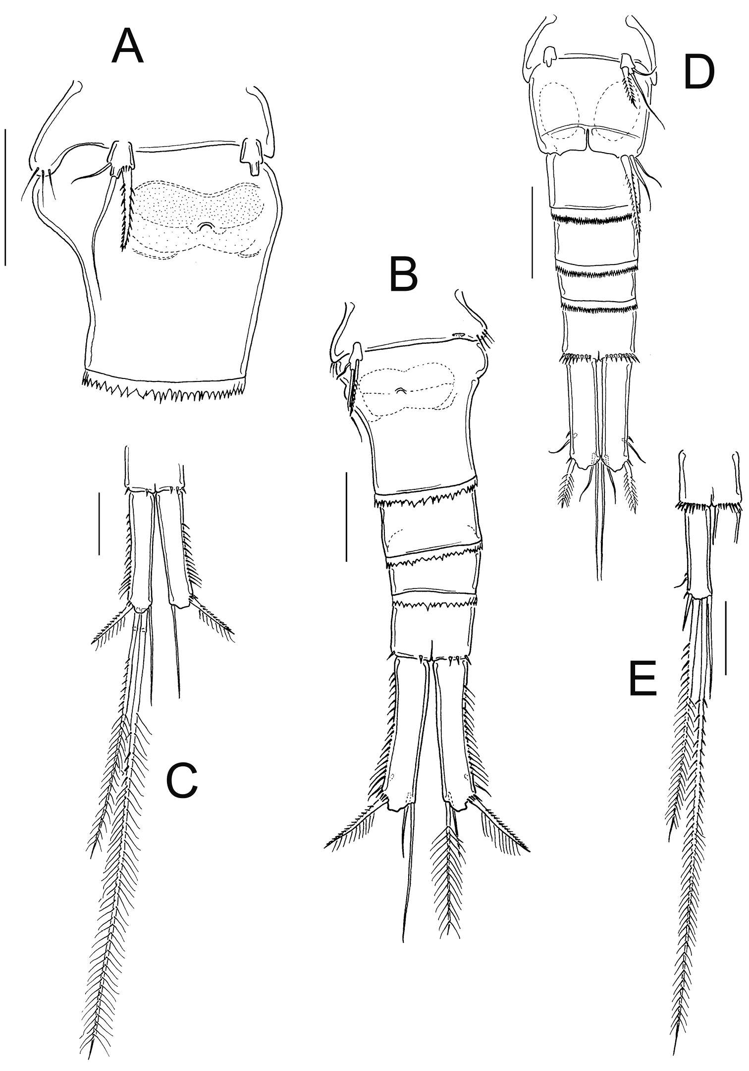
Figure 1.Diacyclops ishidai sp. n., holotype female: A habitus, dorsal view B urosome, ventral view C antennula, dorsal view D antenna, dorsal view. Arabic numerals numbering sensilla and pores consecutively from anterior to posterior end of body, and from dorsal to ventral side (excluding appendages). Scale bars 100 μm.
-
Tomislav Karanovic, Mark J. Grygier, Wonchoel Lee
Zookeys
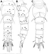
Figure 2.Diacyclops ishidai sp. n., holotype female: A urosome, dorsal view B urosome, lateral view C labrum, anterior view D mandibula, anterior view E maxillula, posterior view (palp armature omitted) F maxillular palp, anterior view G maxilla, posterior view H maxilliped, anterior view. Arabic numerals numbering sensilla and pores consecutively from anterior to posterior end of body, and from dorsal to ventral side (excluding appendages). Scale bars 100 μm.
-
Nancy F. Mercado-Salas, Eduardo Suárez-Morales, Alejandro M. Maeda-Martínez, Marcelo Silva-Briano
Zookeys
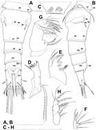
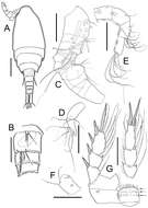
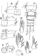
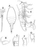
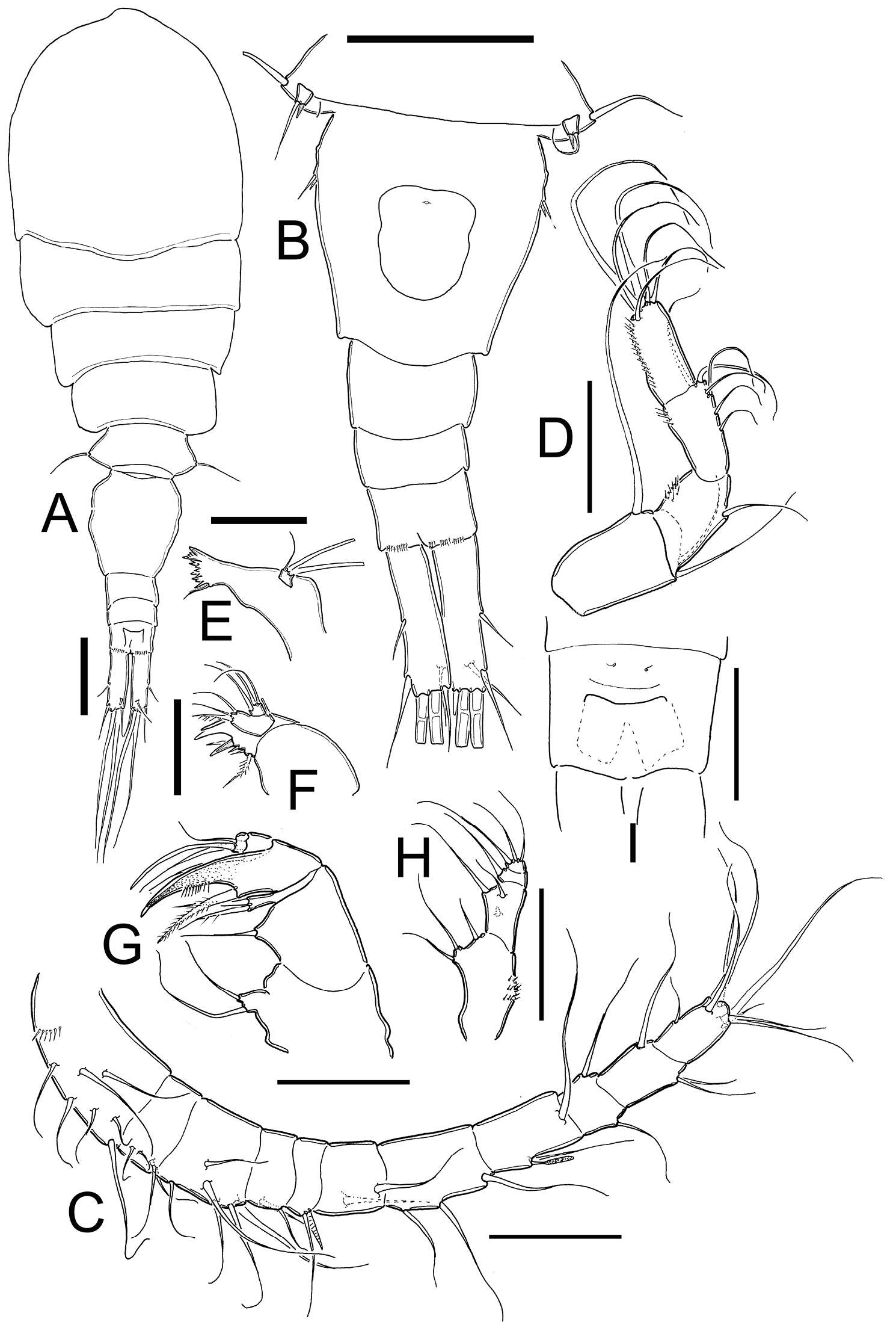
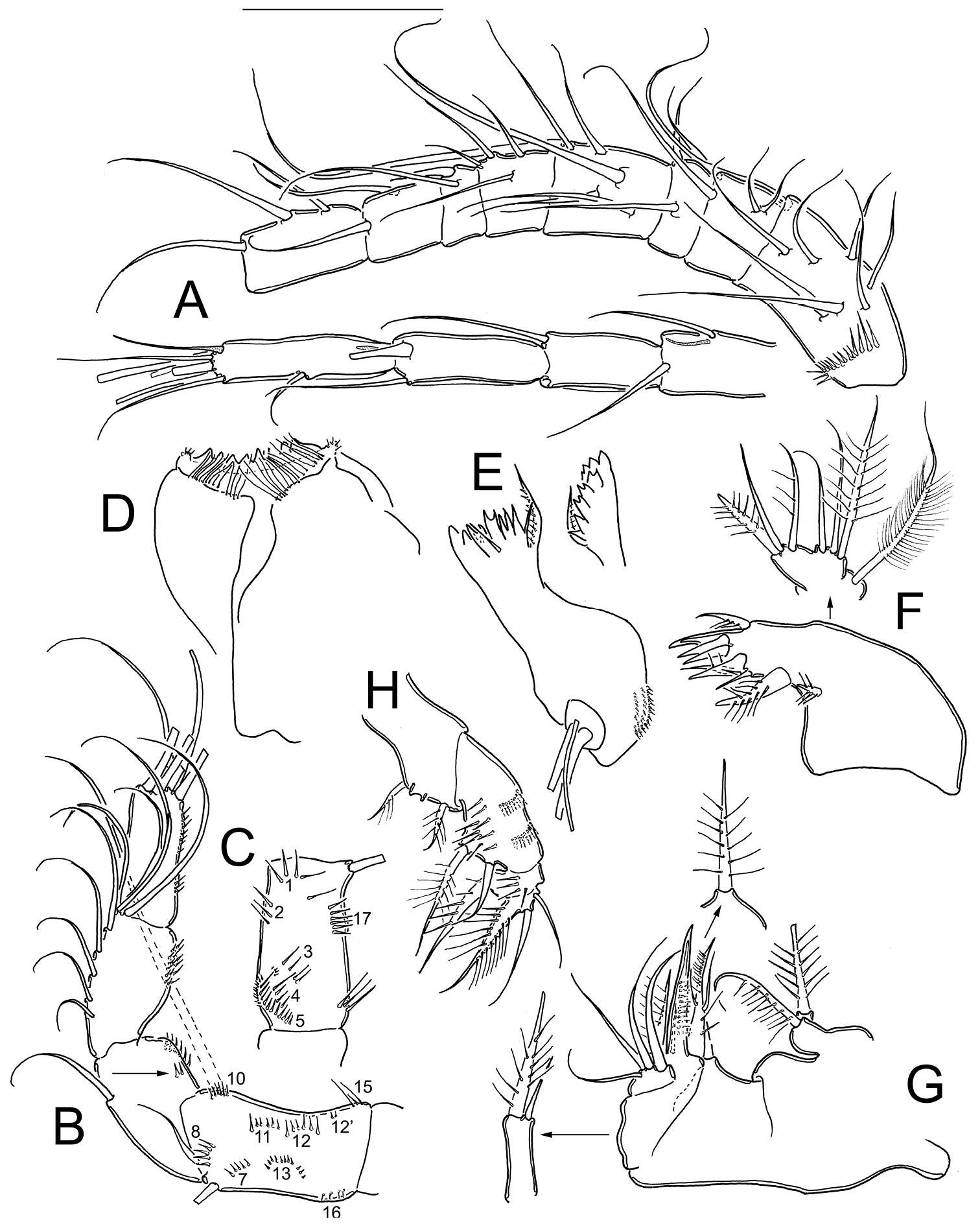
Figure 1.Metacyclops deserticus sp. n., female holotype from Coahuila, Mexico. A habitus, dorsal view B urosome, ventral view C antennule D antenna E mandible F maxillule G maxilla H maxilliped I anal operculum. Scales bars A–B= 100µm; C–I= 50 µm.
-
Nancy F. Mercado-Salas, Eduardo Suárez-Morales, Alejandro M. Maeda-Martínez, Marcelo Silva-Briano
Zookeys
Figure 5.Metacyclops deserticus sp. n., SEM-processed female from Coahuila, México. A leg 1 B endopodite 2 leg 4 C leg 5 D leg 6 E genital double somite, ventral view F caudal ramus, ventral.
-
Martha Angélica Gutiérrez-Aguirre, Nancy Fabiola Mercado-Salas, Adrián Cervantes-Martínez
Zookeys
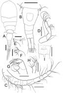
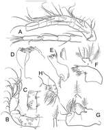
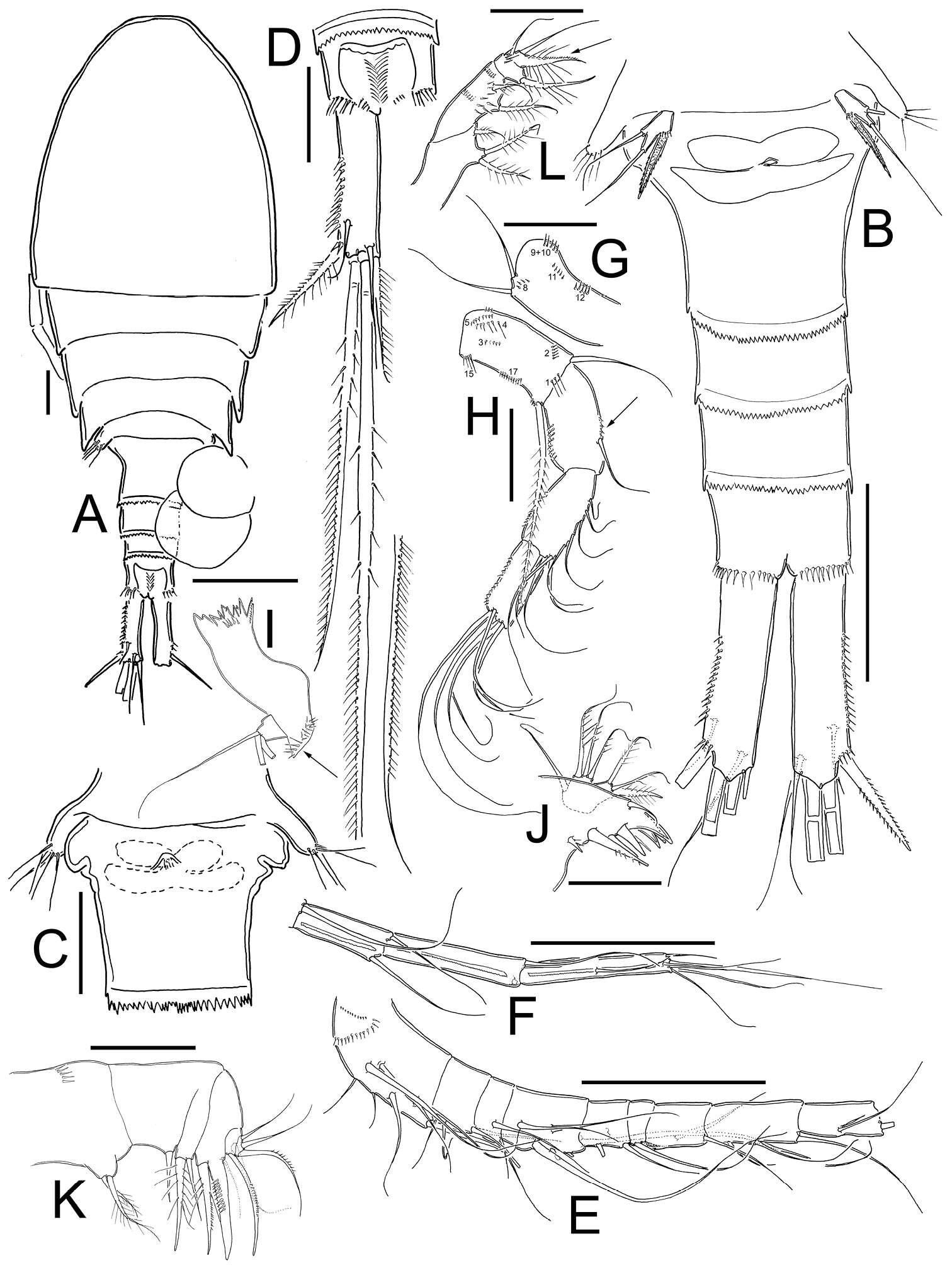
Figure 2.Eucyclops tziscao sp. n. A, C, D paratype B, E–L holotype from Laguna Tziscao, Chiapas. A Habitus, dorsal B Urosome C Genital double-somite, ventral D Anal somite and caudal ramus, dorsal E Antennule, segments 1–9 F Antennule, segments 10–12 G Antenna, caudal H Antenna, frontal I Mandible J Maxillule, caudal K Maxilla, frontal L Maxilliped, frontal. Scales bars: K = 20 µm; A, C, D, G, H, I, J, L = 50 µm; B, E, F = 100 µm.
-
Martha Angélica Gutiérrez-Aguirre, Nancy Fabiola Mercado-Salas, Adrián Cervantes-Martínez
Zookeys
Figure 4.Eucyclops tziscao sp. n. A–B paratype C–G allotype from Laguna Tziscao, Chiapas. A Habitus, dorsal B P5, and P6 C Antennule, segments 1–14 D Antennule, segments 15–16 E Antenna, frontal F Antenna, caudal G P4, caudal. Scales bars: B–G = 50 µm; A = 100 µm.
-
Tomislav Karanovic, Mark J. Grygier, Wonchoel Lee
Zookeys
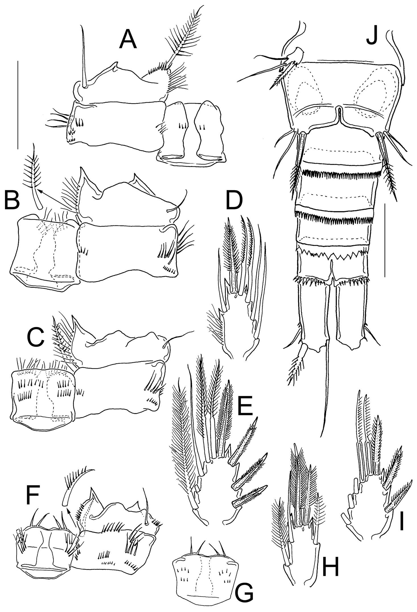
Figure 3.Diacyclops ishidai sp. n., A–E holotype female F paratype female A first swimming leg, anterior view B second swimming leg, anterior view C endopod of third swimming leg, anterior view D fourth swimming leg, anterior view E fifth leg, anterior view F genital double-somite, ventral view. Arabic numerals numbering sensilla and pores consecutively from anterior to posterior end of body, and from dorsal to ventral side (excluding appendages). Scale bars 100 μm.
-
Martha Angélica Gutiérrez-Aguirre, Nancy Fabiola Mercado-Salas, Adrián Cervantes-Martínez
Zookeys
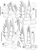
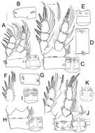
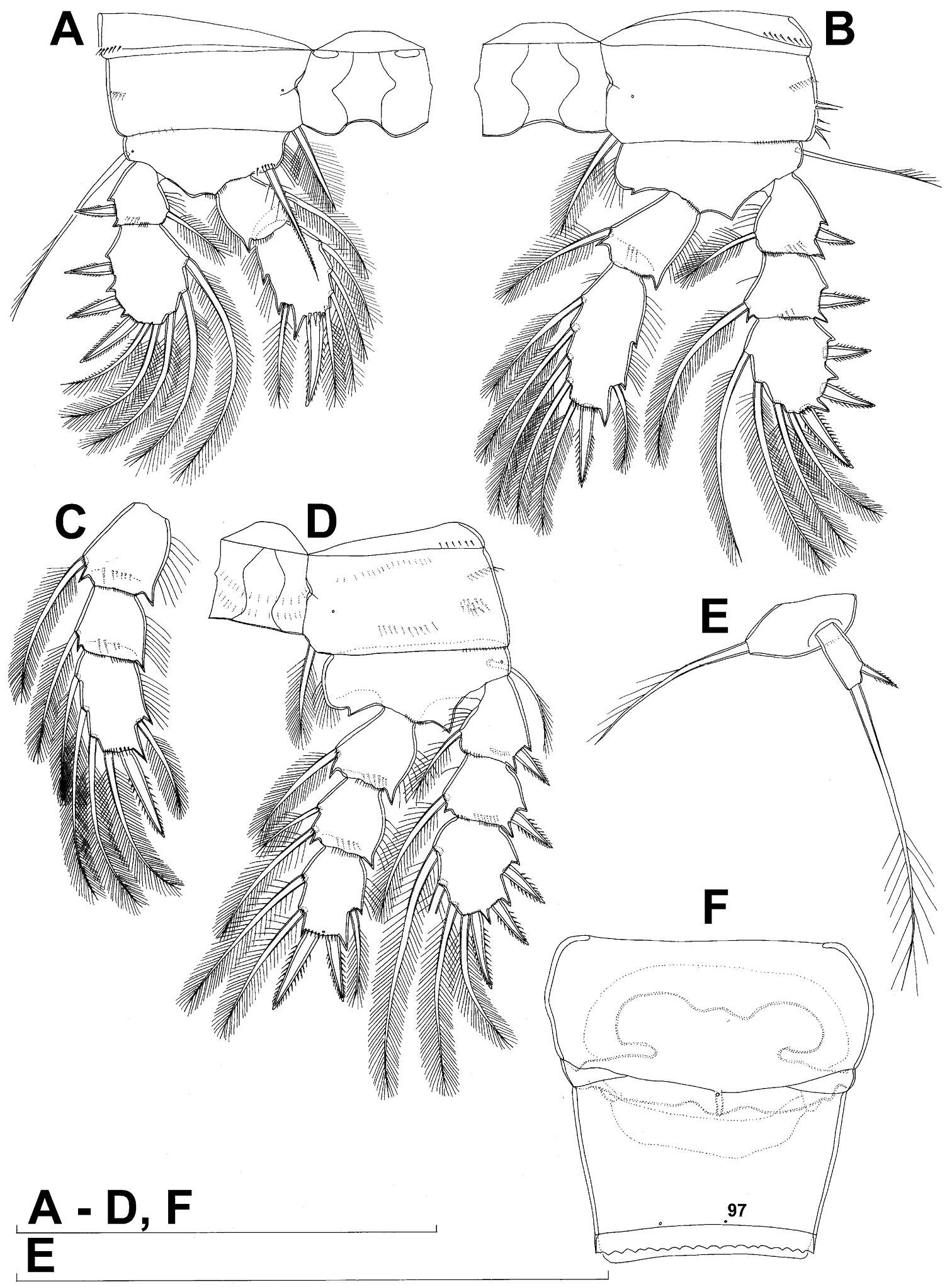
Figure 3.Eucyclops tziscao sp. n. Holotype from Laguna Tziscao, Chiapas. A P1, frontal B Intercoxal sclerite of P1, caudal C P2, frontal D Intercoxal sclerite of P2, caudal E P3, frontal, Exp and Enp separated F Intercoxal sclerite of P3, caudal G P4, caudal H Intercoxal sclerite of P4, frontal I Coxal spine P4 J P5. Scales bars: I= 25µm, J= 50 µm; A–H = 100 µm.
-
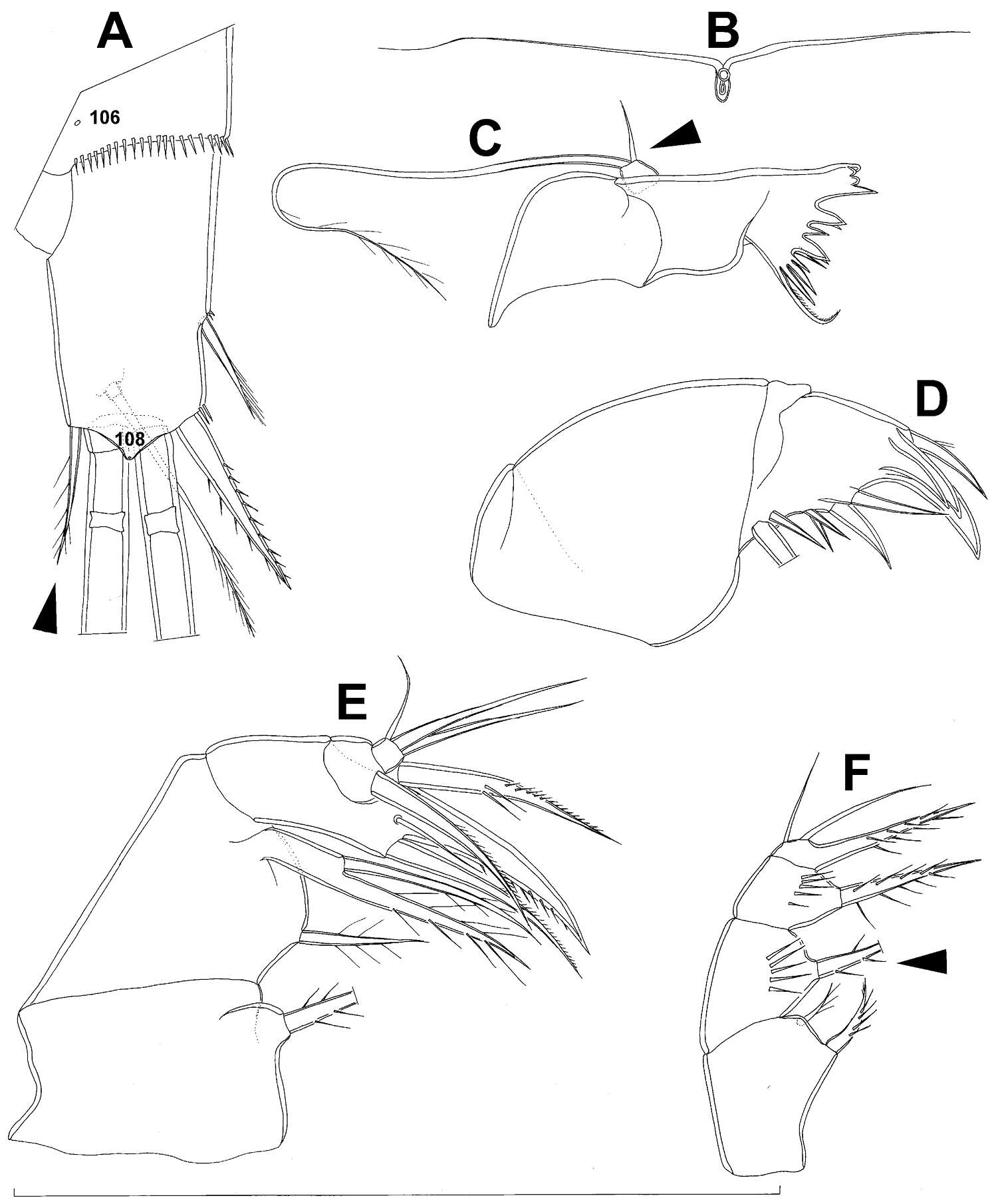
Tomislav Karanovic, Mark J. Grygier, Wonchoel Lee
Zookeys
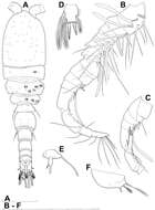
Figure 4.Diacyclops ishidai sp. n., allotype male: A habitus, dorsal view B antennula, flattened and slightly uncoiled, ventral view C middle part of antennula, flattened and uncoiled, dorsal view D third endopodal segment of fourth swimming leg, anterior view E fifth leg, anterior view F sixth leg, ventro-lateral view. Arabic numerals numbering sensilla and pores consecutively from anterior to posterior end of body, and from dorsal to ventral side (excluding appendages). Scale bars 100 μm.
-
Martha Angélica Gutiérrez-Aguirre, Nancy Fabiola Mercado-Salas, Adrián Cervantes-Martínez
Zookeys
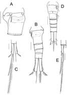
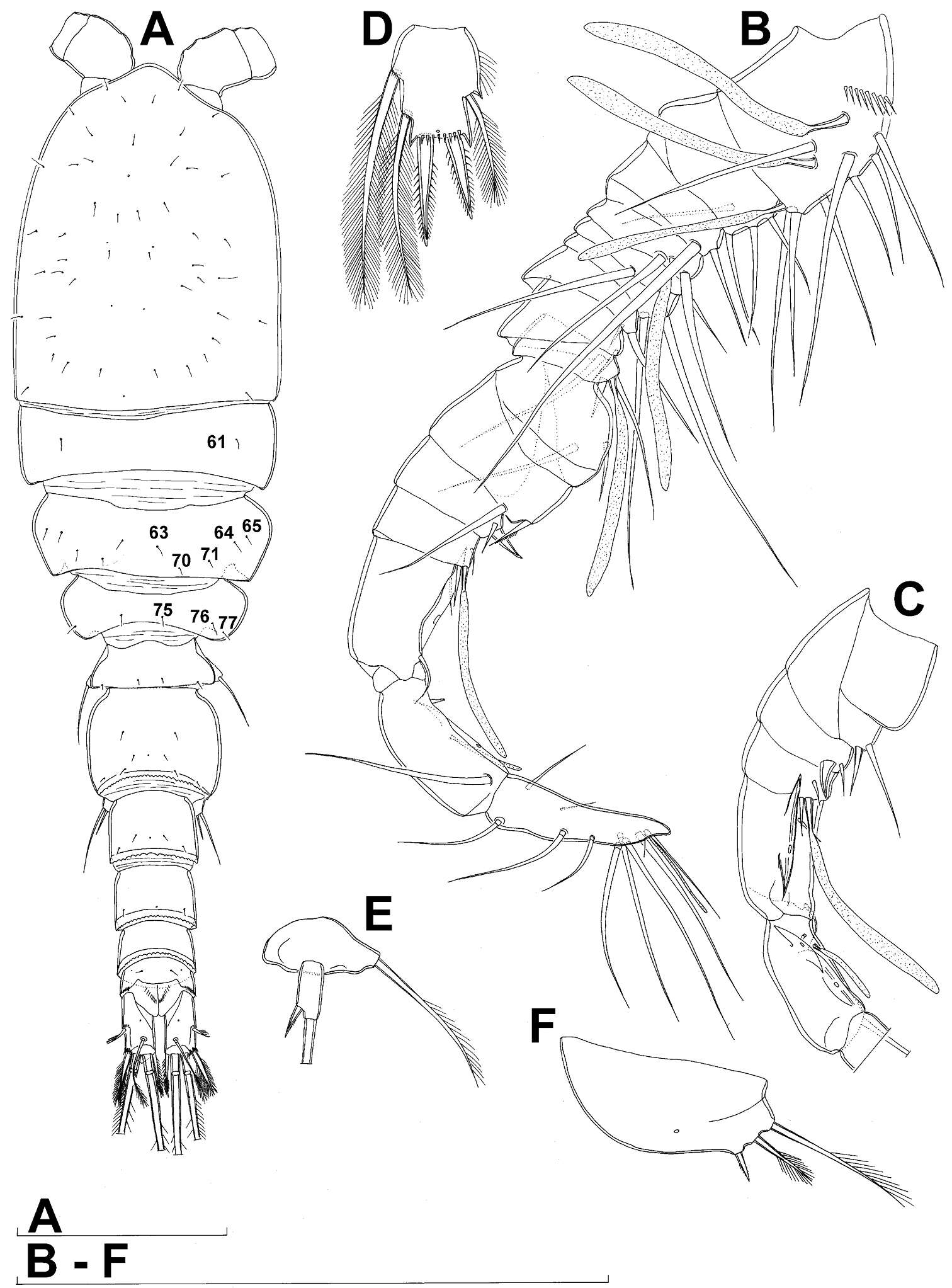
Figure 5.Eucyclops angeli sp. n. A–C paratype D–F holotype from grassland in San Cristóbal de las Casas, Chiapas. A Habitus, dorsal B Second to fourth prosomites, dorsal C Third and fourth prosomites, lateral D Urosome, ventral E Anal somite and one caudal ramus, dorsal F P5. Scale bars 50 µm.
-
Tomislav Karanovic, Mark J. Grygier, Wonchoel Lee
Zookeys
Figure 5.Diacyclops ishidai sp. n., allotype male: A urosome, dorsal view B urosome, lateral view C urosome, ventral view. Arabic numerals numbering sensilla and pores consecutively from anterior to posterior end of body, and from dorsal to ventral side (excluding appendages). Scale bar 100 μm.
-
Martha Angélica Gutiérrez-Aguirre, Nancy Fabiola Mercado-Salas, Adrián Cervantes-Martínez
Zookeys
Figure 9.Eucyclops angeli sp. n. Allotype from grassland in San Cristóbal de las Casas, Chiapas. A Coxa, basis, and intercoxal sclerite of P1, caudal B Coxa, basis, and intercoxal sclerite of P2, caudal C Coxa, basis, and intercoxal sclerite of P3, caudal D Enp3P3 E Exp3P3 F Coxa, basis, and intercoxal sclerite of P4, caudal G Intercoxal sclerite of P4, frontal H Enp3P4 I Exp3P4 J Urosome, ventral. Scale bars 50 µm.
-
All Biocode files are based on field identifications to the best of the researcher’s ability at the time.
-
Tomislav Karanovic, Mark J. Grygier, Wonchoel Lee
Zookeys
Figure 6.Diacyclops ishidai sp. n., allotype male: A cephalothorax, dorsal view B cephalothoracic shield and pleurons of free prosomites, lateral view. Arabic numerals numbering sensilla and pores consecutively from anterior to posterior end of body, and from dorsal to ventral side (excluding appendages). Scale bar 100 μm.
-
Martha Angélica Gutiérrez-Aguirre, Nancy Fabiola Mercado-Salas, Adrián Cervantes-Martínez
Zookeys
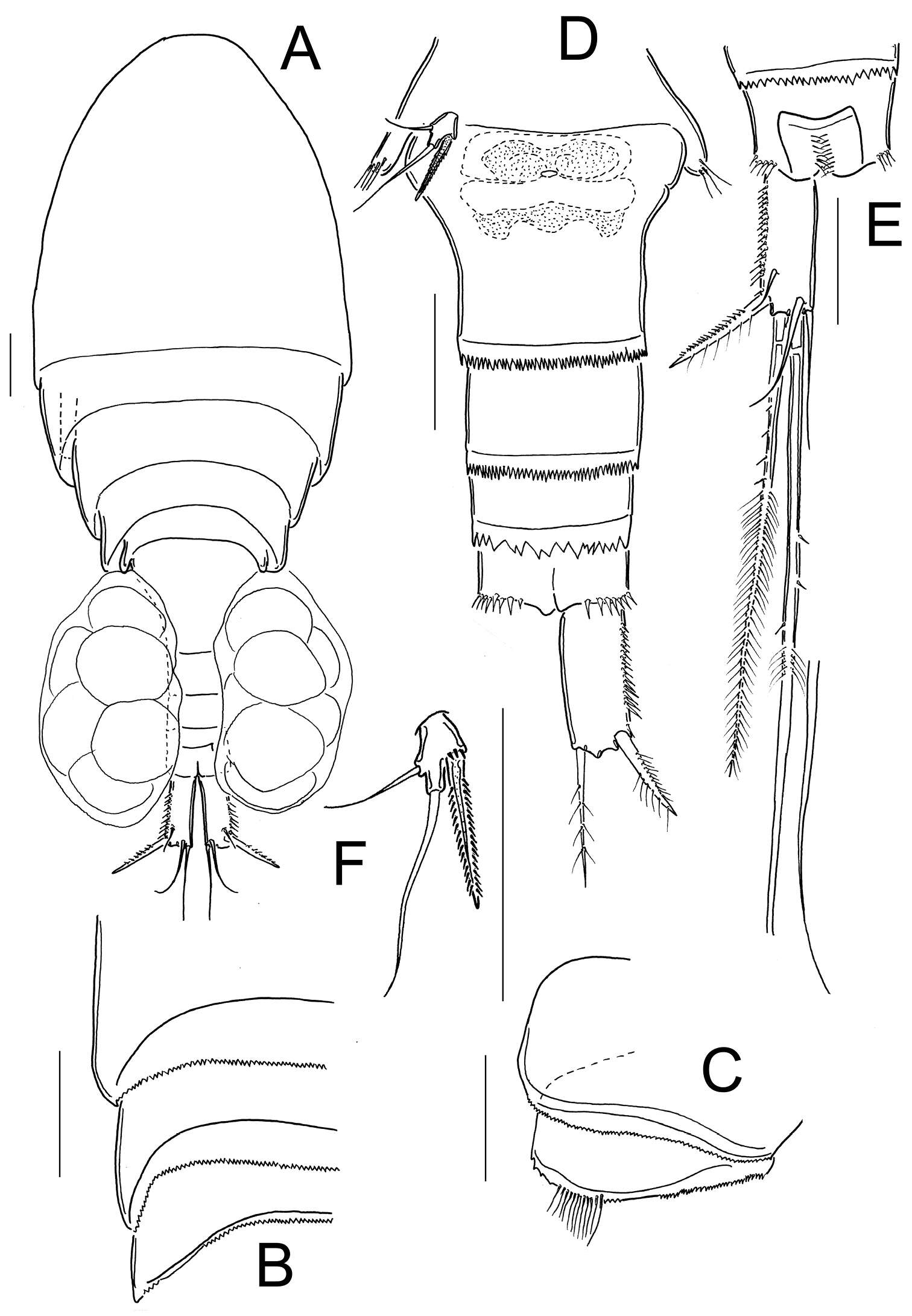
Figure 6.Eucyclops angeli sp. n. Holotype from grassland in San Cristóbal de las Casas, Chiapas. A Antennule B Antenna, caudal C Antenna, frontal D Labrum E Mandible F Maxillule, palp separated G Maxilla, proximal and distal endites of the coxa, separated H Maxilliped, frontal. Scale bar 50 µm.
-
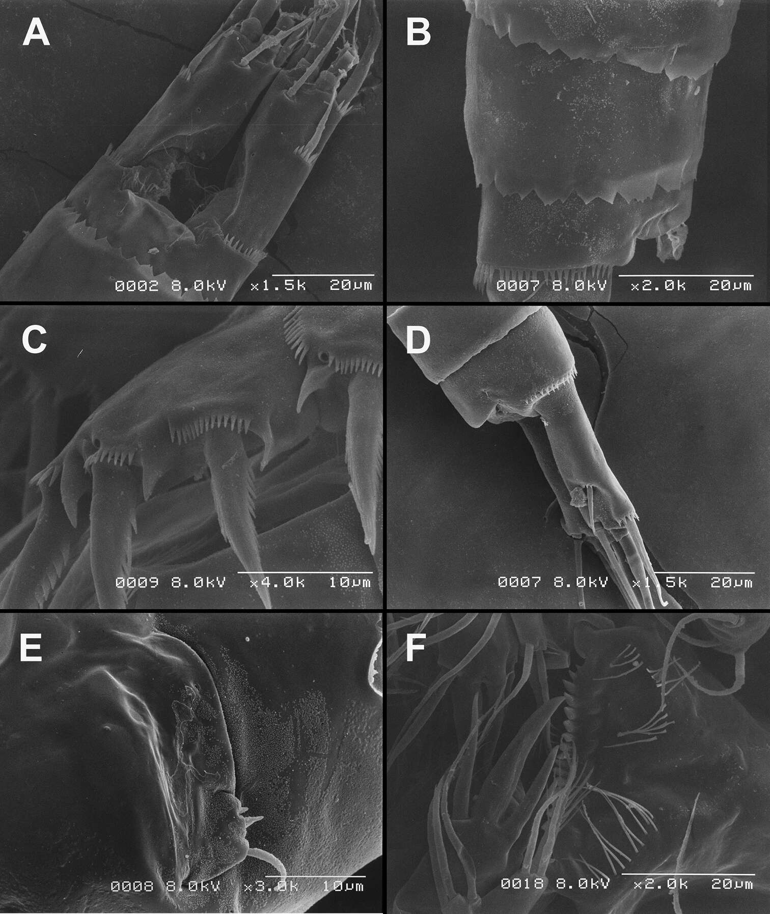
Tomislav Karanovic, Mark J. Grygier, Wonchoel Lee
Zookeys
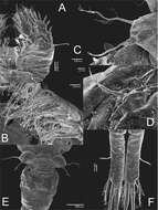
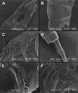
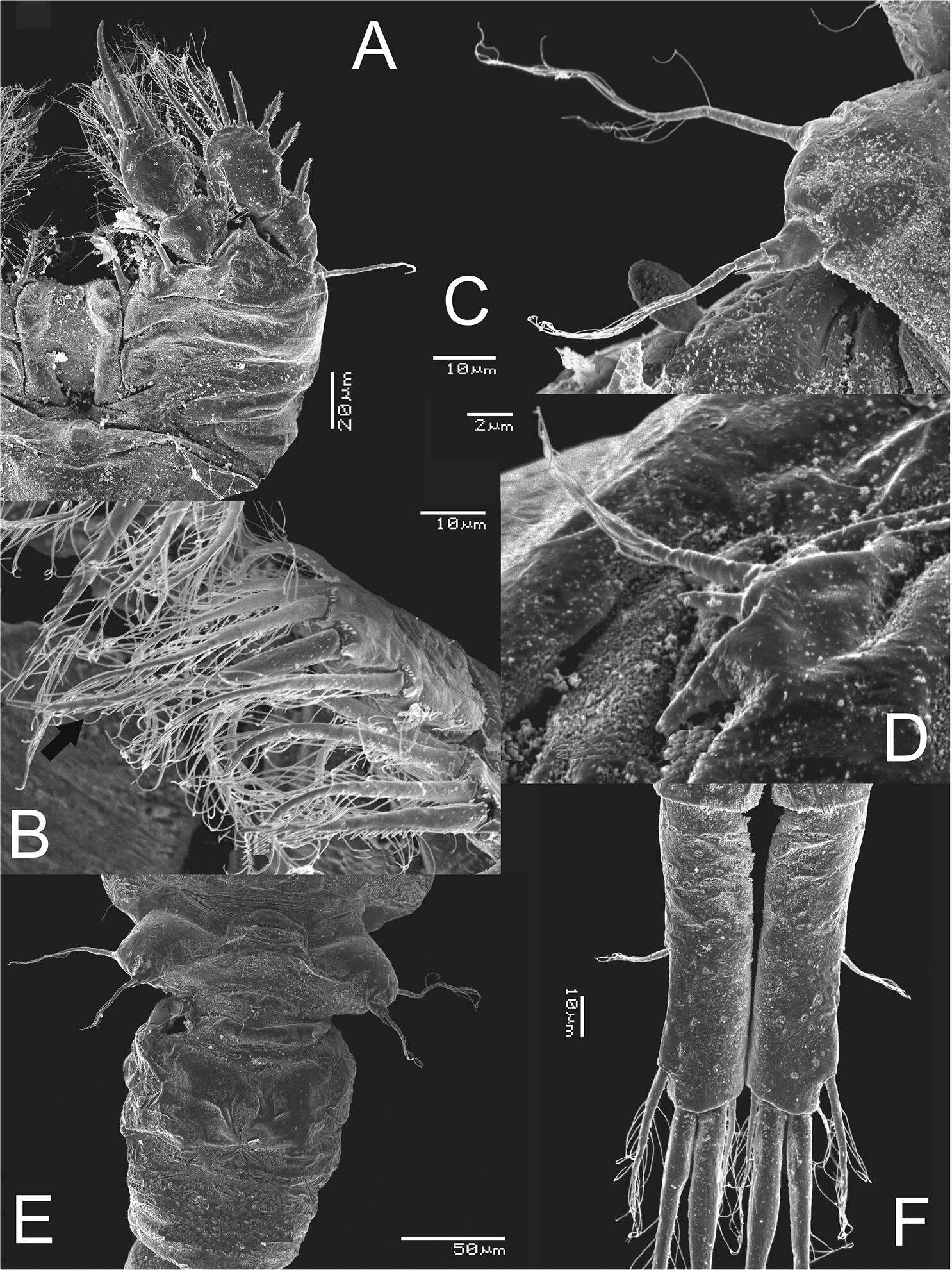
Figure 26.Scanning electron micrographs, A–C Diacyclops ishidai sp. n. D–E Diacyclops parasuoensis sp. n. F Diacyclops suoensis Ito, 1954: A anal somite and caudal rami, dorsal view, paratype female 1 B preanal and anal somites, lateral view, paratype female 2 C last two exopodal segments of second swimming legs, lateral view, paratype female 2 D anal somite and caudal rami, lateral view, paratype female E sixth leg, lateral view, paratype female F labrum and maxillulae, ventral view. Scale bars 20 μm (A, B, D, F) and 10 μm (C, E).
-
Martha Angélica Gutiérrez-Aguirre, Nancy Fabiola Mercado-Salas, Adrián Cervantes-Martínez
Zookeys
Figure 7.Eucyclops angeli sp. n. Holotype from grassland in San Cristóbal de las Casas, Chiapas. A P1, frontal B Coxa of P1, caudal C P2, frontal, Exp separated D Coxa of P2, caudal E Intercoxal sclerite of P2, caudal F Exp3P3 G Coxa of P3, caudal H Coxa, basis, and intercoxal sclerite of P3, frontal I Intercoxal sclerite P3, caudal J P4, caudal; exopod and coxal spine separated K Intercoxal sclerite of P4, frontal. Scale bar 50 µm.
-
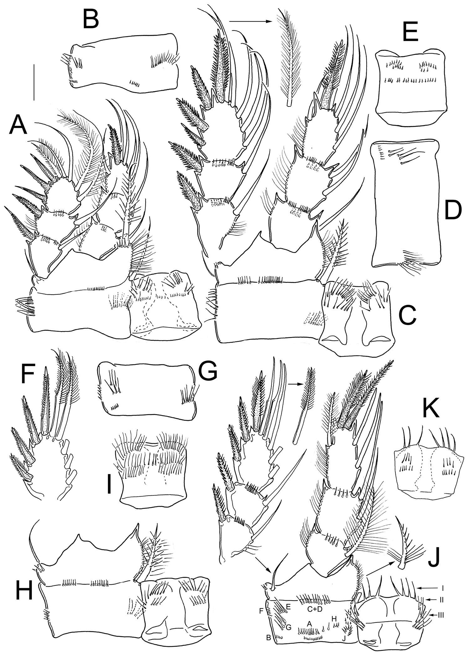
Tomislav Karanovic, Mark J. Grygier, Wonchoel Lee
Zookeys
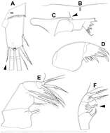
Figure 7.Diacyclops brevifurcus Ishida, 2006, holotype female: A left caudal ramus, ventral view B copulatory pore, ventral view C mandibula, posterior view D maxillula, posterior view (palp broken off) E maxilla, anterior view F maxilliped, anterior view. Arabic numerals indicating sensilla and pores presumably homologous to those in Diacyclops ishidai sp. n. Arrows pointing most prominent specific features. Scale bar 100 μm.
-
Martha Angélica Gutiérrez-Aguirre, Nancy Fabiola Mercado-Salas, Adrián Cervantes-Martínez
Zookeys
Figure 8.Eucyclops angeli sp. n. A–B paratype C–F allotype from grassland in San Cristóbal de las Casas, Chiapas. A Habitus, dorsal B Third, and fourth prosomites, lateral C First to fourth urosomites, lateral D Anal somite and caudal ramus, dorsal E Antennule, last two segments separated F Antenna, caudal G Antenna, frontal. Scale bars 50 µm.
-
Tomislav Karanovic, Mark J. Grygier, Wonchoel Lee
Zookeys
Figure 8.Diacyclops brevifurcus Ishida, 2006, holotype female: A third swimming leg, posterior view B fourth swimming leg, posterior view C fifth leg, anterior view. Arrows pointing most prominent specific features. Scale bars 100 μm.
-
Martha Angélica Gutiérrez-Aguirre, Nancy Fabiola Mercado-Salas, Adrián Cervantes-Martínez
Zookeys
Figure 10.Eucyclops festivus Lindberg, 1955; from pond 3 to Laguna Montebello, Chiapas. A First urosomite, and genital double-somite, ventral B Urosome, ventral C Anal somite and caudal ramus, ventral D Urosome, ventral E Anal somite and caudal ramus, ventral. Scale bars 50 µm. A–C female; D–E male.