Description
provided by Zookeys
Body size of living fissiparous specimens ranged from 7–10 mm in length and 1.5–2 mm in width (Fig. 2). Sexualized specimens were about 13–16 mm in length and about 3 mm in width. Two eyes are present in the middle of the head, and unpigmented auricular grooves are marginally placed just posteriorly to the eyes. The colour is uniformly brown dorsally, and pale ventrally.
Inner and outer pharyngeal musculature is bilayered, i.e. without an extra, third, outer longitudinal muscle layer. The ovaries are hyperplasic, with several scattered masses at a short distance behind the brain, filling up the entire dorso-ventral space. A degenerative condition is clearly evident in the ovaries, in that maturation of the oocytes is regular up to the beginning of the diplotene stage, whereas diplotenic oocytes show progressive cytoplasm vacuolation, followed by collapse of the entire cell content and by cell necrosis.
The anterior portion of the infranucleated oviducts is expanded to form a seminal receptacle that arises in the middle of the ovarian masses at a poorly defined position, dependent upon the hyperplasic condition of the ovaries. The oviducts run ventrally in a caudal direction up to the vaginal area and open asymmetrically into the distal section of the bursal canal. The right oviduct opens dorsally to the left one. The latter opens very close to the point where the canal communicates with the common atrium (Fig. 3). The very abundant shell glands open at the level of the left oviducal opening.
The testes are situateddorsally and extend from just anterior to the ovaries to the posterior end of the body. The testes generally are under-developed in that the majority of germ cells are represented only by spermatogonia (ca. 90%). In only some specimens, and then in only a few follicles, mature sperms are present. However, in all cases anomalies were observed, such as irregularly shaped spermatids and spermatozoa. Vitellaria are located between the testes and the intestinal branches.
The large sac-shaped copulatory bursa is lined by a columnar, glandular epithelium bearing basal nuclei and it is surrounded by a thin layer of muscles. From the mid-posterior wall of the bursa the bursal canal runs in a caudal direction, to the left of the copulatory apparatus. Posteriorly to the gonopore the bursal canal recurves antero-ventrally and, subsequently, opens into the posterior section of the atrium. The bursal canal is lined by a pleated epithelium with cylindrical, infranucleated, and ciliated cells and is surrounded by a thin, subepithelial layer of longitudinal muscles, followed by a thicker layer of circular muscle. Ectal reinforcement is absent (Figs 3, 4C). At its distal section, near the atrium, the bursal canal shows several deep folds.
The moderately developed penis bulb, rich in glands, consists of intermingled longitudinal and circular muscle fibres. It houses an elongated seminal vesicle, which extends through the entire length of the penis bulb. The anterior half of the seminal vesicle is tubular in shape, while its distal, posterior section is considerably expanded.
The vasa deferentia penetrate the antero-lateral wall of the penis bulb and open separately and symmetrically into the seminal vesicle at a position about halfway along the vesicle. No spermiducal vesicles were observed in any of the specimens examined. The seminal vesicle, lined with a flat epithelium and surrounded in its distal, posterior section by layers of circular muscle fibres, opens into the ejaculatory duct via a small diaphragm. The latter, located at the base of the penis papilla, receives the openings of very abundant bulb glands. The blunt penis papilla is lined with an infranucleated epithelium that is underlain with a thin subepithelial layer of circular muscles fibres, followed by a layer of longitudinal muscle fibres.
The ejaculatory duct follows a dorsally displaced course through the penis papilla and has a sub-terminal opening. The spacious lumen of the ejaculatory duct is lined by a cuboidal, infranucleated epithelium that is surrounded by a layer of longitudinal musclesand receives the abundant secretion of penis papillaglands; in the majority of examined specimens the ejaculatory duct contained an empty spermatophore (Fig. 4A, B). Both the bulb glands and the penis papilla glands secrete globules that stain purple in Pasini’s reagent (Fig. 4B). The acentral, dorsally displaced ejaculatory duct makes the penis papilla asymmetrical, with the ventral part being thicker than the dorsal one (Figs 3, 4A, B).
The genital atrium is lined by an infranucleated epithelium that is underlain by a subepithelial layer of circular muscle, followed by a layer of longitudinal muscle fibres. The common atrium communicates with a gonoduct that is lined by a columnar epithelium, which receives the openings of very abundant cement glands; the gonoduct communicates with the ventral gonopore (Fig. 3).
- license
- cc-by-3.0
- copyright
- Giacinta Angela Stocchino, Ronald Sluys, Paolo Deri, Renata Manconi
- bibliographic citation
- Stocchino G, Sluys R, Deri P, Manconi R (2013) Integrative taxonomy of a new species of planarian from the Lake Ohrid basin, including an analysis of biogeographical patterns in freshwater triclads from the Ohrid region (Platyhelminthes, Tricladida, Dugesiidae) ZooKeys 313: 25–43
- author
- Giacinta Angela Stocchino
- author
- Ronald Sluys
- author
- Paolo Deri
- author
- Renata Manconi
Distribution
provided by Zookeys
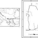
Known from the type locality and, most likely, also from a second Albanian locality, viz. Voskopojë (see below).
Checklist of Tricladida from the Lake Ohrid hydrographic basin.
Taxa Lacustrine habitat Adjacent waters of lake Ohrid Endemic species References Dugesiidae Ball, 1974 Dugesia Girard, 1850 Dugesia superioris _ tributary rivulet – Present paper Dugesia sp.
_ Spring Elešec ? Kenk 1978 Dugesia gonocephala (Dugès, 1830)(?)
_ tributary streams and the effluent Crni Drim River _ Stanković 1960 Schmidtea Ball, 1974 Schmidtea lugubris (Schmidt, 1861)
_ stagnant waters of the Ohrid region; drainage ditch at Teferić _ Stanković 1960; Kenk 1978 Dendrocoelidae Hallez, 1892 Dendrocoelum Örsted, 1844 Dendrocoelum adenodactylosum (Stanković & Komárek, 1927)
littoral, sublittoral, profundal zones littoral cold springs; tributary streams; a tributary of the effluent Crni Drim River _ Stanković and Komárek 1927; Kenk 1978 Dendrocoelum albidum Kenk, 1978
sublittoral zone _ + Kenk 1978 Dendrocoelum cruciferum (Stanković, 1969)
sublittoral zone _ + Stanković 1969; Kenk 1978 Dendrocoelum decoratum Kenk, 1978
sublittoral and profundal zones _ + Kenk 1978 Dendrocoelum dorsivittatum Kenk, 1978
profundal zone _ + Kenk 1978 Dendrocoelum jablanicense (Stanković & Komárek, 1927)
_ Šum Spring; tributary streams + Stanković and Komárek 1927; Stanković 1960; Kenk 1978 Dendrocoelum komareki (Stanković, 1969)
sublittoral zone _ + Stanković 1969; Kenk 1978 Dendrocoelum lacteum (Müller, 1774)
sublittoral and profundal zones stagnant waters _ Stanković and Komárek 1927; Arndt 1938; Stanković 1960; Kenk 1978 Dendrocoelum lacustre (Stanković, 1938)
sublittoral zone _ + Stanković 1938; Stanković 1969; Kenk 1978 Dendrocoelum lychnidicum (Stanković, 1969)
sublittoral zone _ + Stanković 1969; Kenk 1978 Dendrocoelum maculatum (Stanković & Komárek, 1927)
littoral zone tributary streams; littoral springs + Stanković 1960; Kenk 1978 Dendrocoelum magnum (Stanković, 1969)
sublittoral zone _ + Stanković 1969; Kenk 1978 Dendrocoelum minimum Kenk, 1978
profundal zone _ + Kenk 1978 Dendrocoelum ochridense (Stanković & Komárek, 1927)
littoral, sublittoral, profundal zones _ + Stanković 1960; Kenk 1978 Dendrocoelum sanctinaumi (Stanković & Komárek, 1927)
littoral and sublittoral zones tributary streams; littoral springs + Stanković 1960; Kenk 1978 Dendrocoelum sinisai Kenk, 1978
profundal zone _ + Kenk 1978 Dendrocoelum translucidum Kenk, 1978
profundal zone _ + Kenk 1978 Planariidae Stimpson, 1857 Crenobia Kenk, 1930 Crenobia alpina montenegrina (Mrázek, 1904)
_ springs; tributary streams and the effluent Crni Drim River _ Stanković and Komárek 1927; Stanković 1960; Kenk 1978 Phagocata Leidy, 1847 Phagocata maculata (Stanković, 1938)
sublittoral zone _ + Stanković 1938; Stanković 1960; Kenk 1978 Phagocata ochridana (Stanković & Komárek, 1927)
littoral, sublittoral, profundal zones springs and pools + Stanković and Komárek 1927; Stanković 1960; Kenk 1978 Phagocata stankovici (Reisinger, 1960)
sublittoral and profundal zones _ + Reisinger 1960; Kenk 1978 Phagocata undulata (Stanković, 1960)
sublittoral zone _ + Stanković 1960; Kenk 1978 Planaria Müller, 1776 Planaria torva (Müller, 1774)
_ only one specimen at the mouth of the Studenčišta brook _ Stanković 1960; Kenk 1978 Polycelis Ehrenberg, 1831 Polycelis tenuis Ijima, 1884
_ tributary streams _ Stanković 1960; Kenk 1978
- license
- cc-by-3.0
- copyright
- Giacinta Angela Stocchino, Ronald Sluys, Paolo Deri, Renata Manconi
- bibliographic citation
- Stocchino G, Sluys R, Deri P, Manconi R (2013) Integrative taxonomy of a new species of planarian from the Lake Ohrid basin, including an analysis of biogeographical patterns in freshwater triclads from the Ohrid region (Platyhelminthes, Tricladida, Dugesiidae) ZooKeys 313: 25–43
- author
- Giacinta Angela Stocchino
- author
- Ronald Sluys
- author
- Paolo Deri
- author
- Renata Manconi