Giacinta Angela Stocchino, Ronald Sluys, Paolo Deri, Renata Manconi
Zookeys
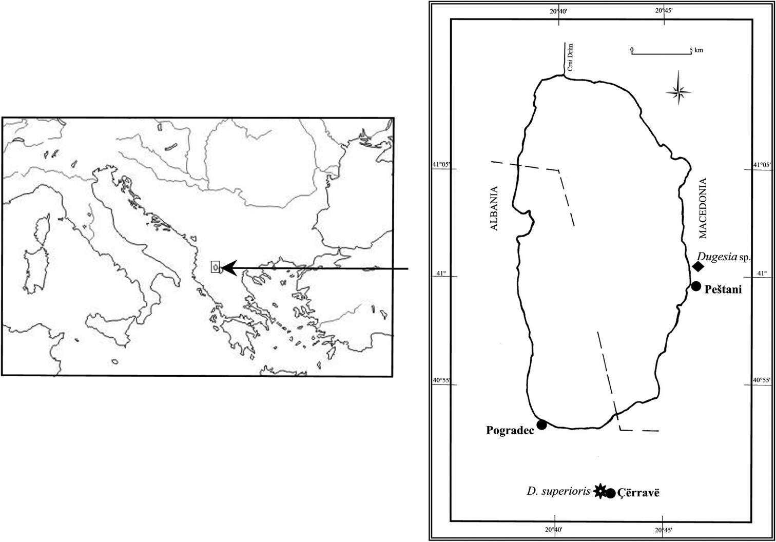
Figure 1.Geographic distribution of Dugesia superioris (indicated by an asterisk) and Dugesia sp. NMNH 55294 (indicated by black diamond) in the Lake Ohrid region.
Giacinta Angela Stocchino, Ronald Sluys, Renata Manconi
Zookeys
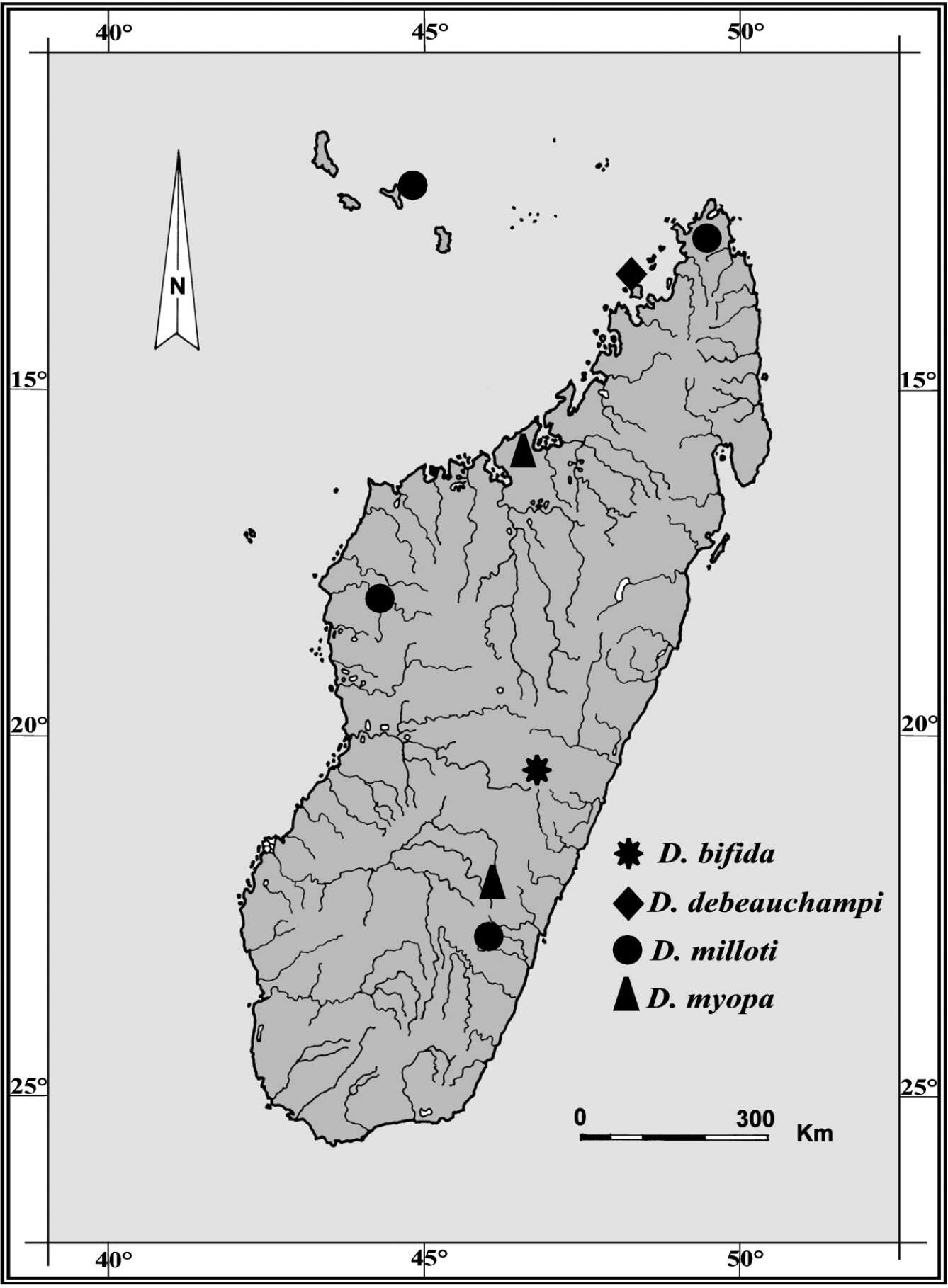
Figure 1.Geographic distribution of Dugesia species recorded from Madagascar and adjacent islands. Type locality of Dugesia bifida in the High Tsiribihina hydrographic basin indicated by an asterisk.