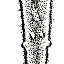
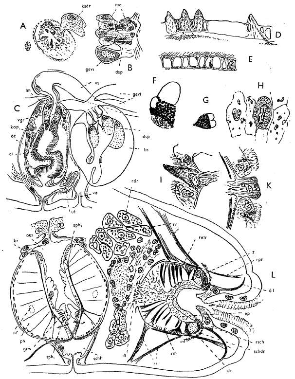
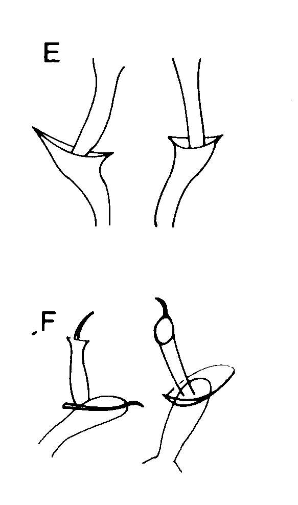
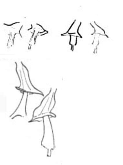
Three drawing at the left (up to down): Organization of the locomotory muscles of the proboscis form the right hand side; terminal organ next to the proboscis and copulatory organ on a squeezed animal. A. Transverse section through the copulatory organ at the level of the outlet of the prostate glands. B. Outlet of the bursal pieces in the ductus communis. C. Sagital reconstruction of the copulatory system. D and E. Optical sections through the epidermis of a live animal, D "toes" of the caudal end. F and G. Eyes of live animals (F: abnormal form). H. Epithelial cells (tangential section). I. "Haftpapille" at the caudal end. K. "Haftpapille" of the dorsal side, rostrally. L. (Figure at the bottom) Sagital reconstruction of the proboscis and pharynx.
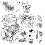
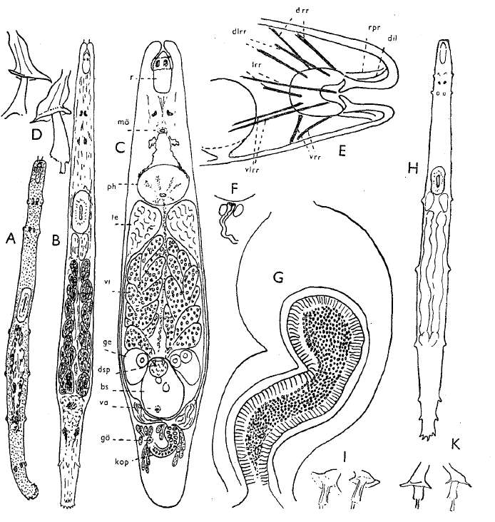
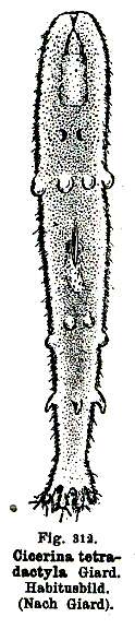
A Free-crawling young animal. B Slightly-squeezed, mature animal C. Organization scheme of a squeezed animal. H. Crawling animal of the North Sea coast.