Comprehensive Description
provided by Smithsonian Contributions to Zoology
Pholoides asperus (Johnson, 1897)
Peisidice aspera Johnson, 1987:184, pl. 9: figs. 56–59; pl. 10: fig. 63.—Moore, 1908:338; 1910:386.—Berkeley and Berkeley, 1941:24; 1942:189; 1948:23, fig. 28.—Hartman, 1939:7; 1948:14; 1968:147, figs. 1–3.—Pettibone, 1953:78, pl. 40: figs. 365–376.—Banse and Hobson, 1968:6.—Lie, 1968:286, 302, 303, 310, 317, 325, 370.—Hartmann-Schröder, 1977:81, figs. 17, 18.
Pareupholoe fimbriata Hartmann-Schröder, 1962a:110, pl. 1: figs. 5, 11, pl. 2: figs. 6, 7, 10, pl. 3: figs. 8, 9, 12 [new synonymy].
Parapholoe tuberculata Hartmann-Schröder, 1965:92, figs. 41–47 [new synonymy].
Peisidice tuberculata.—Hartman and Fauchald, 1971:29.—Hartmann-Schröder, 1977:81.
Pholoe minuta.—Blake, 1975:30, fig. 3A,B [not Aphrodita minuta Fabricius, 1780].
Pholoides aspera.—Fauchald, 1977:67, fig. 16A,B.
Pholoides tuberculata.—Carrasco and Gallardo, 1983:832.
MATERIAL EXAMINED.—SOUTHEAST ALASKA: Behm Canal, vicinity of Yes and Naha Bays, 75–353 m, Albatross sta 4228 and 4235, 1903, 2 specimens (USNM 5722, 5723). Alaska Peninsula, Cold Bay, Leonard Harbor and Canoe Bay, 37–73 m, Alaska King Crab Investigation sta 20–22, 60–61, 70, Sep, Oct 1940, 11 specimens (USNM 21417–21420).
BRITISH COLUMBIA: Nanoose Bay, Newcastle, Head of Princess Louise Inlet, 37 m, Aug 1934, E. and C. Berkeley collectors, 11 specimens (USNM 35015).
WASHINGTON: 48°37′N, 125°32′W, 121 m, Albatross sta 2878, 25 Sep 1888, 1 specimen (USNM 50550). Strait of Juan de Fuca, Washington and Puget Sounds, low water to 193 m, Jun–Aug 1935–1940, 1950, M.H. Pettibone collector, 86 specimens (USNM 25471–25490, 25492–25494, 32361; BMNH ZB 1986.54–56). Puget Sound, 47°10′N, 122°50′W, 22 m, 12 Feb 1963, K. Banse collector, 11 specimens (USNM 36436).
CALIFORNIA: Off Punta Gorda Rock, 22–26 m, D. Henry collector, Jul 1939, 3 specimens (USNM 25491). Santa Barbara Channel and Anacapa Passage, Channel Islands, 62–84 m, Feder and Roberts collectors, 2 Jul 1951, 4 specimens (USNM 50548, 50549). Monterey Bay, 101 m, Albatross sta 4460, May 1904, 6 specimens (USNM 17215). Monterey Bay, 29 m, G.E. MacGinitie collector, 27 Jun 1932, 1 specimen (USNM 35016).
GALAPAGOS ISLANDS: Elizabeth Bay, Albermarle Island, W.L. Schmitt collector, Roosevelt Presidential Cruise sta 20, 26 Jul 1938, 1 specimen (USNM 20507).
PERU: Isla Santa, Bahia Coisco, north of Chimbote, muddy, 9 m, W. Noodt collector, 24 Apr 1956, holotype of Pareupholoe fimbriata (ZMH P-14022).
CHILE: 37°08′S, 73°38′W, 58 m, sta 68, 10 Mar 1960, G. Hartmann-Schröder coll, 10 paratypes of Parapholoe tuberculata (ZMH P-438).
DESCRIPTION.—According to Johnson (1897), specimens from Monterey Bay, in 30 m, 7 mm long, 2 mm wide, 35–38 segments. Another specimen from Monterey Bay (USNM 35016) 8 mm long, 3 mm wide, 41 segments. Specimens from same area (USNM 17215) 4–10 mm long, 1.5–3 mm wide, 34–39 segments. Specimens from Washington (Pettibone, 1953) 5–11.5 mm long, 1.7–4 mm wide, 36–48 segments. Paratypes of P. tuberculata from Chile, 1.5–7 mm long, 0.7–2 mm wide, 17–38 segments. Holotype of P. fimbriata from Peru, 4 mm long, 1.5 mm wide, 27 segments.
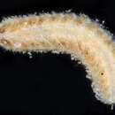
Body subrectangular, tapering slightly anteriorly and posteriorly, flattened ventrally, convex dorsally; middorsum not covered by elytra with globular tubercles and embedded sand grains, forming roughened surface; ventral surface papillate (Johnson, 1897, pl. 9: fig. 56; Berkeley and Berkeley, 1948, fig. 28; Pettibone, 1953, pl. 40: fig. 365). Elytra on large, bulbous elytrophores on segments 2, 4, 5, 7, continuing on alternate segment to end of body; dorsal tubercles on segments without elytra nodular, with small papilla distally (Figures 10A,I,J). Elytra oval to subtriangular, with apex directed toward median line, posterior 4 pairs arranged fanlike; elytra stiff, with concentric rings, thicker on medial side, forming diagonally raised ridge, with embedded sand grains medioposteriorly, thinner near posterior and lateral borders, with long fringes of knobbed papillae on medial, posterior, and lateral margins (Figure 10M; Johnson, 1897, pl. 9: figs. 56, 59; Pettibone, 1953, pl. 40: figs. 365, 368, 369; Hartmann-Schröder, 1965, figs. 41, 46, 47).
Prostomium and first or tentacular segment fused; prostomium subrectangular, wider than long; 2 pairs of closely approximated eyes, anterior pair slightly larger than posterior pair; median antenna with long cylindrical ceratophore on anterior border, long style bulbous subdistally, with long papillae and filiform clavate tip; tentaculophores anterior and lateral to prostomium, each with curved aciculum and projecting conical acicular lobe, 2 bundles of long capillary notosetae: outer dorsal and inner ventral, long tentacular cirrus, similar to median antenna, on dorsal side, and prominent papilla on ventral side (called rudimetary tentacular cirrus by Hartmann-Schröder, 1965); stout palps lateral and ventral to tentaculophores (Figure 10A–E; Johnson, 1897, pl. 9: fig. 56; Hartmann-Schröder, 1965, figs. 41, 42a). Second or buccal segment with first pair of bulbous elytrophores, biramous parapodia, and long papillate ventral buccal cirri on papillate cirrophores lateral to ventral mouth; notosetae numerous, curved, spinose, capillary; neurosetae compound, with rather long spinose blades and spinose shafts; upper and lower lips of mouth papillate (Figure 10A,B,F–H; Hartmann-Schröder, 1965, figs. 41, 42b, 45a–c). Pharynx with 9 pairs of border papillae and 2 pairs of jaws (Figure 10N).
Parapodia with notopodium shorter than neuropodium; notopodium rounded, with low ciliated patches on dorsal side and continuing to ventral sides of elytrophores and dorsal tubercles, latter with small papilla distally; neuropodium with subconical presetal acicular lobe with 2 long papillae distally, postsetal lobe truncate, with numerous short papillae; ventral cirri short, subulate, papillate (Figure 10I,J; Hartmann-Schröder, 1965, fig. 42c). Notosetae numerous, long, strongly curved dorsally changing to slightly curved below, spinose, capillary (Figure 10K; Hartmann-Schröder, 1965, fig. 43). Neurosetae stouter than notosetae, compound, shafts smooth or spinose subdistally, blades short, upper ones slightly longer, smooth or faintly spinose (Figure 10L; Hartmann-Schröder, 1965, fig. 44; 1977, fig. 18). Ventral cirri short, subulate, with few papillae (Figure 10I,J). Pygidium with pair of anal cirri, similar to median antenna.
Young specimen from Peru (holotype of Pareupholoe fimbriata) 4 mm long, 1.5 mm wide, 27 segments. Middorsum with globular papillae (Figure 11A); elytra covered with foreign material and sand grains (Figure 11F; Hartmann-Schröder, 1962a, pl. 1: fig. 11a–d). Usual arrangement of elytrophores and dorsal tubercles (latter called dorsal cirri by Hartmann-Schröder); dorsal tubercles with small distal papilla (Figure 11B,C). Prostomium and tentacular segment fused; median antenna with ceratophore and distal papillate style; lateral tentaculophores with notosetae and single tentacular cirrus (called lateral antenna by Hartmann-Schröder), similar to median antenna (Figure 11A; Hartmann-Schröder, 1962a, pl. 1: fig. 5, pl. 2: fig. 6). Parapodia with notopodia about as long as neuropodia; neuropodial presetal conical acicular lobe with single distal papilla (Figure 11B,C; Hartmann-Schröder, 1962a, pl. 3: figs. 8, 9). Notosetae slender, spinose, capillary, sharply and gradually curved (Figure 11D); stouter compound neurosetae with blades, spinose, long to short, shafts with or without distal spinose rows (Figure 11E).
DEVELOPMENT.—Blake (1975:30) doubtfully referred two planktonic larvae from Tomales Bay, California, a metatrochophore (fig. 3A) and a post-larva (fig. 3B), to Pholoe minuta. He pointed out some differences between his larvae and the larvae described by Cazaux (1968, figs. 1–5) for Pholoe synopthalmica from France. They also show differences from the larvae illustrate by Åkesson (1963, figs. 18, 19) for Pholoe minuta from the Isefjord, Denmark, and for the same species from Ireland illustrated by Heffernan and Keegan (1988, figs. 1–5). The latter group may be considered to be the typical planktotrophic larval forms for species of Pholoe.
The larvae described by Blake (1975:30) are referred herein to Pholoides asperus. The 8-setiger post-larva (800 μ long, 470 μ wide) showed a rounded prostomium, 3 pairs of eyespots, a median antenna with papillae; tentaculophores of the first segment, lateral to the prostomium, with capillary notosetae and a single papillate tentacular cirrus; 7 pairs of biramous parapodia with simple capillary notosetae and compound falcigerous neurosetae; 4 pairs of elytra on lateral side of body on segments 2, 4, 5, 7; a pair of papillate anal cirri similar to the tentacular cirri; pharynx in medial part of segment 2; and large gut filled with oil globules. Its sluggish behaviour and dense nature of the gut contents suggested that it was lecithotrophic and still subsisting on its yolk reserve (Blake, 1975:30).
The 5-setiger post-larva, figured by Sveshnikov (1967:134, fig. 2A) for Pholoe minuta from the Sea of Japan, resembles Pholoides, rather than Pholoe.
DISTRIBUTION.—Eastern Pacific from Southeast Alaska to California, the Galapagos Islands, and Chile, in low water to 353 meters, crawling on stones, crevices, and mixed bottoms.
- bibliographic citation
- Pettibone, Marian H. 1992. "Contribution to the polychaete family Pholoidae Kinberg." Smithsonian Contributions to Zoology. 1-22. https://doi.org/10.5479/si.00810282.532