-
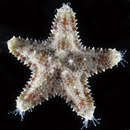
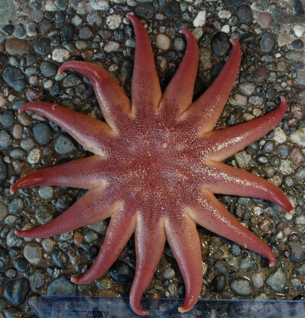
Some S. dawsoni have a color pattern on the aboral surface, but note there are no blue-gray stripes running down the rays.
-
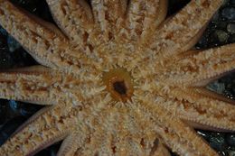
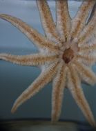
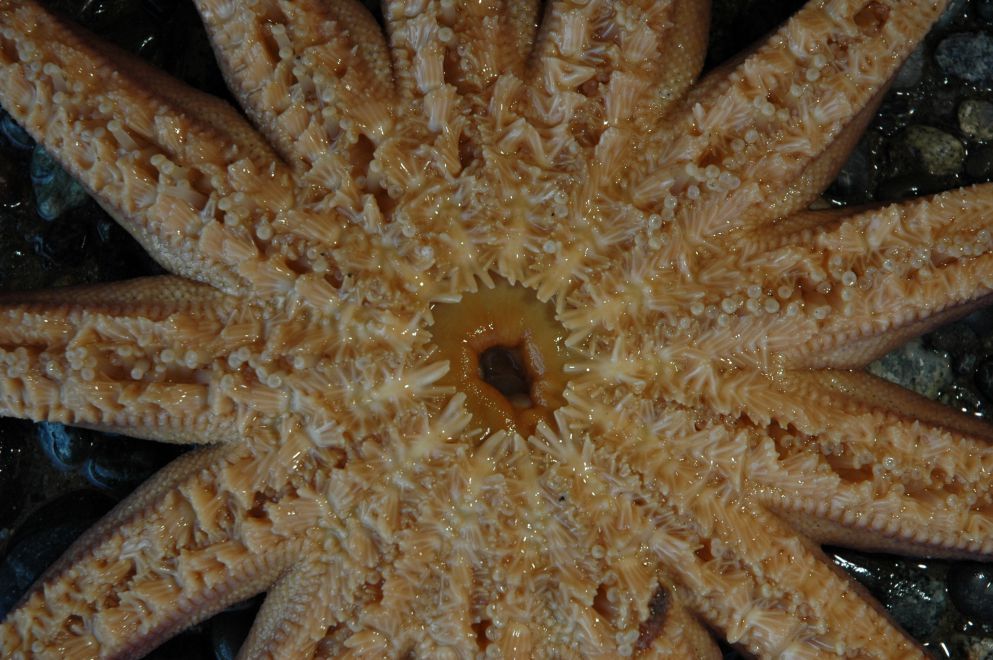
A view of the oral side of S. dawsoni
-
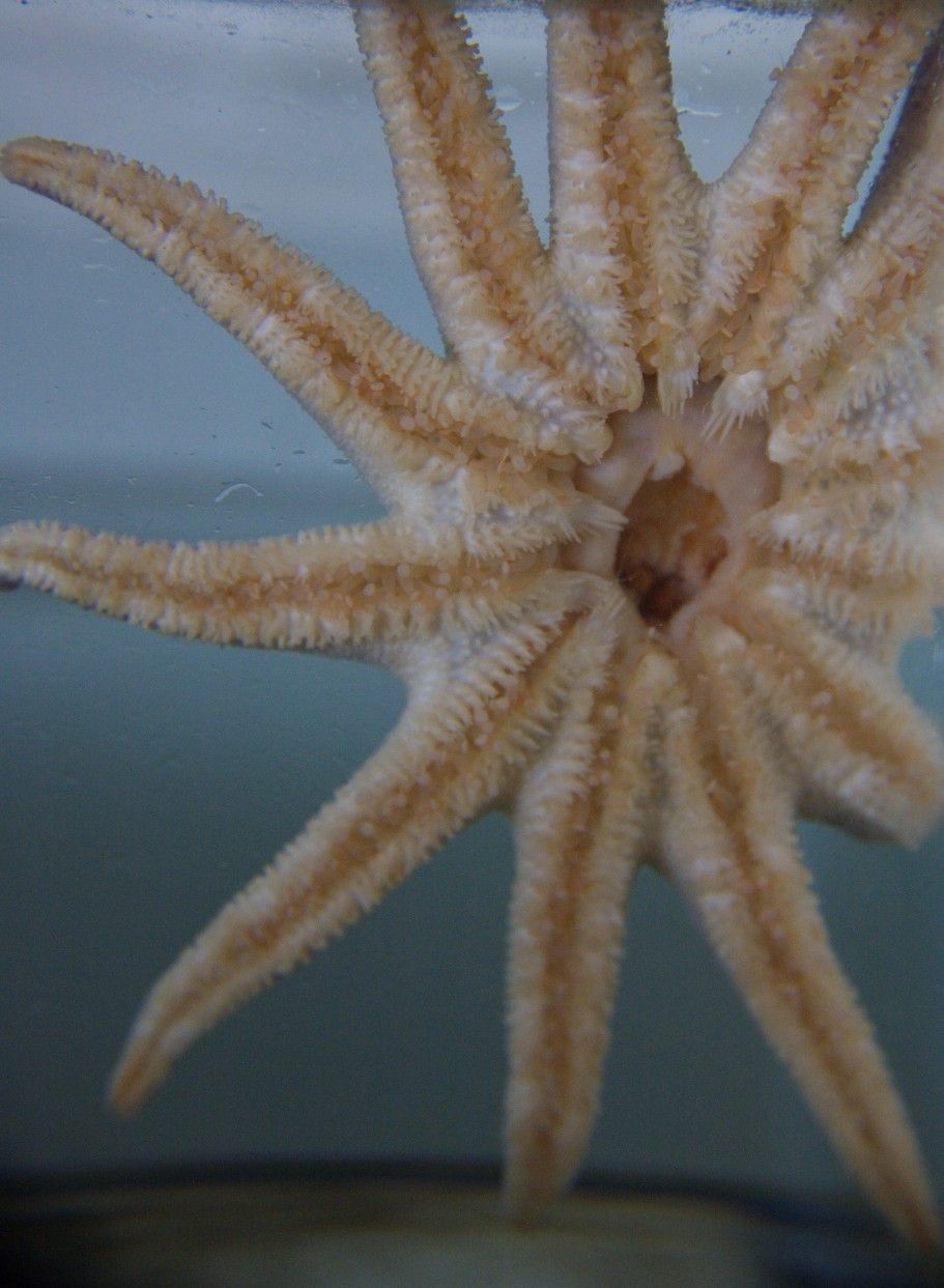
Another view of the open mouth, this time underwater through aquarium glass.
-
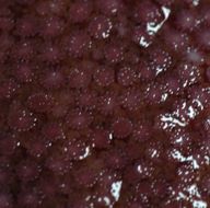
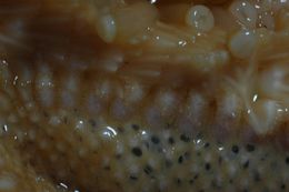
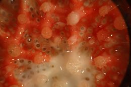
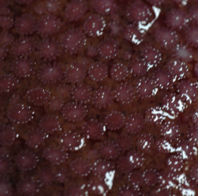
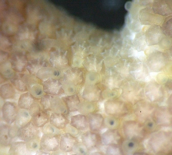
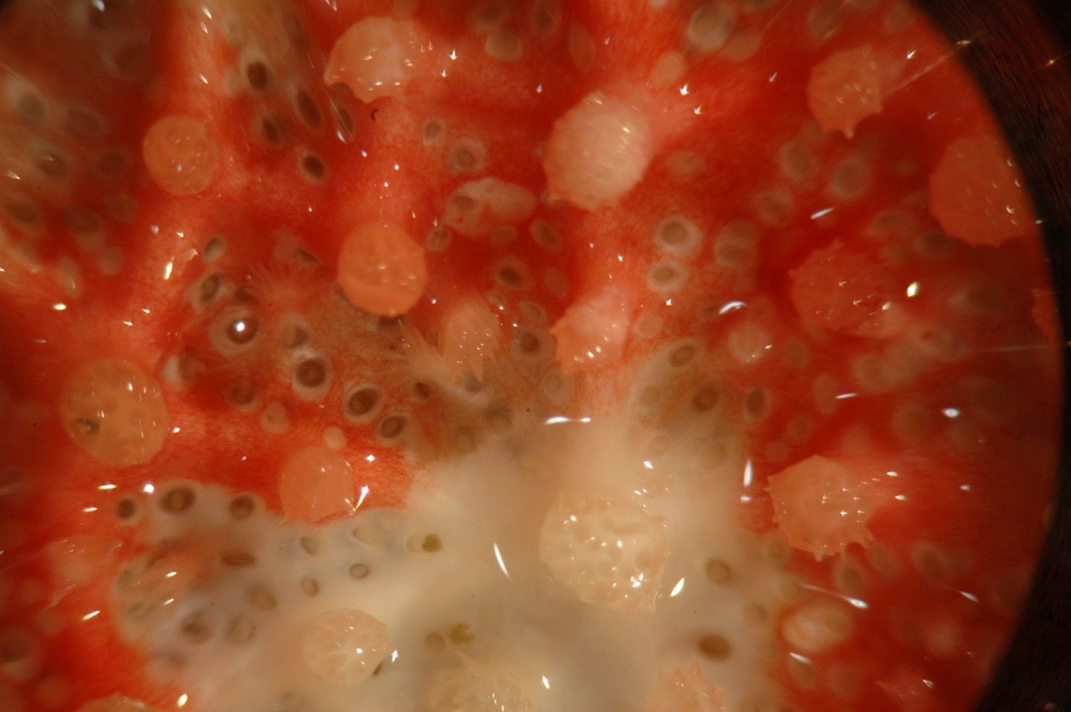
The aboral ossicles or paxillae are well separated. These are magnified.
-
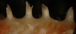
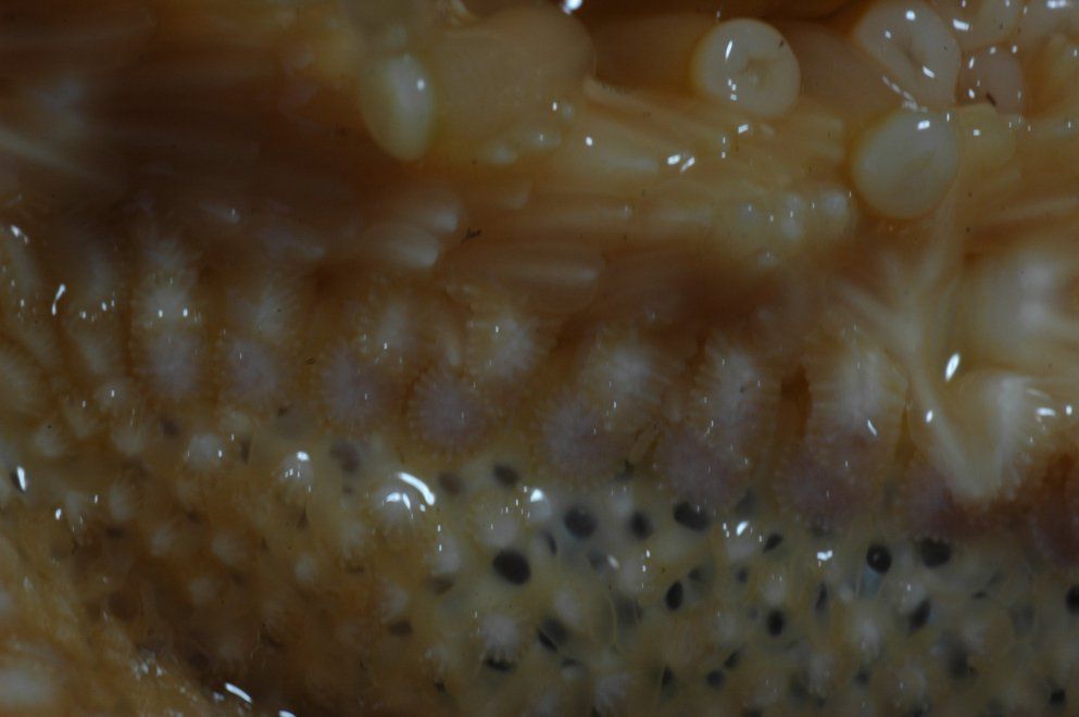
The ossicles along the edge of the ambulacral groove (which is at the top in this photo) are enlarged into marginal plates.
-
This S. dawsoni is swallowing a Leptasterias hexactis that it captured. Notice also the commensal Arctonoesp polychaete scaleworm on the ray.
-
This Solaster dawsoni (left) was found eating this Dermasterias imbricata on the right at low tide. Photo by Brooke Reiswig, July 2006
-
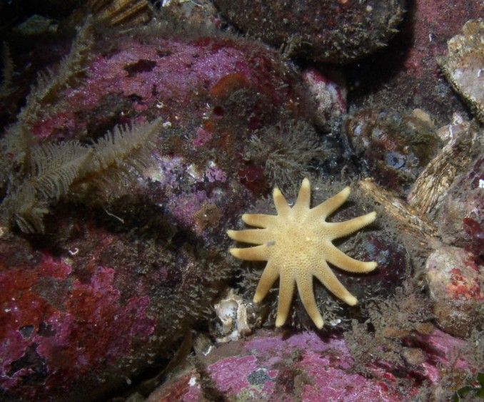
A tiny Solaster dawsoni among hydroids. Underwater photo by Kirt Onthank, August 2007
-
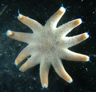
This small individual is about 2.5 cm in total diameter. Photo by Dave Cowles, July 2012
-
The paxillae of the small individual shown above look different from those of adults. The sack-like prejections are papulae. Photo by Dave Cowles, July 2012
-
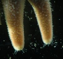
A closeup of the ray tips of the small individual above. Photo by Dave Cowles, July 2012
-
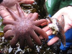
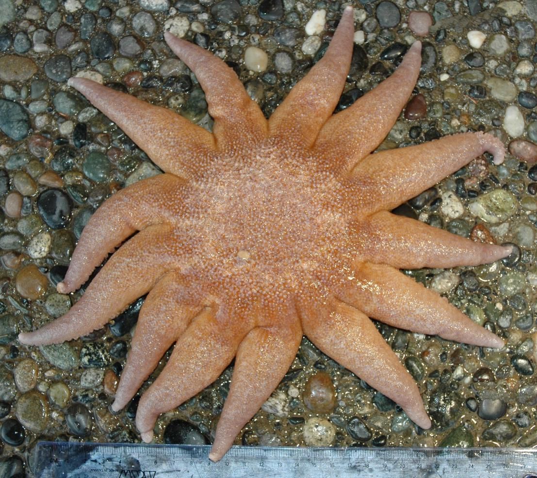
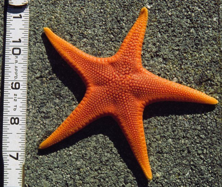
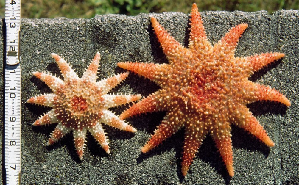
Solaster dawsoni collected from near Northwest Island, WA. Scale is in centimeters (Photo by: Dave Cowles, August 2005)
-
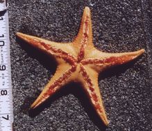
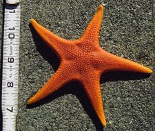
An orange individual from a San Simeon, CA tidepool. About 15 cm diameter. Photo by Dave Cowles, May 1995 These individuals were in the bay at Bodega Marine Lab, California. Photograph by Dave Cowles, August 2010
-
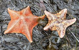
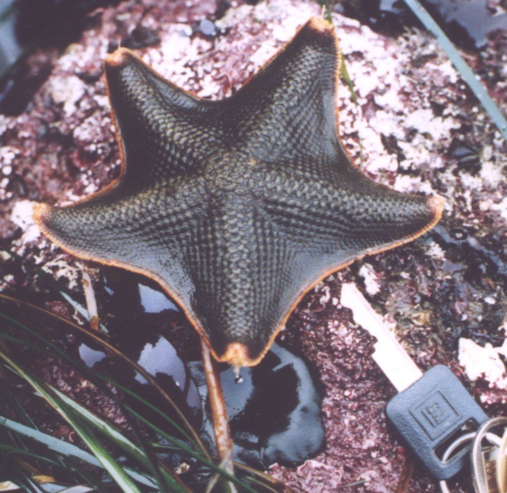
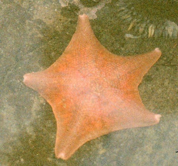
A small Patiria miniata at Cape Flattery, WA (Photo by: Dave Cowles, July 2001)
-
-
Closeup of the aboral surface of Mediaster aequalis. Note the round clusters of ossicles and the madreporite. Photo by Dave Cowles, July 2000
-
A view of the underside. Photo by Dave Cowles, July 2000
-
Mediaster aequalis from 100 m depth, San Juan Channel (Photo by: Dave Cowles, July 2000)
-
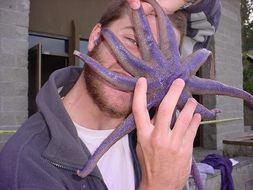
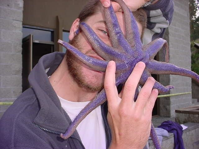
This individual is more blue than usual and appears to be devouring Robbie Wheeling. Note tape on lab in background--the major 2002 lab fire had just occurred.
-
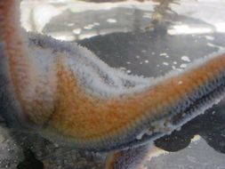
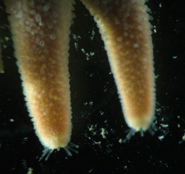
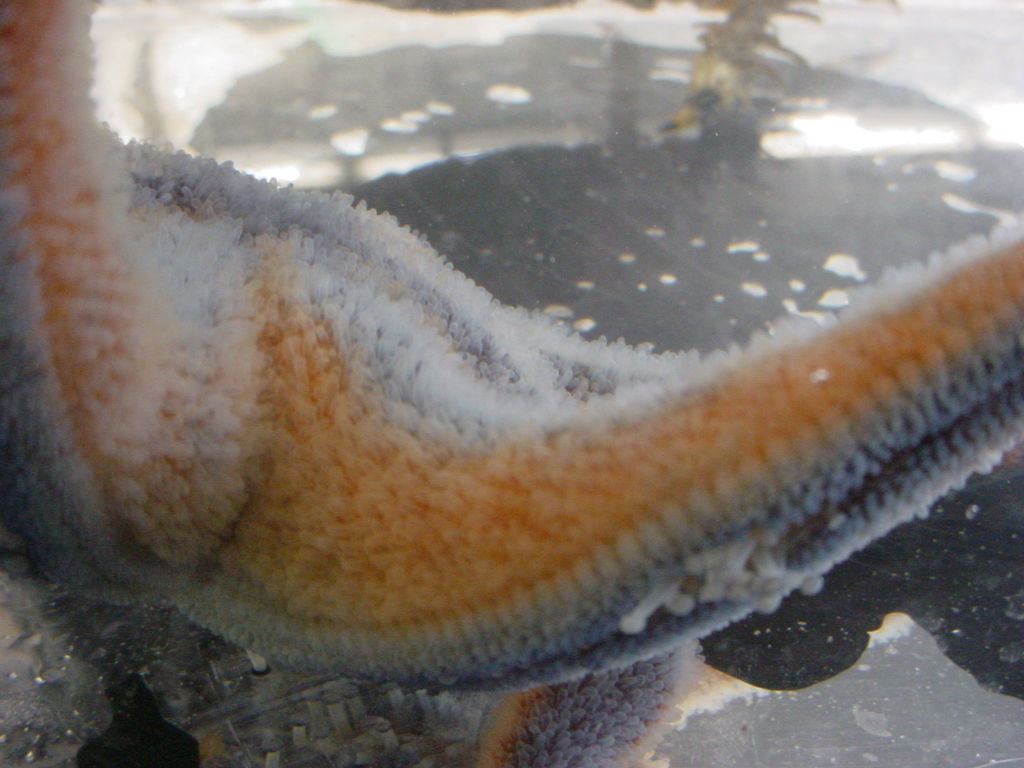
Seasters breathe and rid themselves of wastes via coelomic pouches or papulae that they extrude through their skin when underwater. The extruded papulae give them a fuzzy appearance when underwater. Photo of Solaster stimpsoni by Dave Cowles, Jule 2005.
-
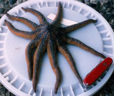
Solaster stimpsoni subtidal from Sares Head, WA. (Photo by: Dave Cowles, 1997)
-
Various color forms can be found. Animals from 100 m depth, San Juan Channel. Photo by Dave Cowles, July 2000
-
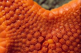
This species has large aboral paxillae, as seen in this closeup.
-
The edges of the rays have long, spiny paxillae.