Platynereis dumerilii gehört zur Gruppe der Borstenwürmer.[2] Die Art wurde zuerst in die Gattung Nereis eingeordnet[3] und später Platynereis zugeordnet.[4] Platynereis dumerilii lebt in gemäßigten bis tropischen Zonen in küstennahen marinen Gewässern, und kann an vielen Orten gefunden werden: Bei den Azoren, im Mittelmeer, in der Nordsee, im Ärmelkanal und im Atlantik bis runter zum Kap der Guten Hoffnung, im Schwarzen Meer, im Roten Meer, im Persischen Golf, im Japanischen Meer, im Pazifik und bei den Kerguelen.[4] Platynereis dumerilii ist heute ein wichtiges Labortier,[5] gilt als lebendes Fossil[6] und wird für viele phylogenetische Untersuchungen als Modellorganismus angesehen. Platynereis dumerilii wird 3 bis 18 Monate alt,[5] die Männchen werden 2 bis 3 cm lang, die Weibchen 3 bis 4 cm.[7]
Platynereis-dumerilii-Würmer bauen Wohnröhren auf ihrem Substrat. Das Substrat können mit Algen bedeckte Hartböden,[8] Seegras,[9][10] Flöße aus pelagischen Sargassum in der Sargassosee[11][12] oder sogar verrottende Pflanzenreste sein.[13] Platynereis dumerilii lebt gewöhnlich in einer Tiefe von 0 bis 5 m[14][15][9][8] und ist daher typisch für flache helle infra-litorale Umgebungen.[14] Aber Platynereis dumerilii wurde auch auf einer Boje in 50 m[16] und auf verrottenden Seetang in 100 m gefunden[17] und kann auch in weniger günstigen Umgebungen leben, wie z. B. in verschmutzten Gebieten in der Nähe von Abwassereinleitungen.[18] Platynereis dumerilii dominiert verschmutzte Gewässer.[19][20] Auch in Bezug auf den pH-Wert zeigt die Art, ungewöhnlich für Meeresorganismen, eine große ökologische Plastizität.[21]
Platynereis dumerilii ist getrenntgeschlechtlich:[22] Bei der Paarung umkreist das Männchen das Weibchen, dabei schwimmt das Weibchen selbst in kleinen Kreisen. Beide geben Eier und Spermien ins Wasser, was durch Sexualpheromone ausgelöst wird. Die Eier werden im Wasser außerhalb des Körpers befruchtet.[23] Platynereis dumerilii hat, wie andere Nereididen, keine segmentalen Gonaden: Die Eizellen reifen frei schwimmend in der Leibeshöhle (Coelom)[22] und färben den Körper des reifen Weibchens gelb.[1]
Platynereis dumerilii entwickelt sich sehr stereotypisch zwischen Gelegen und daher kann die Zeit benutzt werden um das Stadium der Larven von Platynereis dumerilii zu bestimmen. Jedoch beeinflusst die Temperatur die Entwicklungsgeschwindigkeit sehr.[1] Daher gelten die die folgenden Entwicklungszeiten für 18 °C:
Nach 24 Stunden schlüpft aus einem befruchteten Ei eine Trochophora-Larve, die mit 48 Stunden zur Metatrochophora wird.[1] Sowohl Trochophora als auch Metatrochophora schwimmen mit einem Wimpernkranz im Wasser und sind positiv phototaktisch.[24] Die Metatrochophora besitzt neben den Larvalaugen bereits die Anlagen für die komplizierter gebauten definiten Augen des erwachsenen Wurmes.[25][26] Einen Tag später, mit 72 Stunden wird aus der Metatrochophora eine Nektochaeten-Larve. Die Nektochaeten-Larve hat bereits drei Segmente, jedes mit einem Paar Parapodien, die Borsten tragen, welche der Fortbewegung dienen.[1] Die Nektochaeten-Larve kann zwischen positiver und negativer Phototaxis wechseln.[27] Fünf bis sieben Tage nach Befruchtung fangen die Larven an zu fressen und entwickeln sich mit individueller Geschwindigkeit abhängig vom Nahrungsangebot. Nach drei bis vier Wochen, wenn sechs Segmente gebildet worden sind, bildet sich der Kopf mit den Antennen und Mundwerkzeugen.[1]
Platynereis-dumerilii-Larven besitzen zwei Arten von Photorezeptorzellen: Rhabdomerische und ziliäre Photorezeptorzellen.
Die ziliären Photorezeptorzellen befinden sich tief im Larvengehirn. Sie werden nicht durch Pigment beschattet, daher nehmen sie Licht von allen Seiten wahr. Sie gleichen molekular und morphologisch den Zapfen und Stäbchen des menschlichen Auges und exprimieren außerdem ein ziliäres Opsin, das den visuellen ziliären Opsinen der Stäbchen und Zapfen der Wirbeltiere mehr ähnelt als den visuellen rhabdomerischen Opsinen der Wirbellosen. Daraus wird geschlossen, dass das Urbilaterium, der letzte gemeinsame Vorfahre von Weichtieren, Arthropoden und Wirbeltieren, bereits ziliäre Photorezeptorzellen besaß.[28][29][30] Das ziliäre Opsin ist UV-empfindlich (λmax = 383 nm),[31] daher reagieren die ziliären Photorezeptorzellen auf ungerichtetes UV-Licht und lassen die Larven nach unten schwimmen. Dies bildet mit Phototaxis von den rhabdomerischen Photorezeptorzellen der Augen einen farbbasierten Tiefenmesser.[32]
Eine rhabdomerische Photorezeptorzelle bildet mit einer Pigmentzelle ein einfaches Auge.[25] Ein Paar dieser Augen vermittelt Phototaxis in der frühen Trochophora-Larve von Platynereis dumerilii.[24] In der späteren Nektochaeten-Larve wird Phototaxis durch die komplexeren definiten Augen vermittelt.[27] Die definiten Augen exprimieren wenigstens drei Opsine: Zwei rhabdomerische Opsine und ein Go-Opsin.[26][33] Die drei Opsine dort vermitteln Phototaxis alle auf die gleiche Weise durch Depolarisation,[33] obwohl ein Muschel-Go-opsin bekannt ist, das hyperpolarisiert.[34][35]
Das Genom von Platynereis dumerilii ist diploid (2n Chromosomen) und hat einen haploiden Satz von n = 14 Chromosomen.[7][36] Es enthält ungefähr 1 Gbp (Gigabasenpaare) oder 10 9 Basenpaare.[37] Diese Genomgröße liegt nahe dem für andere Tiere beobachteten Durchschnitt. Verglichen mit vielen klassischen wirbellosen molekularen Modellorganismen ist dieses Genom eher groß und daher eine Herausforderung, um genregulatorische Elemente zu identifizieren, die weit von dem entsprechenden Promotor entfernt sein können. Aber es ist intronreich, anders als die Genome von Drosophila melanogaster und Caenorhabditis elegans und ähnelt folglich stärker den Wirbeltier-Genomen einschließlich des menschlichen Genoms.[38]
Platynereis dumerilii gehört zur Gruppe der Borstenwürmer. Die Art wurde zuerst in die Gattung Nereis eingeordnet und später Platynereis zugeordnet. Platynereis dumerilii lebt in gemäßigten bis tropischen Zonen in küstennahen marinen Gewässern, und kann an vielen Orten gefunden werden: Bei den Azoren, im Mittelmeer, in der Nordsee, im Ärmelkanal und im Atlantik bis runter zum Kap der Guten Hoffnung, im Schwarzen Meer, im Roten Meer, im Persischen Golf, im Japanischen Meer, im Pazifik und bei den Kerguelen. Platynereis dumerilii ist heute ein wichtiges Labortier, gilt als lebendes Fossil und wird für viele phylogenetische Untersuchungen als Modellorganismus angesehen. Platynereis dumerilii wird 3 bis 18 Monate alt, die Männchen werden 2 bis 3 cm lang, die Weibchen 3 bis 4 cm.
Platynereis dumerilii is a species of annelid polychaete worm.[3] It was originally placed into the genus Nereis[1] and later reassigned to the genus Platynereis.[4] Platynereis dumerilii lives in coastal marine waters from temperate to tropical zones. It can be found in a wide range from the Azores, the Mediterranean, in the North Sea, the English Channel, and the Atlantic down to the Cape of Good Hope, in the Black Sea, the Red Sea, the Persian Gulf, the Sea of Japan, the Pacific, and the Kerguelen Islands.[4] Platynereis dumerilii is today an important lab animal,[5] it is considered as a living fossil,[6][7][8] and it is used in many phylogenetic studies as a model organism.
Platynereis dumerilii is a small marine ragworm: Males reach a length of 2 to 3 cm, while females reach a length of 3 to 4 cm.[9] Like a number of invertebrate phyla, Platynereis dumerilii has an axochord, a paired longitudinal muscle that displays striking similarities to the notochord regarding position, developmental origin, and expression profile.[10] Its early trochophore larva has a pair of the simplest eyes in the animal kingdom, each eye consists only of a photoreceptor cell and a pigment cell.[11]
P. dumerilii worms have a ciliated surface which beats synchronously to drive locomotion and fluid flow. Larvae have segmental multiciliated cells that regularly display spontaneous coordinated ciliary arrests, which compose the ciliomotor circuitry in the worms. Whole-body coordination of ciliary locomotion is performed by a "stop-and-go pacemaker system".[12]
As the worms develop, they use chaetae, and then parapodia, for locomotion. Unlike other polychaetes, in Platynereis larvae, the parapodia are used only for navigation while the cilia are responsible for propulsive force.[2]
Platynereis dumerilii larvae possess two kinds of photoreceptor cells: Rhabdomeric and ciliary photoreceptor cells.
The ciliary photoreceptor cells are located in the deep brain of the larva. They are not shaded by pigment and thus perceive non-directional light. The ciliary photoreceptor cells resemble molecularly and morphologically the rods and cones of the human eye. Additional, they express an ciliary opsin that is more similar to the visual ciliary opsins of vertebrate rods and cones than to the visual rhabdomeric opsins of invertebrates. Therefore, it is thought that the urbilaterian, the last common ancestor of mollusks, arthropods, and vertebrates already had ciliary photoreceptor cells.[13] The ciliary opsin is UV-sensitive (λmax = 383 nm),[14] and the ciliary photoreceptor cells react on non-directional UV-light by making the larvae swimming down. This forms a ratio-chromatic depth-gauge with phototaxis of the rhabdomeric photoreceptor cells of the eyes.[15]
A rhabdomeric photoreceptor cell forms with a pigment cell a simple eye.[16] A pair of these eyes mediate phototaxis in the early Platynereis dumerilii trochophore larva.[11] In the later nectochaete larva, phototaxis is mediated by the more complex adult eyes.[17] The adult eyes express at least three opsins: Two rhabdomeric opsins and a Go-opsin.[18][19] The three opsins there mediate phototaxis all the same way via depolarization,[19] even so a scallop Go-opsin is known to hyperpolarize.[20][21]
P. dumerilii senses chemicals with four types of organs: The antennae, the palps, the nuchal organs, and the tentacular cirri. These organs detect food and chemical cues such as alcohols, esters, amino acids, and sugars.[22]
Among the four types, the antennae are the primary chemosensory organs and sense a broad range of chemicals, while the palps are specialized on taste, which means they detect food-related chemicals. The cirri are thin thread-like head appendages and are specialized in tactile sensation, but can also give spatial information from were a chemical cue is coming, since a single stimulus can elicit in the left and right cirrus a response at a different times.[22] The cirri also sense light: When they are shaded, the worm retreats rapidly into its tube to protect them. This behavior is called a shadow reflex.[23] The nuchal organ is a singular ciliated pit in P. dumerilii. Among annelids, nuchal organs are conserved and seem to have an important chemosensory function. However, what their exact function is, is still unclear.[22]
The signals from the four chemosensory organs are processed in a lateral region and in the mushroom bodies.[22] The mushroom bodies in annelids resemble those in insects by anatomy, morphology and gene expression. So probably, annelids and insects inherited mushroom bodies from their last common ancestor.[24]
Platynereis dumerilii builds tubes on its substrate. The substrate may be algae-covered hard bottoms,[25] sea grass,[26][27] pelagic Sargassum rafts in the Sargasso Sea,[28][29] or even rotting plant debris.[30] Platynereis dumerilii commonly lives in depths of 0 to 5 meters,[31][32][26][25] and so is typical for shallow bright infra-littoral environments.[31] However, it has been also found on a buoy at 50 meters[33] and on rotting seaweed at 100 m.[34] It may also live in less favorable environments, like at thermal vents[35][36] or polluted areas near sewer outfall pipes.[37] It dominates polluted areas[38][39] and acidic areas with pH values around 6.5[40] fitting the preferred pH value of a subpopulation of late Platynereis dumerilii nectochaete larvae.[41] Larvae feed on plankton, and migrate vertically in the ocean in response to changes in light, causing a daily transport of biomass.[42]
Platynereis dumerilii is dioecious, that means it has two separate sexes.[43] Changes in light are importantly linked to reproduction. The bristle worm is originally found in the Bay of Naples, where it displays reproductive synchrony. The adult worms rise en masse to the water surface a few days after the full moon, during a one- to two-hour dark portion of the night between sunset and moonrise. In the worm’s natural environment, it is important to synchronize spawning to increase the potential for gametes to meet and fertilize. By detecting nighttime lighting in accordance with the lunar cycle, the worms synchronize reproductive activity. Worms that make L-Cry protein are better able to detect appropriate light conditions and synchronize the release of gametes. In addition, the molecule r-Opsin is extremely sensitive to light, and appears to help detect moonrise. Some combination of signals from r-Opsin and L-Cry is believed to help the worms to coordinate rising at a common time to spawn.[44][45][46]
During mating, the male swims around the female while the female is swimming in small circles. Both release eggs and sperm into the water. This release is triggered by sexual pheromones. The eggs are then fertilized outside of the body in the water.[47] Like other Nereidids, Platynereis dumerilii has no segmental gonades, the oocytes mature freely swimming in the body cavity (coelom),[43] and stain the body of the mature female epitoke yellow.[2]
Platynereis dumerilii develops very stereotypically between batches and therefore time can be used to stage Platynereis dumerilii larvae. However, the temperature influences the speed of development greatly.[2] Therefore, the following developmental times are given with 18 °C as reference temperature:
After 24 hours, a fertilized egg gives rise to a trochophore larva. At 48 hours, the trochophore larva becomes a metatrochophore larva.[2] Both trochophore and metatrochophore swim with a ring of cilia in the water and are positively phototactic.[11] The metatrochophore has, beside the larval eyes, already the anlagen for the more complex adult eyes of the adult worm.[16][18] A day later, at 72 hours after fertilization, the metatrochophore larva becomes a nectochaete larva. The nectochaete larva already has three segments, each with a pair of parapodia bearing chaetae, which serve for locomotion.[2] The nectochaete larva can switch from positive to negative phototaxis.[17] After five to seven days, the larvae start feeding and develop on their own speed, depending on food supply. After three to four weeks, when six segments have formed, the head is formed.[2]
Normal development is subdivided into 16 stages.[2] Platynereis dumerilii lives for 3 up to 18 months[5] with an average lifespan of seven months. P. dumerilii reproduces only once,[42] and dies after delivering its gametes.[2]
The genome of Platynereis dumerilii is diploid (2n chromosomes) with a haploid set of n = 14 chromosomes.[9][48] It contains approximately 1 Gbp (giga base pairs) or 109 base pairs.[49] This genome size is close to the average observed for other animals. However, compared to many classical invertebrate molecular model organisms, this genome size is rather large and therefore it is a challenge to identify gene regulatory elements that can be far away from the corresponding promoter. But it is intron rich unlike those of Drosophila melanogaster and Caenorhabditis elegans and thus closer to vertebrate genomes including the human genome.[50]
Bristle worms contain the complex protein haemoglobin, found in vertebrates, annelids (e.g. earthworms), molluscs (e.g. pond snails) and crustaceans (e.g. daphnia). It was once believed that haemoglobin must have evolved multiple times to be a feature of such different species.Comparing bristle worms with other red blooded species suggests that all forms of haemoglobins are derived from a single ancestral gene, cytoglobin.[51][52]
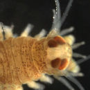
{{cite journal}}: CS1 maint: date and year (link) 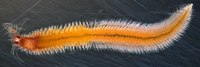 Female epitoke of Platynereis dumerilii: Its body is filled with yellow eggs.
  Male epitoke of Platynereis dumerilii: Its frontal part is filled with white sperm, while its rear is red due to blood vessels.
 Platynereis dumerilii is a species of annelid polychaete worm. It was originally placed into the genus Nereis and later reassigned to the genus Platynereis. Platynereis dumerilii lives in coastal marine waters from temperate to tropical zones. It can be found in a wide range from the Azores, the Mediterranean, in the North Sea, the English Channel, and the Atlantic down to the Cape of Good Hope, in the Black Sea, the Red Sea, the Persian Gulf, the Sea of Japan, the Pacific, and the Kerguelen Islands. Platynereis dumerilii is today an important lab animal, it is considered as a living fossil, and it is used in many phylogenetic studies as a model organism.
Platynereis dumerilii est une espèce de vers annélides marins appartenant à la phylogénie des lophotrochozoaires[2] qui constitue la troisième grande branche des bilatériens (avec les deutérostomiens et les ecdyzozoaires).
Il possède des caractéristiques anatomiques supposées être déjà présentes chez l’Urbilatérien (dernier ancêtre commun aux bilatériens). Il peut être trouvé dans les zones marines côtières tempérées à tropicales. Un dimorphisme sexuel existe chez Platynereis dumerilii : les femelles sont de couleur jaune, tandis que les mâles ont leur côté antérieur blanc et leur côté postérieur rouge à l'âge adulte[3]. Cet animal constitue un bon organisme modèle en génétique et en biologie cellulaire et animale. La longueur de cet organisme est comprise entre 3 et 4 cm chez les femelles, et entre 2 et 3 cm chez les mâles[4].
L’organisation de Platynereis dumerilii est segmentale[5]. Elle comprend :
Platynereis dumerilii est un animal à sexes séparés. Au cours de son développement, il passe tout d'abord par un stade dit juvénile puis il subit une maturation sexuelle observable à travers une modification du phénotype. Les mâles vont perdre leur tube digestif et accumuler des spermatozoïdes tout au long de la maturation sexuelle, observable par la couleur blanche au niveau antérieur. Les femelles, quant à elles, vont avoir une accumulation d'ovules dans la cavité abdominale au cours du temps observable par la coloration jaune de l'animal.
La maturation sexuelle de Platynereis dumerilii est circalunaire, autrement dit elle dépend des différentes phases de la lune. En effet, la proportion d'animaux atteignant la maturité sexuelle est maximale après une nouvelle lune, à contrario elle est minimale durant les périodes de pleine lune. À la suite de la maturation sexuelle, s'engage une danse nuptiale entre le mâle et la femelle. Le mâle tourne autour de la femelle pendant que cette dernière nage de manière circulaire. Le mâle libère ses spermatozoïdes et la femelle ses ovules dans le milieu : la reproduction est dite externe. Après relargage des gamètes mâles et femelles, les deux partenaires meurent d'épuisement. Durant la reproduction sexuelle entre un mâle et une femelle, il est possible d'avoir plusieurs centaines d’œuf.
Après fécondation, mise en place d'une cellule œuf d'environ 160 µm qui se développe en passant par un stade planctonique (ne luttant pas contre le courant), un stade benthique (où elle se retrouvera plus en profondeur), un stade tubicole et un stade pélagique après la maturation sexuelle.
Les larves de Platynereis dumerilii possèdent deux types de cellules photoréceptrices[6] : les cellules photoréceptrices rhabdomériques et ciliaires.
Les cellules photoréceptrices ciliaires sont situées dans le cerveau profond de la larve. Elles ne sont pas ombragées par le pigment et perçoivent ainsi la lumière de manière non directionnelle. On note une ressemblance morphologique et moléculaire entre les cellules photoréceptrices ciliaires et les photorécepteurs présents au niveau de l'œil humain.
De plus, elles expriment une opsine plus proche des opsines ciliaires visuelles des bâtonnets et des cônes des vertébrés que des opsines rhabdomériques visuelles des invertébrés. Par conséquent, on pense que l'urbilaterien, le dernier ancêtre commun des mollusques, des arthropodes et des vertébrés avait déjà des cellules photoréceptrices ciliaires[7]. Une cellule photoréceptrice rhabdomérique forme avec une cellule pigmentaire un œil simple[8].
Après éclosion, les larves ont un comportement particulier durant la phase larvaire. Elles appartiennent au plancton, ensemble d'organismes incapable de lutter contre la houle (le mouvement ondulatoire de la surface de la mer). Cependant la larve n’est pas totalement passive, en effet à 24 heures, elle devient phototactique[9]. Lorsqu’il y a émission d'une source lumineuse à un endroit précis, les larves sont attirées et se déplacent dans la direction lumineuse. Ce comportement est très important pour ces larves qui doivent rester près de la surface de l’eau le plus longtemps possible. Au stade 72 heures les larves deviennent benthiques ; elles tombent dans les fonds marins et deviennent lucifuges, c’est-à-dire qu’elles fuient la lumière. Elles se déplacent alors vers des endroits plus sombres.
La taille du génome de Platynereis dumerilii est d'environ 1 Gpb (giga paires de bases) soit 109 paires de bases. Cette taille de génome se rapproche de la moyenne observée pour les métazoaires eucaryotes. Cependant, en comparaison à de nombreuses espèces de modèles moléculaires d'invertébrés classiques, cette taille de génome est plutôt grande et représente par conséquent un défi pour l'identification d'éléments régulateurs de gènes qui peuvent être situés à des distances considérables du promoteur correspondant.
Il s'agit d'un génome diploïde (2n chromosomes) avec un jeu de n = 14 chromosomes. Les efforts conjoints de séquençage de la communauté scientifique ont généré un génome de référence à couverture élevée, pour cet organisme, dérivé de l'acide désoxyribonucléique des spermatozoïdes des vers mâles de la lignée FL2[4].
Platynereis dumerilii est un organisme modèle en biologie et en génétique car c'est un organisme peu volumineux avec une facilité d'entretien. En laboratoire, l'élevage de platynereis se fait dans des chambres dans lesquelles il est possible de régler la lumière pour reproduire les différentes phases de la lune. L'alimentation se fait à l'aide d'épinards bio (les pesticides étant mortels pour cet organisme). Ils sont conservés dans des bacs d'eau de mer.
Platynereis dumerilii est utilisé dans différents domaines :
Bien qu'il s'agisse d'un organisme modèle assez récent, de par son intérêt en sciences et sa facilité de manipulation, de nombreuses techniques ont été développées pour des analyses au niveau :
Platynereis dumerilii est une espèce de vers annélides marins appartenant à la phylogénie des lophotrochozoaires qui constitue la troisième grande branche des bilatériens (avec les deutérostomiens et les ecdyzozoaires).
Il possède des caractéristiques anatomiques supposées être déjà présentes chez l’Urbilatérien (dernier ancêtre commun aux bilatériens). Il peut être trouvé dans les zones marines côtières tempérées à tropicales. Un dimorphisme sexuel existe chez Platynereis dumerilii : les femelles sont de couleur jaune, tandis que les mâles ont leur côté antérieur blanc et leur côté postérieur rouge à l'âge adulte. Cet animal constitue un bon organisme modèle en génétique et en biologie cellulaire et animale. La longueur de cet organisme est comprise entre 3 et 4 cm chez les femelles, et entre 2 et 3 cm chez les mâles.
Platynereis dumerilii ad Annelida attinet.[2] Primum in genere Nereis posita[1] et postea in Platynereis reposita erat.[3] Platynereis dumerilii in maritimis aquis a zona temperata ad tropicae vivit. Potest inveniri ad Azores, in Mediterraneum, Mare Germanicum, Mare Britannicum, et Atlanticum; ac ad Promunturium Bonae Spei, in Mare Nigrum, Mare Rubrum, Sinum persicum, Mare Iaponicum, Pacifici, ad Kerguelen Insulis.[3] Platynereis dumerilii hodie est animal grave laboratorii,[4] acque quam vivum fossile consideratur[5][6][7], et in multis studiis phylogeneticae quam instar animantem usa est. Platynereis dumerilii 3 ad 18 mensis vetus fit.[4] Mares 2 ad 3 cm longi fiunt, dum feminae 3 ad 4 cm longae fiunt.[8]
Platynereis dumerilii est dioeciosus, id est duabus sexu habeat:[9] Inter coniugium, mas circa feminam natat, et femina in circis parvis natat. Utrique ova et sperma in aquam dimittunt. Hoc a pheromona sexualia inducitur. Ova ergo in aqua extra corpus fertilizatur.[10] Platynereis dumerilii, sicut aliae Nereididae, segmentariam gonades non habet, ova libere natantes in cavitate corporis (coelom) maturat.[9]
Platynereis dumerilii inter individua stereotypice desveloppat, et ideo gradus larvae Platynereidis dumerilii a tempore determinari potest. Autem, temperatura celeritatem valde determinat.[11] Ergo haec tempora desveloppamenti ad temperaturam 18 °C referuntur:
Ad 24 horas post fertilizationem, ovum fit larva trochophora. Ad 48 horas, trochophora fit metatrochophora.[11] Utraeque trochophora et metatrochophora cum anulo ciliorum in aqua natant, et positive phototacticae sunt.[12] Una die postea, ad 72 horas post fertilizationem, metatrochophora fit larva nectochaeta. Ea iam tria segmenta habet, quidque cum pare parapodiorum qua ferrunt chaetas, quae pro motui serviunt.[11] Nectochaeta a photoaxe positiva ad negativam mutare potest.[13] Post quinque ad septem dies, larvae pascere initiant et sua celeritate desveloppant, prout copia cibi permittet. Post tres ad quatuor septimanas, sex segmentis formatis caput formatur.[11]
Larvae Platynereidis dumerilii duo genera cellularum photoreceptorum habent: Cellulae photoreceptorum rhabdomericae et ciliares.
Cellulae photoreceptorum ciliares in cerebro alto larvae sunt. A pigmento non umbrantur et ergo lucem undique percipiunt. Cellulae photoreceptorum ciliares similem sunt moleculariter et morphologicaliter virgis et conis oculi humani. Praeteria opsinum exprimunt quod magis similem est opsinis ciliaribus visualis vertebratorum, quam illis opsinis rhabdomeris invertebratorum. Igitur cogitatur, ut urbilaterium, proavus communis ultimus molluscorum, arthropodorum, et vertebratorum, iam cellulas photoreceptorum ciliares habuerint.[14]
Cellula photoreceptorum rhabdomerica cum cellula pigmentorum format oculum simplicem. Par horum oculorum phototaxim in larva trochophora matura Platynereidis dumerilii mediant.[12] In larva nectochaeta postera, phototaxis a oculis definitis complexioribus mediantur.[13] Oculi definiti exprimunt saltem tria opsina: Duo opsina rhabdomerica et unum Go-opsinum.[15][16] Illa tria opsina omnia ibi phototaxim eodem modo depolarizatione mediant,[16] etsi Go-opsinum concae hyperpolarizare notum est.[17][18]
Platynereis dumerilii ad Annelida attinet. Primum in genere Nereis posita et postea in Platynereis reposita erat. Platynereis dumerilii in maritimis aquis a zona temperata ad tropicae vivit. Potest inveniri ad Azores, in Mediterraneum, Mare Germanicum, Mare Britannicum, et Atlanticum; ac ad Promunturium Bonae Spei, in Mare Nigrum, Mare Rubrum, Sinum persicum, Mare Iaponicum, Pacifici, ad Kerguelen Insulis. Platynereis dumerilii hodie est animal grave laboratorii, acque quam vivum fossile consideratur, et in multis studiis phylogeneticae quam instar animantem usa est. Platynereis dumerilii 3 ad 18 mensis vetus fit. Mares 2 ad 3 cm longi fiunt, dum feminae 3 ad 4 cm longae fiunt.
Platynereis dumerilii is een borstelworm uit de familie Nereididae. Het lichaam van de worm bestaat uit een kop, een cilindrisch, gesegmenteerd lichaam en een staartstukje. De kop bestaat uit een prostomium (gedeelte voor de mondopening) en een peristomium (gedeelte rond de mond) en draagt gepaarde aanhangsels (palpen, antennen en cirri).
Platynereis dumerilii werd in 1834 voor het eerst wetenschappelijk beschreven door Audouin & Milne Edwards.
Bronnen, noten en/of referentiesPlatynereis dumerilii é uma espécie de anelídeo pertencente à família Nereididae.
A autoridade científica da espécie é Audouin & Milne Edwards, tendo sido descrita no ano de 1834.
Trata-se de uma espécie presente no território português, incluindo a sua zona económica exclusiva.
Platynereis dumerilii é uma espécie de anelídeo pertencente à família Nereididae.
A autoridade científica da espécie é Audouin & Milne Edwards, tendo sido descrita no ano de 1834.
Trata-se de uma espécie presente no território português, incluindo a sua zona económica exclusiva.
Научная классификация Царство: Животные Тип: Кольчатые черви Класс: Многощетинковые черви Отряд: Aciculata Подотряд: Phyllodocida Семейство: Нереиды Род: Platynereis Вид: Platynereis dumerilii Латинское название Platynereis dumerilii
Platynereis dumerilii (лат.) — вид многощетинковых червей из семейства нереид.
Червь живёт в умеренной и тропической зонах в прибрежных морских водах в Европе, в Средиземном море и на западном побережье Испании. Platynereis dumerilii является важным лабораторным животным, считается живым ископаемым и служит модельным организмом для многих филогенетических исследований. В лаборатории эти животные достигают в длину 3—4 см и живут в среднем 7 месяцев.
Platynereis dumerilii раздельнополы, яйцеклетки и сперматозоиды выделяются при помощи половых феромонов. Самец обвивает тело самки, в то время как самка двигается, совершая маленькие круги. Оплодотворение происходит вне тела в воде. Гонады отсутствуют, зародышевые клетки созревают, свободно плавая в полости тела (целом).
Из оплодотворённого яйца появляется личинка-трохофора, которая через 48 часов переходит в стадию метатрохофоры. Эти личинки свободно плавают в воде при помощи своих ресничек и положительно реагируют на свет (фототаксис). Наряду с личиночными глазами (Pigmentbecherocellen), появляются также зачатки сложно устроенных глаз червя. Через четыре дня становятся заметны три сегмента животных, каждый из которых имеет пару параподий со щетинками, которые используются для передвижения. Только через три—четыре недели, когда образуется шесть сегментов тела, образуется голова с антеннами и ротовой аппарат.
У личинок имеется два типа фоторецепторов: рабдом и ресничковые фоторецепторов. Ресничковые фоторецепторы похожи по форме на палочки и колбочки человеческого глаза, связь, которая может быть подтверждена обнаружением опсина. Отсюда следует вывод, что эволюционное происхождение этих клеток фоторецепторов очень древнее и является общим для моллюсков, членистоногих и позвоночных[1][2].
Platynereis dumerilii (лат.) — вид многощетинковых червей из семейства нереид.
Червь живёт в умеренной и тропической зонах в прибрежных морских водах в Европе, в Средиземном море и на западном побережье Испании. Platynereis dumerilii является важным лабораторным животным, считается живым ископаемым и служит модельным организмом для многих филогенетических исследований. В лаборатории эти животные достигают в длину 3—4 см и живут в среднем 7 месяцев.