Description
provided by Zookeys
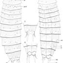
Adult with head, neck and eleven trunk segments (Figs 7A, B, 8A). See Table 4 for measurements, and Table 5 for positions of cuticular structures (sensory spots, glandular cell outlets, and tubules).
Measurements for adult Echinoderes hwiizaa sp. n. (in micrometers). Columns N and SD show sample size and standard deviation, respectively. Abbreviations: (f), female condition of sexually dimorphic character; LD, length of laterodorsal tubule; LTAS, length of lateral terminal accessory spine; LTS, length of lateral terminal spine; LV, length of lateroventral tubule; (m), male condition of sexually dimorphic character; ML, length of midlateral tubule; MSW, maximum sternal width; S, segment length; SW, standard width; TL, trunk length. Character N Range Mean SD TL 8 385–414 400 11.72 MSW-8 9 64–75 69 3.99 MSW-8/TL 8 16.7–19.1% 17.4% 0.75% SW-10 9 52–65 61 4.08 SW-10/TL 8 13.5–16.1% 15.2% 0.89% S1 8 38–47 41 2.53 S2 8 30–41 36 3.66 S3 9 28–32 30 1.56 S4 9 31–33 32 0.71 S5 9 31–38 34 1.79 S6 9 37–43 39 1.66 S7 9 43–48 46 1.94 S8 9 46–56 52 3.45 S9 9 46–56 50 3.2 S10 9 43–52 46 3.42 S11 9 34–46 38 4.16 LV 5 9 18–25 20 2.1 LV 7 9 15–22 34 2.42 ML 8 9 16–22 19 2.36 LV 8 9 16–22 19 1.81 LV 9 9 13–19 17 1.71 LD 10 (m) 5 15–21 18 2.39 LD 10 (f) 4 10–14 12 2.06 LTS 9 46–53 51 2.11 LTS/TL 9 11.6–13.5% 12.7% 0.59% LTAS (f) 4 21–29 26 3.58
Summary of location of cuticular structures, tubules, and spines in Echinoderes hwiizaa sp. n. Abbreviations: (f), female condition of sexually dimorphic character; gco1, type 1 glandular cell outlet; gco2, type 2 glandular cell outlet; LD, laterodorsal; ltas, lateral terminal accessory spine; lts, lateral terminal spine; LV, lateroventral; (m), male condition of sexually dimorphic character; MD, middorsal; ML, midlateral; PD, paradorsal; pe, penile spine; SD, subdorsal; si, sieve plate; SL, sublateral; ss, sensory spot; tu, tubule; VL, ventrolateral; VM, ventromedial. Position MD PD SD LD ML SL LV VL VM segment 1 gco1 ss ss gco1 ss 2 ss gco2 ss, gco2, ss gco2 ss, gco1 3 ss ss ss gco1 4 gco2 ss gco1 5 ss ss gco2 tu gco1, ss 6 ss gco2, ss gco1, ss 7 ss ss gco2 tu ss gco1 8 ss gco2 tu tu gco1 9 ss ss ss si tu ss gco1 10 ss tu ss gco1 11 ss ltas (f), pe (m) lts
Head consists of retractable mouth cone and introvert (Figs 9A, B, 10). Mouth cone with inner oral styles and nine outer oral styles. Exact number and arrangement of inner oral styles not observed. Each outer oral style composed of rectangular basal part and triangular distal part. Basal parts of outer oral styles alternate in size: five large in odd sectors of introvert, and four small in even sectors (Fig. 9A). Posterior to basal part of each outer oral style, two spinose hairs project anteriorly, covering outer oral style (Fig. 9A). Introvert composed of seven rings of scalids and one ring of trichoscalids (Figs 9B, 10). Ring 01 includes ten primary spinoscalids with basal sheath and long, smooth end piece (Fig. 9B). Each basal sheath with three fringes. Proximal fringe extends into three long projections, like a trident, covering next fringe. Middle basal fringe with two lateral projections, overlapping end piece. Distal fringe with five to seven threads projecting between two projections of middle fringe. End piece of primary spinoscalids is longest unit. Rings 02 and 04 with 10 spinoscalids, and rings 03 and 05 with 20 spinoscalids. Spinoscalids of rings 02–05 similar in length. Rings 06 and 07 could not be examined in detail, but at least seven relatively short spinoscalids present in ring 06, and 13 leaf-like scalids in ring 07. Six trichoscalids present each attached with trichoscalid plate in sectors 2, 4, 5, 7, 8, and 10.
Neck with 16 placids (Figs 7A, B, 8B, 10). Midventral placid broadest (ca. 17 μm at basal width and ca. 11 μm at tip width); remaining placids with similar size (ca. 11 μm at basal width and ca. 5 μm at tip width).
Segment 1 consists of complete cuticular ring with pachycyclus at anterior margin (Figs 7A, B, 8B). Non-bracteate cuticular hairs densely cover entire segment (Fig. 7A, B). Paired rounded subdorsal and laterodorsal sensory spots located close to anterior margin of the segment (Figs 7A, 9C). Rounded ventromedial sensory spots centered between anterior and posterior margins (Fig. 7B). Type 1 glandular cell outlets situated anteriorly in middorsal and lateroventral positions (Fig. 7A, B). Posterior part of the segment with pectinate fringe with very short tips (Fig. 7A, B).
Segment 2 also with complete cuticular ring (Fig. 7A, B), with thick pachycyclus at anterior margin (Fig. 8B). All cuticular surface, except anterior and posterior areas covered with bracteate cuticular hairs (Figs 7A, B, 8B, 9C). Oval sensory spots in middorsal, two pairs in laterodorsal, and pair in ventromedial positions (Figs 7A, B, 8B, 9C). Type 2 glandular cell outlets in subdorsal, laterodorsal, and ventrolateral positions (Figs 7A, B, 9C). All type 2 glandular cell outlets of this segment and segment 4–7 situated slightly anterior to sensory spots. In LM observation, type 2 glandular cell outlets show oval or box shaped structure, whereas in SEM observation, they show single large pore (Fig. 9E). Type 1 glandular cell outlets placed close to anterior margin in ventromedial position on this and following eight segments (Fig. 7A, B). Posterior margin of segment with pectinate fringe with longer tips than on preceding segment (Figs 7A, B, 9C).
Segment 3 and following eight segments consist of one tergal and two sternal plates (Fig. 7A, B). Each plate with thicker pachycycli in anterior areas and articulate areas with other plates. Cuticular hairs on this and following seven segments bracteate, covering entire segment except in anterior, posterior, and paraventral areas (Fig. 7A, B). Sensory spots in subdorsal, laterodorsal, and sublateral positions (Figs 7A, B, 8C). Pectinate fringes as on segment 2.
Segment 4 with pair of laterodorsal sensory spots and paired subdorsal type 2 glandular cell outlets (Fig. 7A, B). Pectinate fringes as on segment 2.
Segment 5 with lateroventral tubules (Figs 7B, 8C, 9D). Paired sensory spots in subdorsal, laterodorsal, and ventromedial positions (Figs 7A, B, 8C). Pair of type 2 glandular cell outlets located in midlateral position (Figs 7A, B, 8C). Tips of pectinate fringes similar in length, and longer than those on three preceding segments on this and following four segments.
Segment 6 with paired subdorsal, midlateral, and ventromedial sensory spots (Figs 7A, 8D, 9D). Pair of type 2 glandular cell outlets present in midlateral position (Figs 7A, B, 8D).
Segment 7 with lateroventral tubules (Figs 7B, 8D, 9D). Middorsal and paired laterodorsal and ventrolateral sensory spots present (Figs 7A, B, 8D). Type 2 glandular cell outlets in midlateral position (Figs 7A, B, 8D, 9D).
Segment 8 with midlateral and lateroventral tubules (Figs 7A, B, 8D, 11B). Paired sensory spots in subdorsal position (Fig. 7A). Paired type 2 glandular cell outlets in laterodorsal position, close to midlateral tubules (Figs 7A, B, 8D).
Segment 9 with lateroventral tubules (Figs 7B, 8E, 11A, B). Paired paradorsal, laterodorsal, midlateral, and ventrolateral sensory spots present (Figs 7A, B, 8E, 11A, B, C). Sieve plates with narrow, oval sieve area and single posterior pore present in sublateral position (Figs 7A, B, 8D, E, 11B, C).
Segment 10 with thin laterodorsal tubules in males, and short, thin, hook-shaped laterodorsal tubules in females (Figs 7A, C, 11D). Paired subdorsal and ventrolateral sensory spots situated close to posterior margin of the segment (Figs 7A–D, 11D). Posterior margin ends as pectinate fringe with short tips.
Segment 11 with short and thick lateral terminal spines ending in blunt tip (Figs 7A–D, 8E, 11D). Pair of short lateral terminal accessory spines present only in females (Figs 7A, B, 8E, 11D), and three pairs of penile spines present only in males (Figs 7C, D, 8F, 11B). Cuticular hairs absent. Paired sensory spots situated in subdorsal position (Figs 7A, C, 11D). Tergal plate projects laterally and ends in short, pointed tergal extensions (Figs 7A–D, 8F, 11D).
- license
- cc-by-3.0
- copyright
- Hiroshi Yamasaki, Shinta Fujimoto
- bibliographic citation
- Yamasaki H, Fujimoto S (2014) Two new species in the Echinoderes coulli group (Echinoderidae, Cyclorhagida, Kinorhyncha) from the Ryukyu Islands, Japan ZooKeys 382: 27–52
- author
- Hiroshi Yamasaki
- author
- Shinta Fujimoto