Description
provided by Zookeys
Field characters: All pine-feeding Chionaspis reported here, including Chionaspis heterophyllae and Chionaspis pinifoliae are indistinguishable by eye in the field. See the description above for Chionaspis brachycephalon Vea.
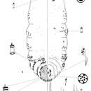
Slide-mounted adult female (Figure 4): spindle-shaped and elongate, lobed laterally and broader posteriorly (generally broadest at metathorax), length of holotype 1.55 mm, range (n=16) 1.15 mm – 1.65 mm; maximum width of holotype: 0.65 mm; range (n=16) 0.475 – 0.875 mm, maximum width at metathorax, rarely on first abdominal segment.
Pygidium: Lobes. Posterior margin with 3 pairs of definite lobes (L1, L2 and L3), fourth pair (L4) of lobes appear as series of low, sclerotized points; paraphyses absent. L1 separated by space 0.3 – 1 (0.6) times width of lobes, with a thick, protruding, U-shaped yoke uniting L1, lateral margins of lobes parallel, slightly diverging near apex, medial margin parallel-sided. L1 usually entire (1 minute notch may be present); L2 bilobed, smaller than L1, medial lobule always larger, both lobules entire; L3 bilobed, lateral lobule usually obsolete, or, when present, shorter than medial lobule but about equal in width. Gland spines. Gland spine formula 2-2-2 (microduct formula 2-2-2), with 1 short gland spine near each body margin on abdominal segment 5; without gland spines between median lobes. Ducts. Large macroducts in submedian area of segments 5 and 6 (with 4 – 6 (5) on segment 5 and 3 – 6 (4) on segment 6); in submarginal areas of segment 5 (with 5 – 8 (6)); in marginal area of segments 5 to 7 (with 2 – 3 (2) on segment 5, 2 on segment 6 and 1 on segment 7); absent on segment 8. Largest macroduct on segment 7 (between L1 and L2) 15 – 22.5 (20) μm long. Pygidial microducts always on venter in submarginal areas of segment 5 to 7, with 1 – 2 (1) duct on segment 5, 2 ducts on segment 6 and 1 duct on segment 7; pygidial microducts absent from dorsum. Pores. Perivulvar pores with 5 loculi, in 5 groups, 1 median with 8 – 17 (8) pores, 2 anterolateral with 20 – 27 (23) pores, 2 posterolateral with 17 – 26 (20) pores. Anal opening. Located 6.1 – 11.2 (9) times length of anal opening from base of median lobes, diameter 12.5 – 17.5 (15) μm. Setae. Dorsal setae: 2 setose on L1, 1 setose (~ 11 m) between lobules of L2 and L3. Ventral setae: 1 small on L1, 1 marginal at base of each gland spine cluster and 1 in submarginal area of each segment, 2 in submedian area of segment 6, small and short; 2 pairs of setae in a row anterior to the vulva.
Prepygidium: Gland spines. Near each body margin from segment 1 or 2 to 4, absent from mesothorax and metathorax; with 0 – 3 (0) on segment 1, 1 – 7 (3) on segment 2, 2 – 4 (3) on segment 3 and 1 – 2 (2) gland spine on segment 4 with 2 microducts extending, short and protruding from margin. Gland spines from segment 1 to 3 the smallest and never protruding from margin. Ducts. Macroducts of 2 sizes; largest macroducts in submedian and submarginal areas of abdominal segments 4 and 3. Small macroducts in submedian area of either or both of segments 3 and 4, in marginal areas from meso- or metathorax to segment 3. Prepygidial microducts sparsely present on venter and dorsum from segment 1 to 4.
Cephalothorax: Microducts sparsely present on venter and dorsum.Perispiracular pores primarily with 3 loculi, anterior spiracles with 5 – 8 (6) pores, posterior spiracles with 2 – 5 (3) pores. Eyes represented by small sclerotized area, located on body margin at level near anterior clypeolabral shield. Antennae each with 1 long seta and 2 shorter setae, distance between two antennae 65 – 135 (85) μm.
- license
- cc-by-3.0
- copyright
- Isabelle M. Vea, Rodger A. Gwiazdowski, Benjamin B. Normark
- bibliographic citation
- Vea I, Gwiazdowski R, Normark B (2013) Corroborating molecular species discovery: Four new pine-feeding species of Chionaspis (Hemiptera, Diaspididae) ZooKeys 270: 37–58
- author
- Isabelle M. Vea
- author
- Rodger A. Gwiazdowski
- author
- Benjamin B. Normark