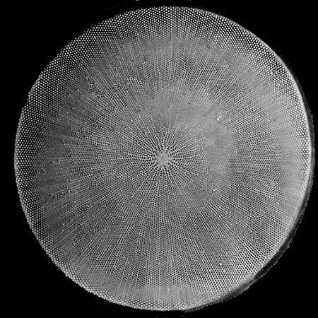
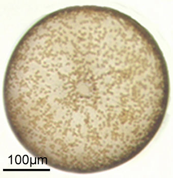
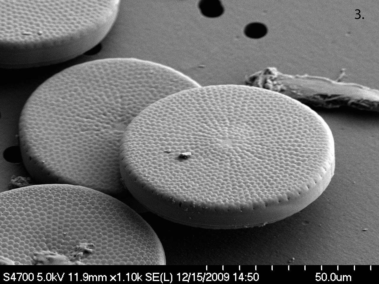
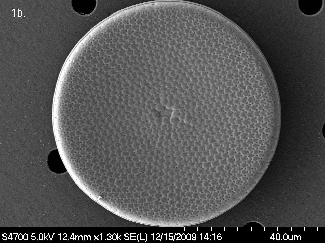
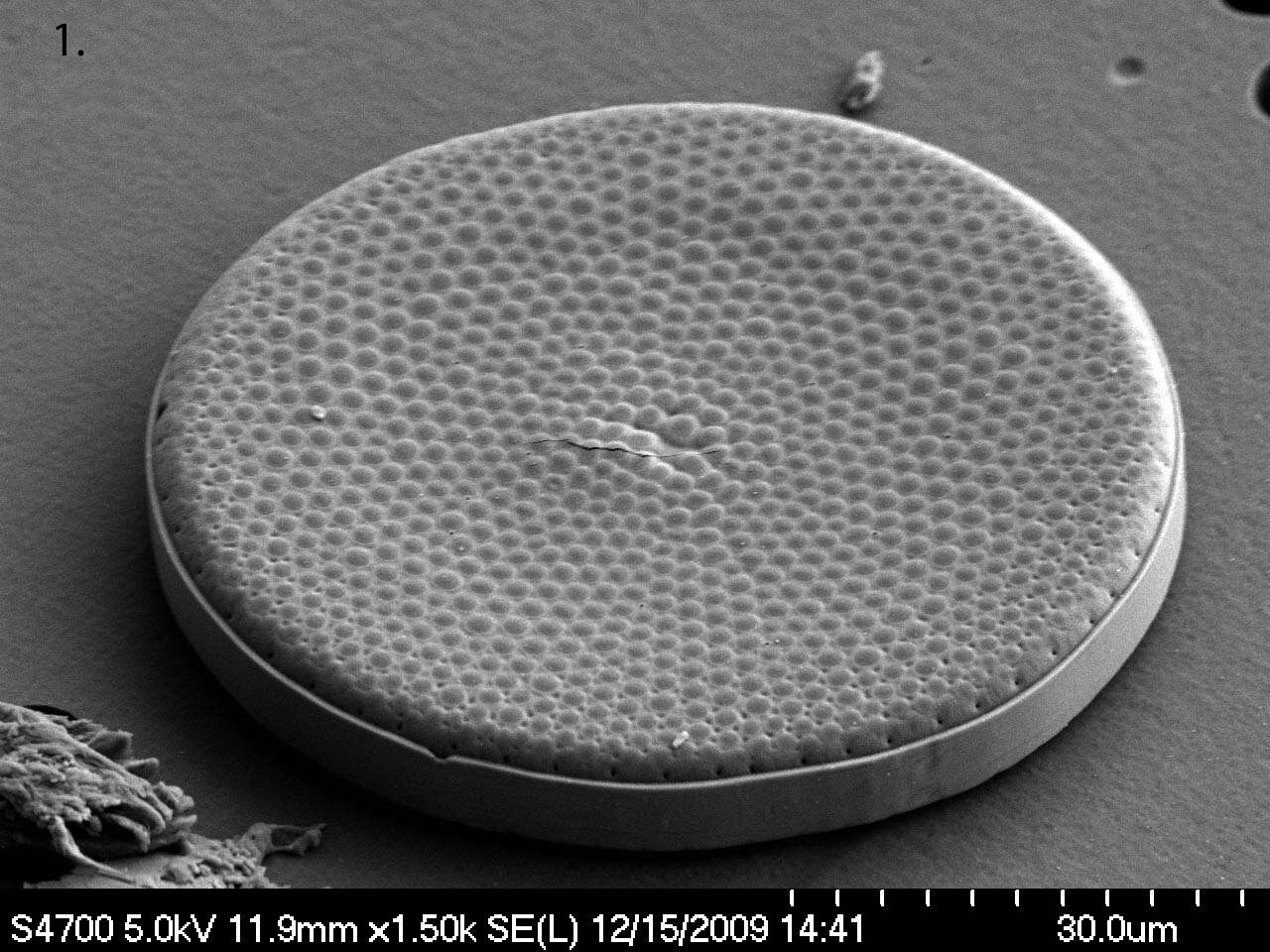
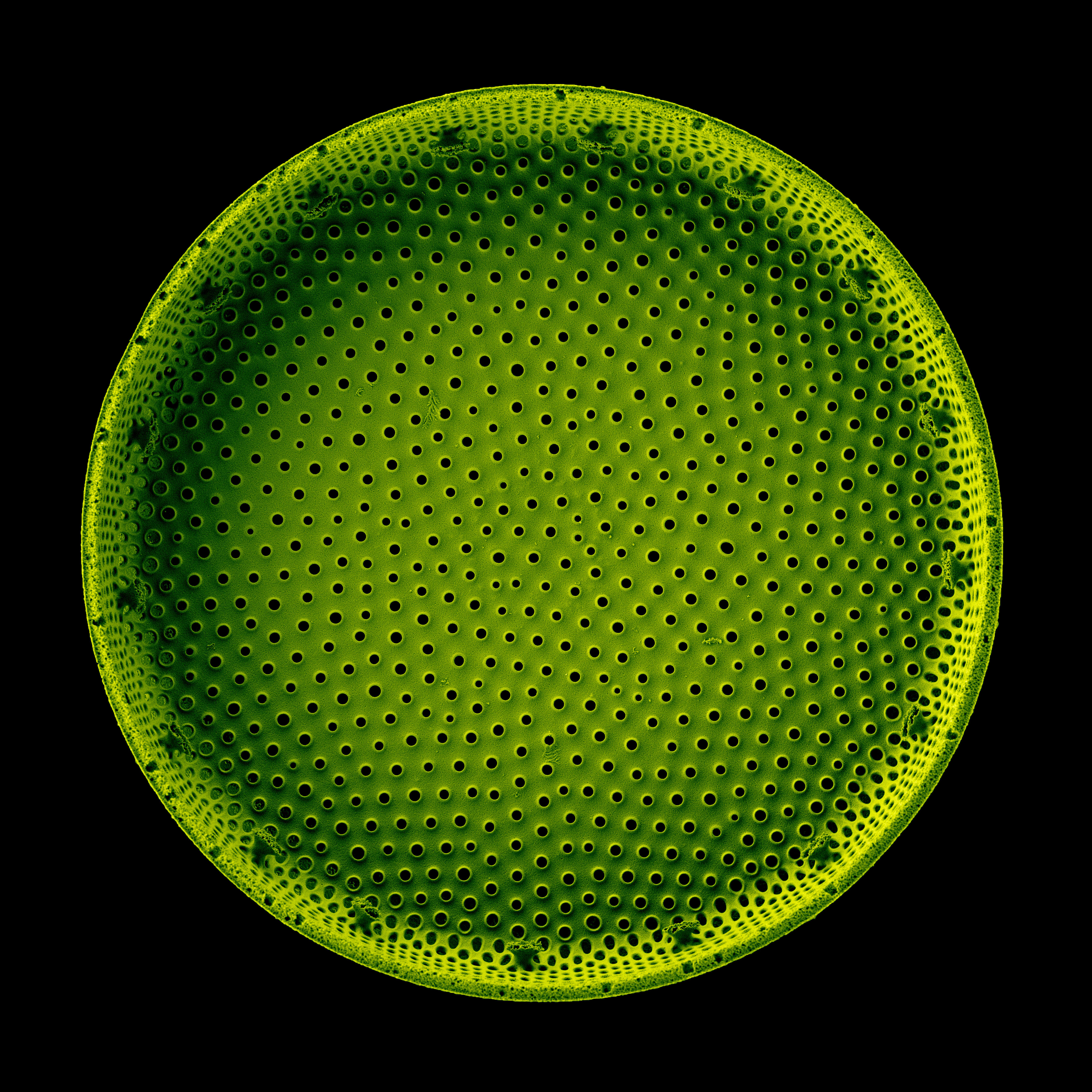
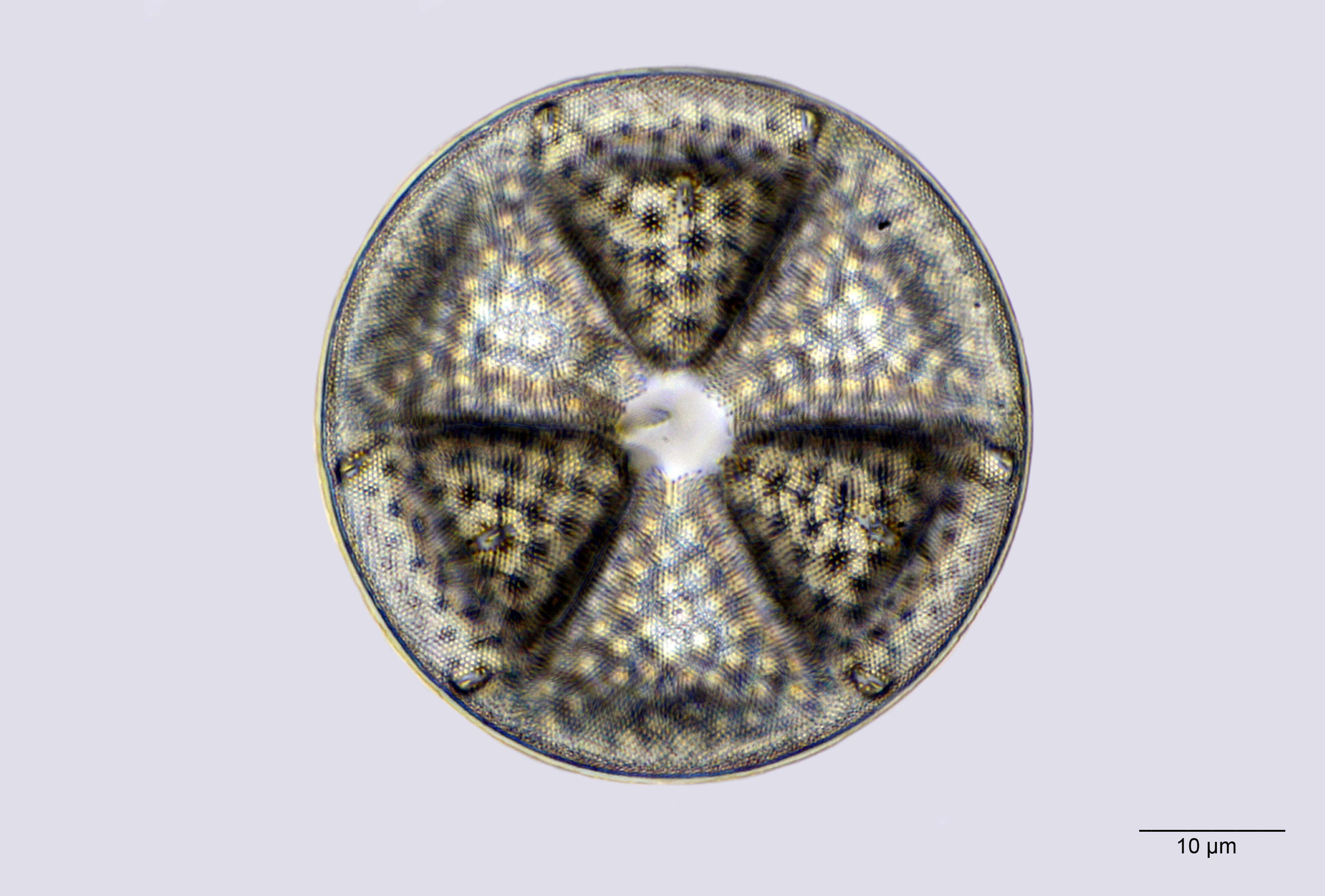
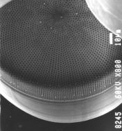
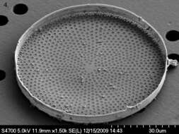
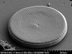
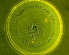
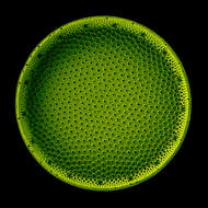
Summary.mw-parser-output table.commons-file-information-table,.mw-parser-output.fileinfotpl-type-information{border:1px solid #a2a9b1;background-color:#f8f9fa;padding:5px;font-size:95%;border-spacing:2px;box-sizing:border-box;margin:0;width:100%}.mw-parser-output table.commons-file-information-table>tbody>tr,.mw-parser-output.fileinfotpl-type-information>tbody>tr{vertical-align:top}.mw-parser-output table.commons-file-information-table>tbody>tr>td,.mw-parser-output table.commons-file-information-table>tbody>tr>th,.mw-parser-output.fileinfotpl-type-information>tbody>tr>td,.mw-parser-output.fileinfotpl-type-information>tbody>tr>th{padding:4px}.mw-parser-output.fileinfo-paramfield{background:#ccf;text-align:right;padding-right:0.4em;width:15%;font-weight:bold}.mw-parser-output.commons-file-information-table+table.commons-file-information-table,.mw-parser-output.commons-file-information-table+div.commons-file-information-table>table{border-top:0;padding-top:0;margin-top:-8px}@media only screen and (max-width:719px){.mw-parser-output table.commons-file-information-table,.mw-parser-output.commons-file-information-table.fileinfotpl-type-information{border-spacing:0;padding:0;word-break:break-word;width:100%!important}.mw-parser-output.commons-file-information-table>tbody,.mw-parser-output.fileinfotpl-type-information>tbody{display:block}.mw-parser-output.commons-file-information-table>tbody>tr>td,.mw-parser-output.commons-file-information-table>tbody>tr>th,.mw-parser-output.fileinfotpl-type-information>tbody>tr>td,.mw-parser-output.fileinfotpl-type-information>tbody>tr>th{padding:0.2em 0.4em;text-align:left;text-align:start}.mw-parser-output.commons-file-information-table>tbody>tr,.mw-parser-output.fileinfotpl-type-information>tbody>tr{display:flex;flex-direction:column}.mw-parser-output.commons-file-information-table+table.commons-file-information-table,.mw-parser-output.commons-file-information-table+div.commons-file-information-table>table{margin-top:-1px}.mw-parser-output.fileinfo-paramfield{box-sizing:border-box;flex:1 0 100%;width:100%}} Description: English: Highly porous hierarchical structure of a Coscinodiscus oculus-iridis (Ehrenberg) Ehrenberg 1840 diatom shell. Colored SEM image acquired with TESCAN CLARA SEM. Sample without any conductive layer, 1keV electron landing energy, field of view 40 μm, E-T detector. Русский: Иерархическая высокопористая структура панциря диатомовой водоросли Coscinodiscus oculus-iridis (Ehrenberg) Ehrenberg 1840. Изображение получено с помощью сканирующего электронного микроскопа TESCAN CLARA с последующей колоризацией. Образец без токопроводящего покрытия, энергия электронов 1кэВ, поле обзора 40 um, E-T детектор. Deutsch: Poröse hierarchische Struktur einer Coscinodiscus oculus-iridis (Ehrenberg 1840) Diatomeenschale. Eingefärbtes REM-Bild, aufgenommen mit TESCAN CLARA SEM (Scanning Electron Microscope), Probe ohne leitende Schicht, 1 keV, Sichtfeld 40 μm, E-T-Detektor. Polski: Wysoce porowata hierarchiczna struktura skorupy okrzemki Coscinodiscus oculus-iridis (Ehrenberg) Ehrenberg 1840. Kolorowy obraz SEM uzyskany za pomocą TESCAN CLARA SEM. Próbka bez warstwy przewodzącej, energia lądowania elektronu 1 keV, pole widzenia 40 μm, detektor ET. Date: 14 September 2021. Source:
File:Coscinodiscus oculus-iridis (Ehrenberg) Ehrenberg 1840 diatom shell. Colored SEM image.png. Author:
Pavel.Somov. Other versions:
500x120px:
Coscinodiscus oculus-iridis (Ehrenberg) Ehrenberg 1840 diatom shell. Colored SEM image.png png version. Licensing[
edit].mw-parser-output.responsive-license-cc{clear:both;text-align:center;box-sizing:border-box;width:100%;justify-content:space-around;align-items:center;margin:0.5em auto;background-color:#f9f9f9;border:2px solid #e0e0e0;border-spacing:8px;display:flex}.mw-parser-output.responsive-license-cc div{margin:4px}.mw-parser-output.rlicense-text div{margin:0.5em auto}@media screen and (max-width:640px){.mw-parser-output.responsive-license-cc{flex-flow:column}.mw-parser-output.rlicense-text{order:1}} This file is licensed under the
Creative Commons Attribution-Share Alike 4.0 International license. You are free: to share – to copy, distribute and transmit the work to remix – to adapt the work Under the following conditions: attribution – You must give appropriate credit, provide a link to the license, and indicate if changes were made. You may do so in any reasonable manner, but not in any way that suggests the licensor endorses you or your use. share alike – If you remix, transform, or build upon the material, you must distribute your contributions under the
same or compatible license as the original.https://creativecommons.org/licenses/by-sa/4.0CC BY-SA 4.0 Creative Commons Attribution-Share Alike 4.0 truetrue


_Ehrenberg_1840_diatom_shell._Colored_SEM_image.png)
_Ehrenberg_1840_diatom_shell._Colored_SEM_image.png&action=edit&redlink=1)