Comprehensive Description
provided by Smithsonian Contributions to Zoology
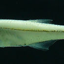
Xenurobrycon heterodon
MATERIAL EXAMINED.—ECUADOR: KU 17925, holotype, male, SL 16.2 mm; KU 17926, paratypes, 1 male, 1 female, SL 16.3–17.2 mm; USNM 219389, paratypes, 1 male, 1 female, SL both 17.0 mm; USNM 219390, paratypes, 1 male, 1 female, SL 16.4–16.7 mm; Pastaza: Río Bobonaza at Sarayacu, 1°16′S, 78°31′W, G.R. Smith and J.D. Lynch, 12–22 Jul 1968. FMNH 94940, paratypes, 12 adults, SL 17.5–18.5 mm; MEPN (uncatalogued), paratypes, adults, SL 16.8–18.2 mm; Napo: Río Aguarico, tributary of Río Napo, 0°5′S, 76°31′W, D. Stewart, M. Ibarra, and R. Barriga, 20 Oct 1983.
PERU: USNM 219391, paratypes, 12 (including 2 cleared and stained) juveniles to adults, SL 13.8–16.7 mm; FMNH 87544, paratypes, 12 juveniles to adults, SL 14.1–16.4 mm; MCZ 54255, paratypes, 12 juveniles to adults, SL 13.9–16.2 mm; Huanuco: Río Pachitea, across river from Hotel Puerto Inca in Puerto Inca, 9°19′S, 75°4′W, D. and T. Greenfield and G. Glodek, 29 Jul 1975.
DIAGNOSIS.—Premaxilla (Figure 65) with 6 to 11 ( = 8.5) teeth, some exserted anteriorly from others to varying degrees, including 1 or 2 exserted “tusk” teeth. Teeth not in well-defined rows. Premaxilla with medial “cluster” of 3 or 4 teeth of which 1 or 2 are offset anteriorly. This accompanied by a more laterally situated oblique row of 6 to 8 teeth of which medial 1 or 2 are tusk teeth. Exserted teeth conical; nonexserted teeth usually conical but sometimes with 1 or 2 small supplemental cusps. Maxilla with single row of 5 to 7 ( = 5.5) conical teeth, anterior 1 or 2 of these exserted, forming “tusk” teeth. Dentary with single row of 9 to 12 ( = 10.4) teeth; anterior 3 or 4 often with 3, sometimes 4, cusps. Adipose fin absent. Adult males with following features: pouch scale approximately teardrop shaped (Figures 12, 30). Eighteenth principal caudal-fin ray (counting ventrally from dorsal principal caudal-fin ray) without platelike bilateral projections but with each ray half in this region bearing a narrow ventrolateral ridge as in young, maturing males of X. macropus (figure 11; (compare with Figure 12). Bony hooks sometimes present on large unbranched and 9 or 10 anterior branched anal-fin rays, their presence probably seasonal and associated with breeding. (See Fink and Weitzman, 1974:22), and p. 89 regarding seasonality in characid fin hooks).
DESCRIPTION.—See Tables 3 and 4 for morphometrics and meristics. Measurements are given separately for males and females for four characters. Body moderately elongate, sides compressed. Greatest depth about midway between nape and dorsal-fin origin or sometimes at dorsal-fin origin. Predorsal body profile gently convex to tip of snout. Body profile slightly elevated at dorsal-fin origin, slightly concave along dorsal-fin base and nearly straight to origin of dorsal procurrent caudal-fin rays. Dorsal-fin origin nearer to caudal-fin base than to snout tip. Ventral body profile gently convex from anterior tip of lower jaw to pelvic-fin origin, somewhat concave in region of pelvic-fin insertion. Body profile between pelvic-fin insertion and anal-fin origin slightly convex in males, slightly concave in females. Profile gently concave at base of anal fin and nearly straight from posterior termination of anal fin to origin of ventral procurrent caudal-fin rays.
Head and snout moderately elongate. Lower jaw protruding slightly beyond upper jaw or jaws equal. Mouth angled anterodorsally from mandibular joint to tip of snout. Maxillary bone extending posteriorly approximately to a point on a vertical line drawn ventrally from anterior border of eye.
Dorsal-fin rays ii, 7 in nearly all specimens, ii, 6 in one specimen from Río Bobonaza, Ecuador, and in one specimen from Río Aguarico, Ecuador. Last dorsal-fin ray not split to its base. Adipose fin absent. Anal-fin rays iii, 13 to iii, 14, usually iii, 14; branched rays distributed as in Tables 3 and 4. Last anal-fin ray split to its base. Anal fin with a strongly lobed anterior portion in males, much less lobed in females. Anal fin of male from Río Bobonaza, Ecuador, with bilateral antrorse bony hooks on largest anterior unbranched ray and 9 to 10 anterior branched rays; total number of rays with hooks 11 in 3 males and 10 in 1 male. Males from Río Aguarico, Ecuador, similar to those from Río Bobonaza. Males from Río Pachitea, Peru, without hooks on anal fin but with bilateral small, irregularly surfaced eminences, apparently remnants of hooks, distributed as are hooks on Ecuadoran specimens.
Pectortal-fin rays i, 8 in holotype; range i, 7 to i, 9 for all specimens, usually 8 branched rays. Distal tips of pectoral-fin rays extending posteriorly to pelvic-fin origin in females and well beyond that point in males due to anterior placement of pelvic fin in males.
Pelvic-fin rays i, 6 in all specimens. Pelvic fin sexually dimorphic, greatly elongate in males. Male pelvic-fin origin much nearer pectoral-fin origin than anal-fin origin. Female pelvic-fin origin about equidistant between pectoral-fin origin and anal-fin origin. Compare snout to pelvic-fin origin ratios in Tables 3 and 4. Female pelvic-fin rays not elongate, without hooks, much like pelvic-fin rays of most female glandulocaudine characids. Pelvic fin of males arched with interradial membranes broad as described for X. macropus. As in X. macropus, fins form inverted boat-shaped canopy when spread with forceps. Males from Río Bobonaza and Río Aguarico, Ecuador, with bony hooks like those of X. macropus; males from Río Pachitea, Peru, without hooks but cleared and stained specimen with irregular eminences and rough spots, apparently hook remnants, which are especially obvious distally. (The hypothesis that hooks on these fishes are seasonal and were recently either shed or, less likely, resorbed in Peruvian specimens is corroborated by the presence of an unattached but well-developed hook caught in membrane of left pelvic fin of a cleared and stained specimen, 16.7 mm SL). Pelvic-fin rays of cleared and stained male 16.4 mm SL from Río Bobonaza, Ecuador, with mostly antrorse pelvic-fin hooks as follows: anterior large unbranched ray with 31 hooks distributed over 17 ray segments, with about 9 hooks on long basal ray segment and more distal segments with 1 to 3 hooks each. Second ray, branched, bears 43 hooks distributed over 21 segments of unbranched and branched portions of ray. Third ray bears 45 hooks distributed over 21 segments of branched and unbranched portions of ray. Fourth ray bears 37 hooks over 18 segments of branched and unbranched portions of ray. Fifth ray, unbranched, bears 21 hooks distributed on 10 segments. Sixth ray, unbranched, bears 34 hooks on 17 segments, and seventh ray, unbranched, bears 33 hooks on 16 segments.
Pelvic girdles of males and females similar to girdles of X. macropus, except that in cleared and stained male from Río Pachitea, Peru, posteromedian processes are separated from each other by only about 0.7 mm or a distance just over length of pelvic girdle instead of 1.2 mm or a distance of more than length of pelvic bone as in X. macropus.
Caudal fin with principal rays in all specimens; caudal fin of females as in most other characids. Male caudal fin (Figure 12) similar to that described for X. macropus (Figures 11, 25, 47). Hooks very reduced to absent in males from Río Pachitea, Peru, but apparent hook remnants appear similar in distribution to hooks on male specimens from Río Bobonaza, Ecuador. Rays of dorsal and ventral caudal-fin lobes modified as in X. macropus, except that ventral branched ray lacks ventrolaterally projecting, bilateral laminae of bone. Concavity on ventral surface of this ray is present. Consequently, this ray with sharp ventrolateral borders in position of bilateral lamelle. (Compare Figures 12 of X. heterodon and Figure 11 of X. macropus.)
Hypural skeleton of both males and females as in X. macropus.
Caudal squamation of female as in X. macropus. Caudal squamation of male as in X. macropus, except for certain chracteristics of pouch scale. Pouch scale approximately teardrop-shaped; its posterior field pointed with only a small portion of its distal border free of striated radii (Figure 30).
Morphology of presumed caudal gland of male as in X. macropus, except that no groove occurs in tissue of eighteenth principal caudal-fin ray. Caudal musculature of male as in X. macropus except that muscle bundles are less robust. Caudal musculature of female as in X. macropus.
The hypothesis proposed below for X. macropus regarding the functioning of a caudal “pump” applies to X. heterodon as well. Presumably the flattened margins of the eighteenth principal caudal-fin ray of X. heterodon do not deflect the pheromone-bearing water in the same manner as the bilateral platelike lamellae of X. macropus.
Scales cycloid with very few radii along posterior margins except in caudal region and along narrow dorsal and ventral body surfaces. Lateral line incomplete. Scales mostly missing in specimens from Río Pachitea, Peru; one male specimen with 4 perforated lateral-line scales and 32 scales in lateral series, excluding large caudal scale. Scale counts could be made on two specimens of each sex from Río Bobonaza, Ecuador. Holotype with 6 perforated lateral-line scales and 35 scales in lateral series including large caudal-fin scale. Usually about 34 lateral series scales. (See Tables 3 and 4 for distribution of perforated lateral-line and lateral series scale counts). Scale rows between dorsal-fin origin and pelvic-fin origin 8 in all specimens. This count could be made in only one specimen from Río Pachitea, but counts of pockets could be made. Scale rows around caudal peduncle 13 in all specimens. These scales missing in specimens from Río Pachitea, but counts of pockets could be made. As in other species of Xenurobrycon, a median ventral scale row is absent at the narrowest region of the caudal peduncle. Predorsal scales 15 in holotype; 14 to 16, usually 15 in other specimens (Tables 3, 4).
Teeth of jaws unicuspid, bicuspid, or tricuspid depending on position in jaws (Figure 65 and description below). Maxillary and dentary teeth in single row. Premaxilla with some teeth offset anteriorly to varying degrees, including 1 or 2 exserted “tusk” teeth; the resulting configuration not accurately described as either 1 row or 2 rows as those terms are usually applied in characid morphology. Teeth of holotype (values not in parentheses) as follows: Teeth of premaxilla and maxilla conical. Premaxilla with second and fourth tooth from midline offset anteriorly and projecting slightly anteriorly. Two (2 or 3) “tusk” teeth exserted from oral cavity (Figure 65) and with their bases dorsal to fourth tooth from midline. Lateral to 2 (2 to 3) “tusk” teeth are 5 (5 to 8, usually 5 or 6 in other specimens) additional teeth along lateral ramus of premaxilla, each of which in succession is directed less anteriorly and more ventrally than its anteromedial neighbor. Together with “tusk” teeth these form an oblique row across lateral portion of premaxilla. Maxilla with 7 (5 to 7, usually 5 or 6) teeth also forming a sharply angled row; anterior 2 teeth exserted and each of remaining 5 teeth directed, in succession, less anteriorly and more ventrally than its anteromedial neighbor. Dentary with 11 (9 to 12, usually about 10) teeth; medial 4 of these tricuspid and much larger than remaining 5 to 8. Smaller, lateral 5 to 8 teeth unicuspid and decreasing in size in an anterior to posterior sequence.
Premaxillary tooth counts in specimens from all localities, n = 20, range = 6 to 11, = 8.5, SD = 1.890. Premaxilla with 3 or 4 teeth “clustered” on its medial portion, usually with 3 (as in holotype). Remaining premaxillary teeth forming transverse row on lateral portion of premaxilla; usually 2 (occasionally only 1) of these teeth exserted. Premaxilla sometimes with multicuspid teeth: two males from Río Bobonaza with medial premaxillary tooth biscuspid, one female with 2 medial premaxillary teeth bicuspid, one female with 3 medial premaxillary teeth tricuspid, and cleared and stained female with left side having 5 of 6 nonexserted teeth bicuspid and right side having medial tooth bicuspid and laterally adjacent tooth tricuspid. Maxillary tooth counts as follows: n = 26, range = 5 to 7, = 5.5, and SD = 0.982. All maxillary teeth unicuspid in all specimens, forming an angled row, with 2 exserted “tusk” teeth anteriorly. Dentary tooth counts as follows, n = 32, range = 9 to 12, = 10.4, SD = 0.982. Dentary usually with medial 4 teeth (3 teeth in some) tricuspid, larger than remaining teeth. Some males and females with 5 tricuspid dentary teeth, with one of smaller teeth also being tricuspid.
Statistical comparison by use of two-tailed Student's t test using square-root transformations of numbers of teeth on each jaw bone for two population samples, one from Río Aguarico, the the other from Río Bobonaza (both in Ecuador), of X. heterodon showed no significant differences at the 0.05 level. Patterns of tooth arrangement in specimens from Río Aguarico almost the same as in specimens from Río Bobonaza, with the following exceptions: premaxilla always with 4, never 3, teeth “clustered” medially. Maxilla occasionally with 1 rather than 2 exserted “tusk” teeth. In both sexes, all premaxillary teeth usually unicuspid (cleared and stained male with medial tooth bicuspid on one side). Dentary usually with 3 (rarely 4) medial teeth tricuspid and larger than remaining teeth. Lateral cusps in specimens from Río Aguarico not as prominent as those found in population sample from Río Bobonaza, Ecuador.
See Tables 3 and 4 for vertebral counts including Weberian apparatus and terminal compound centrum and for gill raker counts.
Branchiostegal rays in three cleared and stained specimens 4 (3 on one side of one specimen). In three specimens 3 rays originate from anterior ceratohyal and 1 ray from border between anterior ceratohyal and posterior ceratohyal. In one specimen all 4 rays originate from anterior ceratohyal.
COLOR IN ALCOHOL.—Color pattern (Figure 5) similar to that of X. macropus; no consistent differences detected.
SEXUAL DIMORPHISM.—Tables 3 and 4 present morphometric data for specimens of X. heterodon arranged by sex and locality. Unequivocal differences occur in the same characters in X. heterodon as in X. macropus, including distance from snout to pelvic-fin origin (shorter in males), depth of caudal peduncle (greater in males), length of pelvic fin (much greater in males), and length of anterior anal-fin lobe (greater in males).
Analysis of covariance by sex using log transformed data for 19 morphometric characters was done for specimens from the Río Aguarico, FMNH 94940 and MEPN (uncataloged) (Table 4). Coefficients of determination were somewhat greater than found for Xenurobrycon macropus because of a large sample size, but again, correlation was not good, especially in the males with little variation in adult size and sexual maturation at varying sizes within that restricted range. (For discussion of male maturation size, see p. 38.) Intercepts, but not slopes, again showed significant differences (P<0.01 determined in a two-tailed test) between males and females for two characters which also differed in Xenurobrycon macropus, a greater dorsal-fin height and a longer anal-fin base in males than in females. As in X. macropus, hooks are absent in fins of females whereas males appear to have them in pelvic, anal, and caudal fins at least during breeding season. Complex glandular, osteological, and muscular modifications of caudal-fin region of males are absent in females.
POPULATION VARIATION.—Sample size is too small from Río Bobonaza, Ecuador, and Río Pachitea, Peru, to permit population comparison. Also, these populations were not in breeding condition as were the specimens from the Río Aguarico, Ecuador. In addition, the former two population samples were not in as good condition (scales mostly gone) as the specimens from the Río Aguarico. No population comparison was made.
ETYMOLOGY.—The name heterodon is from the Greek heteros (other or different) and odus or odon (tooth). The name refers to the mixture of tricuspid, bicuspid, and conical teeth found in this species.
Putative Pheromone Pumping Mechanisms in Glandulocaudine Fishes
Several putative pheromone pumping mechanisms of two general functional categories are found in the caudal region of glandulocaudine males. The first category, a diaphragm scale pump, has two major variations and occurs in most of the glandulocaudines. One of these two is a passive pump without much obvious muscular specialization; this pump appears to be the primitive form within the xenourobryconin glandulocaudines. A considerable amount of difference in caudal pump scale morphology is displayed by the passive pump system of various species and genera. Iotabrycon has a unique form of this system with obvious specialized pouch scale and musculature. The latter is not directly attached to the bellows of the pump and therefore the pump appears passively driven. The other major variation, known only in the minute subgroup B xenurobryconins, includes obvious specialized musculature and appears to be an active pump directly driven by specialized muscles attached to the diaphragm, the pouch scale, of the bellows.
The second category of pump, morphologically very different, has a valve and is formed of modified fin rays, associated scales, and soft tissue. This fin-ray pump is known only in Mimagoniates and evolved independently of the diaphragm pumps. One glandulocaudine genus, Glandulocauda, lacks a pump but does have glandular tissue distributed on the caudal-fin ray membranes.
Historical Summary
Eigenmann (1914:34) first defined the glandulocaudines and in part recognized them by the possession of “peculiar glandular scales or pouches covered by united scales” in the males. Eigenmann did not further discuss this feature. Little more was said about the caudal glands and scales until Nelson (1964a:70–74) hypothesized a pheromone function for them. For example, Böhlke (1958a:43) hypothesized that the glandulocaudines were polyphyletic, and of the caudal gland stated that all species “possess some sort of specialized scaly structure at the base of the caudal fin.” Kutaygill (1960) described the histology of the secondary sexual characters of Corynopoma riisei but recognized no pumping mechanism. Géry (1964:8–12) described in some detail the osteology of the caudal-fin rays of “Mimagoniates cf. microlepis” but did not suggest a caudal pumping mechanism.
Nelson (1964a:70–74) described and illustrated the gross superficial anatomy of ten species in eight glandulocaudine genera and (page 142) hypothesized a sexual pheromone function for the glandular tissue. Nelson (1964a: 142) also suggested that in Coelurichthys tenuis (Mimagoniates lateralis of the present paper), a caudal-fin movement that he termed “Dusting” appears to be
ideally suited to wafting an odorous substance from the caudal gland to the region of the female's head, and inspection of the caudal morphology in this species reveals that it would be fitted for the purpose. Two fin rays are modified to form opposing halves of a tube and are so arranged that the edge of one fits inside the other (fig. 3,F), so that any differential movements of the two fin rays will cause them to operate as a bellows. [He also postulated that the caudal gland] produces a substance which when directed toward the female by this bellows during Dusting, increases the probability that she will Pair.
Weitzman (1975:409) briefly described courtship activity of Corynopoma riisei and Pterobrycon myrnae. In the original unpublished English version of this article he reported that during “Extending” (defined under the term “Extending Paddle” for Corynopoma by Nelson, 1964a:95), among other activities, the male of both these species “Tail Beats” toward the female. Tail-beating was described for Mimagoniates microlepis and Mimagoniates lateralis by Nelson (1964a:90). Tail-beating in Corynopoma riisei is different from “Twitching” and “Shaking” described by Nelson (1964a:90) and also observed by us. In tail-beating by Corynopoma riisei the male “flaps” his tailfin. fin toward the female but with less of the V-shaped body action described for Mimagoniates species as illustrated by Nelson (1964a:87, fig. 6). During this activity the dorsal and anal fins are also maximally extended, and in Corynopoma the opercle is extended toward the female; in Pterobrycon elongate, specialized epaxial scales are extended toward the female. The remarks about tail-beating were eliminated from the German text by the editor. However, this extending and tail-beating activity is illustrated by Weitzman (1975) for Corynopoma riisei (bottom figure, page 406) and for Pterobrycon myrnae (bottom figure, page 408). It is hypothesized here that during tail-beating the male propels one or more pheromones toward the female's head. Nelson (1964a:91, fig. 106) also illustrated a male Corynopoma in tail-beating activity. No accurate record of the number of beats per second is available, but the number is relatively low, only a few per second. Nelson (1964a:90) states that “Dusting” (a specialized form of tail-beating?) is confined to Mimagoniates (= his Coelurichthys) among the glandulocaudines he examined; he did not record tail-beating for Corynopoma. The “Dusting” activity described for Mimagoniates lateralis by Nelson (1964a:89, figs. 8c, 94) is also probably a specialized form of tail-beating. Tail-beating in several behavior patterns occurs not only in glandulocaudines but in other characiforms as well. We have observed it during lateral display in several species of Nannostomus (Lebiasinidae) and Hyphessobrycon (Characidae). Sometimes the beating is in head-to-facing-tail position; at other times it may be in tail-to-tail and head-to-head position. The behavior needs further study, but in the case of the glandulocaudines it seems suited for propelling a pheromone toward the female's head. We expect that tail-beating activity associated with courtship and pheromone delivery to the female may occur in a variety of specialized forms in various glandulocaudine taxa.
Nelson (1964a: 142) states: “A superficial inspection of courtship in Corynopoma, Pseudocorynopoma, Glandulocauda [inequalis] [=Mimagoniates inequalis of the present study], or C. microlepis [here = Mimagoniates microlepis] reveals no striking features that might be correlated with caudal morphology.” He goes on to point out that a more penetrating analysis does reveal a correlation. In a letter from Nelson to the senior author dated 22 January 1963, Nelson suggests a function for the caudal gland.
[In] Mimagoniates, especially barberi [= lateralis of the present report], the male fans the female's head with his caudal as with a feather duster [hence “Dusting,” of Nelson, 1964a: 94]. Similar things occur in other species, but are much less pronounced, so much that I at first missed it completely in Corynopoma. My idea is that the gland secretes an excitatory substance, which he wafts toward the female during courtship. The elaborate pouches prevent unnecessary waste and as a result of the specialized caudal movements of courtship may act then as a bellows.
Clearly Nelson here conceived of an economical mechanism for production of a sexual pheromone in the male and a method for its transport to the female. We believe his hypothesis has much merit and explore it below in conjunction with a more detailed review of the anatomy of caudal structures.
Atkins and Fink (1979) described and discussed the histology of the glandular caudal tissue in Corynopoma riisei. They hypothesized a sexual pheromone substance in the caudal gland and noted that males placed in aquaria without females soon have hypotrophied glands, but that when they are returned to aquaria containing females the glands again become hypertrophied. Liley (1982:28, 32) and Liley and Stacey (1983:10, 12), based on the literature, have briefly called attention to possible sexual pheromone function in the caudal glands of glandulocaudine characids. Weitzman (1982, 1985) has noted the existence of and briefly described the passive and active putative pheromone diaphragm pumps in glandulocaudines, especially the muscular pump of the xenurobryconins.
Mahnert and Géry (1984:503) briefly described parts of the osteology of male Xenurobrycon macropus and on page 505 noted that the base of the eleventh caudal-fin ray (= their eighth ray of the ventral caudal-fin lobe) is movable and bears strong musculature. They indicate that this musculature extends to neighboring fin rays and assumed that movement of the base of the eleventh ray might be involved with expelling the glandular secretion. They presented no concept of a muscular diaphragm pump.
Caudal Pump Morphologies and Putative Function
THE DIAPHRAGM SCALE PUMP
All glandulocaudine species, except those in the genera Planaltina, Acrobrycon, and Diapoma, have the caudal gland and associated structures confined to the males. The passive diaphragm scale pump consists of an epidermal invagination between the fin rays and the squamation, forming a sac or pouch usually located at the base of the ventral caudal-fin lobe. The sac is posteriorly or posteroventrally open to surrounding water, and glandular tissue occurs in the vicinity of the opening, especially on the fin-ray side of the pouch. In Argopleura and other xenurobryconins there appears to be a large amount of glandular tissue just inside the pouch opening on the medial wall of the pouch. Among species and groups of glandulocaudines there is much variation in the placement of the pouch and in the form, size, and location of the scales covering its lateral surface. For example, the relatively primitive xenurobryconin glandulocaudine Agropleura has the pouch mostly covered by a terminal lateral-line scale and a scale from a row just ventral to the lateral-line scale row (Figures 8, 9). Other glandulocaudines, such as Corynopoma riisei (Figure 16) and Gephyrocharax atricaudatus (Figure 17), have more ventrally placed scales on the lateral wall of the pouch. Diapoma speculiferum (Figure 15) has several somewhat hypertrophied scales covering the lateral wall of the pouch instead of one large scale and additional smaller scales.
This type of pheromone-associated organ may function in a similar manner in all glandulocaudine species that possess it. We hypothesize that it works as a passive pump in association with tail-beating. If one takes a freshly killed male Corynopoma, Hysteronotus, or Gephyrocharax and bends the tail while the fish is held under water, the scale or scales on the lateral wall of the pouch, which are on the concave side of the caudal fin, buckle outward, drawing water into the pouch. Releasing the tail, allowing it to align with the long axis of the fish, causes the scale to resume a flatter shape, collapsing the pouch and forcing water out over the glandular tissue. The apparatus thus appears to act like a bellows without a valve and only one opening. If the pouch and scale operate this way during tail-beating, the motion of the tail and the subsequent passive pumping action of the caudal pouch may increase the rate or focus the direction of the dissemination of pheromone substances into the water around the tail fin. During tail-beating in Corynopoma and Pterobrycon, the male bends his tail predominantly toward the female and bends it more slowly and to a lesser extent in the reverse direction. The pouch on the side of the fish away from the female would be much less active during tail-beating because there is little tail bending in the direction that would expand that pouch. This presumably could conserve pheromone substances on the side of the male away from the female during tail-beating. Thus glandulocaudine males may physically activate their caudal glands only, or at least to a greater extent, on the side facing the female. Corynopoma and Pterobrycon irregularly will alternate sides facing the female during courtship and tail-beating. Although not well described in the systematic literature, males of some glandulocaudine species may have part of the dorsal and ventral tail lobes separate due to interruption of the interradial membrane. Thus the fin lobes may be moved independently. This feature, tail-beating, and pheromone propagation may be related in some synergistic way to increase the efficiency of pheromone communication, but the production of pheromone and the detailed function of these morphological features remain speculative.
Another, more elaborate, kind of diaphragm-scale pumping mechanism is found in Iotabryconpraecox (Figures 2, 10, 23, 44, 45, 68). This pumping mechanism is similar to that just described, but the opening of the sac and the associated large scale are uniquely complex (Figures 10, 23). The glandular tissue is located around the opening of the pouch but does not extend into the elongate posteroventral process of the scale. The pouch is primarily located deep to the major fimbriated area of the scale and the pouch opening occurs along the ventral fimbriated border of the main body of the scale. The parallel fimbriae of the dorsal scale lobe lateral to the pouch (apparently hypertrophied scale lamellae separated by radii, compare Figures 23, 32) are joined by membrane so that the lateral wall of the pouch is very expandable in a lateral direction. This species has its dorsal and ventral caudal-fin lobes largely connected by a membrane. Exactly how the caudal fin of Iotabrycon operates in tail-beating is unknown, but presumably the structure is used during courtship to somehow bring pheromone substances to the female. For example, the hypertrophied epaxial and hypaxial muscle mass on the dorsal caudal-fin lobe may increase the force of caudal movement (e.g., sculling). This increase in turn may compensate for the greater water resistance that may affect the caudal pump of this small fish (see the following discussion of water resistance in the small pumps of subgroup B xenurobryconins).
A second type of caudal pheromone diaphragm pump is found in Xenurobrycon, Scopaeocharax, and Tyttocharax. Its physical appearance has been described above in some detail for Xenurobrycon macropus (p. 77–83). Also, various aspects of the structure have been described above under apomorphies 27–37, subgroup B; 47–50, Xenurobrycon; 55–59, subgroup C; and 61, Scopaeocharax.
Parts of the structure are extensively illustrated here for various species of Xenurobrycon, Scopaeocharax, and Tyttocharax, but reference to Figures 11–14, 24, 30, 46, 47 are especially helpful in understanding the structure. Briefly, the epidermal pouch is located medial to the middle and posteroventral regions of the enlarged pouch scale and opens to the surrounding water along the fimbriated posteroventral border of the scale. Part of this scale flares slightly laterally. In that vicinity the fimbriae are especially fine and the membrane between them highly distensible. The greatest concentration of hypertrophied gland cells occurs around and just inside this opening as in Argopleura. The scale is bound to fin rays 10 and 11 by one or two ligaments. Specialized interradialis-b muscle fibers originate along the base and unciform process of fin-ray 11 and insert in roughly a semicircle near the border of the posterior three-quarters of the pouch scale. Interradialis-a muscle fibers originate near the bases of fin rays 12–17 and insert on the unciform process of ray 11 (Figures 11–14, 46, 47). The distal portions of the dorsal-lobe caudal-fin rays pass to one side or the other of the ventral-lobe caudal-fin rays (Figure 67).
We propose that many of these modified structures of the male caudal fin may serve as a pumping mechanism increasing the rate of dissemination of a pheromone or pheromones into the surrounding water from glandular tissue situated in and around the mouth of the pouch or sac. We suggest that the boundary layer effect, in which the velocity of flowing water decreases the closer the water is to a substrate, reaching virtually zero at the boundary (Leyton, 1975:5), may be associated with the evolution of the directly driven muscular pump. Water that flows through relatively small tubes or into small sacs, such as the small pumps of subgroup B xenurobryconins, is limited to those boundary layers where greater energy is required to increase water velocity. Greater energy would not be needed in the larger pumps of the larger glandulocaudines. In addition, a relative increase in water turbulence, which occurs at orifices of small size (Leyton, 1975:19), may produce additional water flow retardation. Thus, in fishes the size of the minute xenurobryconins, the energy needed to make water flow in the caudal pump may be such that the passive mechanism in the form present in larger glandulocaudines would not be effective. Additional energy may be needed, and in subgroup B xenurobryconins may be supplied by the modified interradialis muscles. The relatively large size of the pump and diaphragm scale in relation to body size possessed by miniature glandulocaudines compared to those present in the larger glandulocaudines also may compensate for decreased flow and increased water turbulence.
The caudal pump may function in the following manner: the fan-shaped interradial muscle bundle, interradialis-b, when contracted, may cause the scale to buckle laterally and thus expand the volume of the sac internal to the scale. This would cause water to enter the sac and pass over the associated glandular tissue. Relaxation of this muscle presumably would allow the natural resilience of the scale to bring the scale back to its undeformed state, forcing water out of the sac and again over the glandular tissue. The scale presumably acts as a movable diaphragm, pumping water in and out of the sac and over the glandular tissue. Other movements used in either tail-beating or head-to-tail lateral display activity may aid in pumping water into and out of the caudal sac. The posteroventrally directed interradialis muscle, “interradialis-a” (Figures 46, 47), may act to stabilize the sigmoid ray (principal ray 11; p. 82) during contraction of the fan-shaped interradialis-b, as well as to control a pivoting or rocking motion of the sigmoid ray around its articulation with hypural 2. The extensive interradialis fibers of dorsal caudal-fin rays 7 and 10 probably cause exaggerated sculling movements, with rays 7 to 10 passing to one or the other side of the less mobile rays of the ventral caudal-fin lobe. The radii of the large scale may facilitate bending of the scale during normal swimming as well as during pumping action. The platelike projections on each side of the eighteenth principal caudal-fin ray in Xenurobrycon macropus may act as deflecting devices directing water bearing the pheromone towards the female.
THE FIN-RAY PUMP
The second type of glandulocaudine pump, the one mentioned by Nelson (1964a:142) on page 95, involves modifications of scales of the base of the dorsal caudal-fin lobe and modification of principal caudal-fin rays 8 to 13 and especially 11 and 12 (Figure 19 of Mimagoniates microlepis). This caudal mechanism will be treated in more detail in a paper on Glandulocauda and Mimagoniates by Menezes and Weitzman in preparation. Briefly, the mechanism consists of a series of about 5 to 11 dorsal lobe scales which together form a single flap extending posteriorly and ventrally lateral to the proximal parts of principal caudal-fin rays 8 to 12. A broadly open pouch or pocket lies between this flap and fin rays 8 through 11. The opening occurs along the entire ventral border of these scales. In histological sections apparent secretory cells form heavy concentrations in the scale pouch or fold. Glandular cells are also widely distributed on the fin but appear most concentrated around these modified scales and associated fin rays.
A second pouch or pocket occurs between the highly modified seven to twelve or so basal segments of the ray halves of principal caudal-fin rays 11 and 12. These segments are expanded and curved so that their opposing faces form the concave walls of the pouch. Rays 9, 10, and 13 are also modified in support of this pouch or tubular pump. Glandular tissue is not apparent inside this fin-ray pouch but does occur around its posterior opening. The structure is bilateral. Each pouch is open along the lateral borders of the fin-ray segments. This opening is divided into an elongate anterior portion and a smaller posterior portion by the expanded segments of ray 11. In life, the soft tissue around the openings restrict their extent, but they do remain continuous with each other. During ventilation of the pouch the narrow central part between the major openings is the first area to close during compression and the last to open during expansion. When the caudal fin is flexed away from the pouch being observed, the volume of the contents of the pouch decreases and the anterior portion of the opening closes. When the fin is flexed toward the pouch, the volume of the pouch expands and the anterior portion of the opening expands widely. The posterior portion of the opening remains open during all flexing activity. Nelson referred to the fin-ray mechanism as a bellows, and the pump is a type of bellows, one with a valved opening in the proximal and middle portions. The anterior portion of the opening closes as the pouch is compressed and opens as the pouch expands, while the smaller posterior portion of the opening remains open at all times. Therefore, we would suggest that the predominant flow of water is into the anterior portion of the opening and out of the posterior portion of the opening. A small amount of water might enter through the posterior opening only when the pouch is being expanded. The interradialis muscles are well developed on all fin rays and the other caudal musculature is quite robust but not greatly modified. Direct muscular action on the fin rays as well as the general bending of the fin during tail-beating may be involved in driving this pump.
Glandulocauda, apparently the closest relative of Mimagoniates, does not have a caudal fin ray pump and appears the more primitive morphologically. The fin-ray pump of Mimagoniates apparently evolved independently of the diaphragm scale pump present in other glandulocaudines.
Summary
The Xenurobryconini is corroborated as monophyletic by five synapomorphies, providing a new diagnosis. The tribe includes Argropleura from the Magdalena and Cauca basins in Colombia, Iotabrycon from the Guayas basin in Ecuador, and Xenurobrycon, Scopaeocharax (new), and Tyttocharax all from the Amazon basin. Xenurobrycon is also found in the Paraguay-Paraná basin.
Subgroups and genera of the Xenurobryconini are corroborated by apomorphies. Seventy-one apomorphies were used in this treatment, many based on secondary sexual characters of the males and many based on reductive characters associated with small size in the adults of Iotabrycon, Xenurobrycon, Scopaeocharax, and Tyttocharax. A key to the groups and genera is presented.
The phylogenetic analysis, in part, is a study of the congruence of innovative characters, such as features associated with the caudal gland, and reductive characters, such as various losses of portions of the laterosensory canal system. Both kinds of evolutionary change may be associated with miniature size and with the hydrodynamics of reduced water flow in small tubes, such as the laterosensory canals and caudal pheromone pumps. We find that the two kinds of characters usually are congruent and corroborate a single phylogenetic hypothesis. However, most of the homoplasies that we detected are reductive laterosensory system characters and we suggest that these characters, apparently related to size at maturity, maybe reversible and therefore labile in the studied taxa.
Xenurobrycon macropus from the Paraguay-Paraná basin is redescribed and two species, Xenurobrycon pteropus from Fonte Boa, Amazonas, Brazil, and Xenurobrycon heterodon from the upper Amazon basin in Ecuador and Peru, are described as new. A key to the species is included.
The distribution of the Xenurobryconini is reviewed and an area cladogram of the major subgroups is presented. Three areas of endemism are discussed, the Magdalena plus the San Juan basins of Colombia, the Guayas basin of Ecuador, and the Amazon plus the Paraguay–lower Paraná basins.
Caudal pheromone pumping mechanisms are described for several glandulocaudine groups. There are two major types, one of which has several forms. One type consists of a diaphgram pump. The diaphragm is formed of a large scale or scales lateral to a sac or pouch. The pouch has a single posterior opening surrounded by glandular tissue that presumably produces a pheromone. In most instances the pump may work passively as a result of tail-beating during courtship. In one miniature xenurobryconin, Iotabrycon, this passive pump has become associated with highly modified caudal musculature. In three miniature xenurobryconin genera, Xenurobrycon, Scopaeocharax, and Tyttocharax, the pump appears to be directly driven by modified interradialis muscles, which are inserted along most of the posterior border of the scale diaphgram. This we call an active muscular pump. It may be driven in association with tail-beating or perhaps indepenent of tail-beating.
An entirely different type of caudal pheromone pump is present in the nonxenurobryconin glandulocaudine genus Mimagoniates. This pump is an elongate chamber continuously open laterally and formed primarily by modified fin rays rather than scales. The anterior portion of the chamber opening appears to act in an intake valve, and the posterior portion always remains open. We postulate that this pump works by taking water into the anterior portion of its opening, closing that opening (and its connection with the posterior opening), compressing the resulting chamber and forcing water out the posterior opening. Glandular tissue is associated with the posterior opening as well as with the caudal fin rays and is especially concentrated medial to a flap of tissue formed of modified scales just dorsal to the pump mechanism.
In Appendix 3 hypotheses of the monophyly of the Glandulocaudinae are reviewed and it is concluded that insufficient information is currently available to either support or refute a hypothesis of the monophyletic origin of the subfamily. There do, however, appear to be monophyletic groups within the subfamily.
Several hypotheses associated with the concept of internal fertilization in all glandulocaudines were advanced by Nelson (1964a). These are reviewed in Appendix 3 and it is noted again that insufficient information is available to corroborate or reject these hypotheses for all mebmers of the subfamily. Nelson (1964a) suggested that in glandulocaudines internal fertilization and a habitus of feeding at the water's surface are evolutionarily associated. New evidence suggests that the glandulocaudines may not be surface-dependent for food any more than other tetragonopoterine characids. Furthermore, most glandulocaudines are not known to be internally fertilized and the hypothesis that internal fertilization is typical of the group appears to have little corroboration.
Nelson (1964a) also proposed that internal fertilization in glandulocaudines evolved in association with wet-season flooding of their habitats. Based on habitats that are currently occupied by many glandulocaudines and that are subject only to ephemeral flooding, there seems to be little or no evidence for many species of the group to support Nelson's hypothesis.
Resumo
A idéia de que Xenurobryconini forma um grupo monofilético é corroborada por cinco sinapomorfias que possibilitam uma nova diagnose do grupo. A tribo inclui Agropleura, que ocorre nas bacias do Magdalena e do Cauca na Colômbia; Iotabrycon, da bacia do Guayas no Equador; e Xenurobrycon, Scopaeocharax (novo), e Tyttocharax, todos da bacia Amazônica. Xenurobrycon também ocorre na bacia Paraguai-Paraná.
Os subgrupos e gêneros de Xenurobryconini são corroborados por apomorfias. Setenta e uma apomorfias foram usadas neste contexto, muitas baseadas em caracteres sexuais secundários dos machos e outras em caracteres “reductivos” associados ao pequeno tamanho dos adultos de Iotabrycon, Xenurobrycon, Scopaeocharax, e Tyttocharax. É apresentada uma chave para os grupos e gêneros reconhecidos.
A análise filogenética é, em parte, um estudo da cogruência dos caracteres “inovadores,” tais como características associadas á glândula caudal, e caracteres “reductivos,” como por exemplo, o desaparecimento de várias partes do sistema lá-tero-sensorial. Os dois tipos de mudança evolutiva podem estar associados à redução de tamanho (“miniaturização”) e com a hidrodinâmica do fluxo reduzido de água em tubos pequenos, como os canais látero-sensoriais e as bombas de feromônio da região caudal. Nós, achamos que os dois tipos de caracteres em geral são congruentes e corroborarm uma única hipótese filogenética. Entretanto, a maioria das homoplasias que nós detectamos são caracteres “redutivos” do sistema látero-sensorial e sugerimos que estes caracteres, aparentemente relacionados ao tamanho na maturidade, podem ser reversíveis e portanto lábeis nos taxons estudados.
Xenurobrycon macropus da bacia do Paraguai-Paraná é redescrito e duas espécies, Xenurobrycon pteropus de Fonte Boa, Amazonas, Brasil, e Xenurobrycon heterodon da parte superior da bacia Amazõnica (Equador e Perú), são descritas como novas. É incluida uma chave para espécies.
A distribuição de Xenurobryconini é revista e um cladograma de áreas dos subgrupos principais é apresentado. Três áreas de endemismo são discutidas: as bacias dos rios Magdalena e San Juan na Colômbia, a bacia do Guayas no Equador e as bacias do Amazonas e Paraguai-Paraná.
Mecanisomos de bombeamento de feromônio na região caudal são descritos para vários grupos de Glandulocaudinae. Há dois tipos principais, um dos quais tem várias formas. Um tipo consiste de uma bomba tipo diafragma. O diafragma é formado por uma ou mais escamas mais desenvolvidas que recobrem uma bolsa ou saco de posição lateral em relação às escamas. A bolsa tem uma única abertura posterior circundada por tecido glandular que, ao que tudo indica, produz um feromônio. Na maioria dos casos a bomba parece trabalhar passivamente como resultado do batimento da cauda durante o processo de cortejamento. Em um Xenurobryconini, Iotabrycon, esta bomba passiva tornou-se associada com uma musculatura caudal altamente modificada. Em três gêneros de Xenurobryconini, Xenurobrycon, Scopaeocharax, e Tyttocharax, a bomba parece estar directamente relacionada com músculos interradialis modificados que se inserem ao longo da maior parte da margem posterior da escama que funciona como diafragma. Este tipo especial é denominado bomba muscular ativa. Aparentemente funciona em associação com o batimento da cauda ou talvez independente dele.
Um tipo totalmente diferente de bomba caudal associada a produção de feromônio, está presente no gênero Mimagoniates. Esta bomba consiste de uma câmara alongada continuamente aberta lateralmente e formada primariamente por raios modificados ao invés de escamas. A parte anterior da abertura parece agir como uma válvula e a parte posterior sempre permanece aberta. Nós postulamos que esta bomba funciona sugando água para a parte anterior de sua abertura fechando-a (bem como a conexão com a abertura posterior) e comprimindo a câmara que se forma, forçando desta forma a água para o exterior pela abertura posterior. Tecido glandular existe em associação com a abertura posterior e com os raios da nadadeiras caudal e está especialmente concentrado medialmente a uma pequena aba de tecido formada por escamas modificadas situadas dorsalmente à bomba propulsora.
No Apêndice 3, duas hipóteses sobre o monofiletismo dos Glandulocaudinae são revistas e concluise que apesar de atualmente nâo existir informação suficiente para apoiar uma hipótese de origem monofilética, parece existir grupos monofiléticos dentro da subfamilia.
Várias hipóteses associadas com um conceito de fecundaçâo interna para todos os Glandulocaudinae foram propostas por Nelson (1964a). Estas são revistas no Apêndice 3 e é notado novamente que não existe informação suficiente para corroborar ou rejeitar tais hipóteses para todos os membros da subfamilia. Nelson (1964a) sugeriu que em Glandulocaudinae a fecundação interna e o hábito de alimentar-se na superficie são evolutivamente associados. Novas evidencias indicam que o grupo depende da superficie como fonte de alimento em grau comparável ao de outros caracídeos da subfamilia Tetragonoptrinae. Além disto, a maioria dos Glandulocaudinae parece não possuir fecundação interna e a hipótose de que este fenômeno é típico do grupo parece ter pouca corroboração.
Nelson (1964a) também admitiu que a fecundação interna nos Glandulocaudinae evoluiu em associação com a inundação de seus “habitats.” Com base nos ambientes atualmente ocupados por muitos Glandulocaudinae e que são sujeitos apenas a inundações efêmeras, parece haver pouca ou nenhuma evidência que apoie esta hipótese de Nelson para muitas espécies do grupo.
Appendix 1
Glandulocaudine Specimens Examined
Specimens of more than 40 species and 17 genera of glandulocaudines used for comparative purposes in this study are listed below. For all these genera and for most of the species, alizarin red S and sometimes alizarin and alcian blue preparations were available. All are labeled C and S. Specimens of Xenurobrycon used in the comparative analysis are listed in the species accounts.
Acronyms used below for institutions are defined in the acknowledgments, except the following, which indicate collections in which the specimens were formerly housed: IUM (Indiana University Museum), CM (Carnegie Museum), SU (Stanford University Natural History Museum). When the term male or female is used it means the specimens are sexually mature specimens of their respective sex. When the term adult is used it means that sexually mature specimens of both sexes are present.
Acrobrycon ipanquianus (Cope). CAS 39060 (IUM 16193), C and S, 7 adults, SL 61.0–97.7 mm, from 21 specimens, SL 61.0–100.0 mm; Peru: Cuzco (bridge at San Miguel, Rio Uru-bamba, about 13°25′S, 71°32′W; C.H. Eigenmann, Nov 1918.
Argopleura chocoensis (Eigenmann). USNM 249871, C and S, paratypes, 1 male and 1 female, SL 48.5, 45.5 mm, and USNM 76943, paratypes, 20 adults, SL 35.5–52.8 mm; Colombia: Choco (Istmina, upper Rio San Juan, 05°09′N, 76°39′W); C.H. Eigenmann, 19–20 Mar 1912. CAS 39030 (IUM 12939), C and S, paratypes, 1 male and 1 female, SL 42.5, 44.5 mm, plus 15 adults, SL 42.5–50.7 mm; same locality and collection data as preceding.
Argopleura diquensis (Eigenmann). FMNH 56272, holotype, male, SL 46.3 mm; Colombia: Atlantico (Soplaviento, town on Dique de Cartagena between Cartagena and Calamar, 10°24′N, 75°08′W); C.H. Eigenmann, 11–13 Jan 1912. Following paratypes with same locality and collection data as preceding: FMNH 526273, 6 adults, SL 34.4–46.5 mm; FMNH 69690, 5, adults, SL 33.8–45.2 mm; CAS 39013, 7 adults, SL 35.3–46.9 mm.
Argopleura magdalenensis (Eigenmann). USNM 236097 (from FMNH 56264, formerly CM 5064), paratypes, 50 adults, SL 33.0–48.5 mm; Colombia: Cundinamarca (Rio Magdalena, Girardot, 04°18′N, 74°48′W); C.H. Eigenmann, 9–11 Feb 1912.
Argopleura sp. ANSP 127516, C and S, 1 male and 1 female, SL 41.9, 42.3 mm, from 32 specimens, SL 30.7–44.1 mm; Colombia: Caldas (Rio Mercedes, tributary of Río Miel, Río Magdalena drainage, about 05°43′N, 74°45′W); J.E. Böhlke, W. Saul, W. Smith-Vaniz, 23 Mar 1973.
Argopleura sp. USNM 235922, C and S, 1 male and 1 female, SL 44.6, 45.1 mm, and USNM 220369, 11 adults, SL 41.9–45.9 mm; Colombia: Cundinamarca (Rio Magdalena basin, Rio Calandaima, tributary of Rio Bogota between Viota and Mesitai del Colegio, about 04°28′N, 74°31′W); F. Flores, 9 Aug 1977. USNM 220369, 11 adults, SL 41.5–47.4 mm; same locality and collection data as USNM 235922.
Corynopoma riisei Gill. USNM 235925, C and S, 1 immature, 1 female, 4 maturing males, 3 sexually mature males, SL 27.8–35.7 mm, and USNM 231951, 52 juveniles to adults, SL 21.1–34.7 mm; Trinidad (Cemetiere Road, below bridge on road between Point Fortin and Chatham, 10°07′N, 61°44′W); R. Bruce, 15 Mar 1982. USNM 231949, 43 juveniles to adults, SL 19.0–36.9 mm; Trinidad (central region, Tunapuna River at El Quemada Road, about 10°38′N, 61°24′W); R. Bruce, 25 Feb 1982. USNM 235923, C and S, 3 males, 1 female, SL 36.8–41.2 mm, and USNM 235924, 12 adults, SL 34.3–45.3 mm; Venezuela: Sucre (Los Barrancas, Rio Manzanares, between Cumaná and Cumanacoa, 10°22′N, 65°05′W); F. Mago-Leccia, S. Weitzman, and party, 2 Mar 1977. USNM 221171, C and S, 2 males, 1 female, SL 35.6–40.5 mm, and USNM 216893, 20 juveniles to adults, SL 19.6–41.5 mm; Venezuela: Carabobo (hacienda Monte Sacro, Rio Chirgua, about 09°34′N, 67°45′W); M.V. Ramirez, 24 Sep 1959. CAS(IUM) 13180, paratypes of Stevardia aliata Eigenmann, 3 males, 3 females, SL 42.5–47.3 mm; Colombia: Meta (Rio Negro near Villavicencio, Rio Negro tributary to Rio Meta, about 04°10′N, 73°43′W); M. Gonzales, 1914. CAS(IUM) 13720, 20 juveniles to adults, SL 19.5–48.2 mm; Colombia: Meta (Barrigon, town next to Río Humea, tributary to Rio Meta and flowing from Villavicencio, 04° 10′N, 73°01′W); M. Gonzales, Mar 1914.
Diapoma speculiferum Cope. USNM 236094, C and S, 3 males, 2 females, SL 29.2–32.9 mm, and USNM 221160, 5 juveniles to adults, SL 20.5–28.1 mm; Brazil: Rio Grande do Sul (Rio Forqueta at Marqués de Souza, 29°19′S, 52°05′W); H. Britski, N. Menezes, S. Weitzman, M. Weitzman, R. LaCorte, 20 Sep 1977. USNM 221155, 1 male, 1 female, SL 44.3, 45.5 mm; Brazil: Rio Grande do Sul (Rio Forqueta, Fäo, on road between Lajeado and Passo Fundo, 29°12′S, 52°14′W); N. Menezes, S. Weitzman, M. Weitzman, 7 Dec 1979. USNM 221154, 10 juveniles to adults, SL 21.6–33.3 mm; Brazil: Rio Grande do Sul (Arroio Grande where it passes under highway [BR116] between Pelotas and Jaguarào, ~1 km from city of Arroio Grande, 32°14′S, 53°50′W); N. Menezes, S. Weitzman, M. Weitzman, 14 Dec 1979.
Diapoma terofali (Géry). USNM 235927, C and S, 1 male, 1 female, SL 40.6, 41.3 mm, and USNM 236275, 2 adults, SL 38.7, 39.2 mm; Uruguay: Artigas (laguna of Arroyo Catalan Chico, about 30°49′S, 56°22′W); R. Vaz-Ferreira, J. Soriano, 13 Jan 1960. ANSP 139721, paratype, 1 adult, SL 48.3 mm; Argentina: Buenos Aires (Canal “El Cazader,” Río Lujan, tributary to Río de la Plata), J. Foerster, 12 Sep 1962. (See Appendix 2 for generic referral).
Gephyrocharax atricaudatus (Meek and Hildebrand). USNM 236086, C and S, 1 male, 1 female, SL 42.8, 47.4 mm; Panama (creek by road 13 km N of Cerro-Azul, 09°07′N, 79°16′W); H. Loftin, W. Kosan, 27 Aug 1962. USNM 78533, paratypes, 33 juveniles to adults, SL 23.4–44.1 mm; Panama: Canal Zone (Frijoles, 09°10′N, 79°48′W); S.E. Meek, S.F. Hildebrand, 14 Mar 1911.
Gephyrocharax caucanus Eigenmann. FMNH 56012, holotype, female, SL 48.8 mm, and USNM 81921, paratypes, 3 females, SL 44.5–50.1 mm; Colombia: Caldas (Cartago, Río Viejo, Rio Cauca drainage, 04°45′N, 75°55′W); C.H. Eigenmann, 22–23 Feb 1912. USNM 120156, 1 male, SL 36.3 mm, and USNM 121289, 1 male, SL 39.6 mm; Colombia: Vale de Cauca (“upper Rio Cauca tributaries,” ca 03°30′N, 76°30′W); C. Miles, Oct 1942.
Gephyrocharax chocoensis Eigenmann. FMNH 56016, holotype, male, SL 48.2 mm, and FMNH 56017, paratypes, 10 adults, SL 38.6–51.6 mm; Colombia: Choco (Istmina, upper Rio San Juan, 05°09′N, 76°39′W); C.H. Eigenmann, 19–20 Mar 1912.
Gephyrocharax intermedius Meek and Hildebrand. FMNH 8945, holotype, male, SL 44.2 mm, and USNM 78556, paratypes, 26 juveniles to adults, SL 24.1–37.4 mm; Panama: (Rio Chame, about 08°34′N, 79°53′W); S. Meek, S. Hildebrand, 14 Feb 1912. Note: A label in USNM 78556 in Hildebrand's handwriting states: Rio Chame, Chame, Panama.” Apparently the type locality is therefore the Rio Chame at the town of Chame, although not listed this way in Meek and Hildebrand (1916:278) and Hildebrand (1938:254).
Gephyrocharax major Myers. CAS(IUM) 17289, “cotypes,” 23 adults, SL 42.0–54.1 mm; Bolivia: Beni (Rio Beni, Rurrenabaque, 14°27′S, 67°34′W); N.E. Pearson, Oct 1921.
Gephyrocharax melanocheir Eigenmann. FMNH 56049, holotype, male, SL 34.7 mm; FMNH 69554, paratypes, 9 adults, SL 30.5–39.8 mm; and FMNH 56050, paratypes, 6 adults, SL 28.8–35.9 mm; Colombia: Cundinamarca (“Bernal Creek” (=vernal creek?), tributary to Rio Magdalena, near Honda, about 05°12′N, 74°32′W); C.H. Eigenmann, 28 Jan 1912.
Gephyrocharax sinensis Dahl and Medem. ICN 749, 80 juveniles to adults, SL 16.2–41.6 mm; Colombia: Cordoba (“upper” Río Sinu), G. Dahl, Dec 1950.
Gephyrocharax sp. USNM 236104, C and S, 3 males, 2 females, SL 34.2–39.8 mm; Panama: (Rio Tebarico, ~4.8 km W of Llano de Piedra, 07°40′N, 80°35′W); H. Loftin, E. Tyson, 30 Sep 1961.
Gephyrocharax valencia Eigenmann. USNM 257494, C and S, 1 male, 1 female, SL 33.3, 33.9 mm; Venezuela: Guarico (Rio Guarico, 6 km SW of Calabozo, 08°53′N, 67°28′W); F. bond, 13 Feb 1938. USNM 121325, 5 adults, SL 31.0–37.1 mm; Venezuela: Aragua (Rio Guarico drainage, between Sebastion and San Casimiro, between 09°54′N, 67°11′W and 10°00′N, 67°01′W); L.P. Schultz, G. Zuloaga, B. Phelps, 12 May 1942.
Gephyrocharax venezuelae Schultz. USNM 236087, paratypes, C and S, 2 males, 1 female, SL 30.5–34.8 mm, and USNM 121366, paratypes, 122 juveniles to adults, SL 20.4–38.0 mm; Venezuela: Zulia (Rio Negro, tributary to Rio Santa Ana, below mouth of Rio Yasa, a tributary of Rio Negro, 09°49′N, 72°32′W); L.P. Schultz, 2 Mar 1942.
Gephyrocharax whaleri Hildebrand. USNM 106513, holotype, male, SL 39.0 mm; USNM 235926, paratypes, C and S, 1 male, 1 female, SL 39.5, 41.9 mm; and USNM 109276, 39 juveniles to adults, SL 19.1–48.5 mm; Panama (Rio Chame and several nearby smaller coastal streams crossing road between Campaña and La Venta, about 08°35′N, 79°52′W); S.F. Hildebrand, F. Whaler, 10 Mar 1937.
Glandulocauda melanogenys Eigenmann. USNM 236093, C and S, 1 female, 1 male, SL 39.6, 40.2 mm; Brazil: Sào Paulo (small mountain stream near Alto da Serra (now Campo Grande), small railroad station ~3.5 km before railroad reaches Paranapiacaba from Sào Paulo, headwaters of Rio Tietê, about 23°40′S, 46° 19′W); H. Britski, N. Menezes, R. LaCorte, S. Weitzman, M. Weitzman, 7 Oct 1977. FMNH 54891, holotype, male, SL 38.0 mm; FMNH 54892, paratypes, 7 males, 8 females and juveniles, SL 23.3–40.2 mm; FMNH 15025, paratype, 1 male, SL 31.3 mm; FMNH 15026, paratype, 1 male, SL 35.0 mm; USNM 177724, paratypes, 1 female, 1 male, SL 32.3, 39.9 mm; CAS(IUM) 13287, 1 male, 1 female, SL 29.0, 34.5 mm; Brazil: Sào Paulo (Alto da Serra, coordinates as above); J.D. Haseman, 25 Jul 1908. MZUSP 26890, 2 males, 2 females, SL 26.0–41.5 mm; Brazil: Sào Paulo, 2–3 km N of Alto da Serra, headwaters of Rio Tietê, about 23°50′S, 46° 19′W); R. Castro, 30 Nov 1979. USNM 236415, 3 males, 4 females, SL 37.6–42.6 mm, and MZUSP 26892, 3 males, 1 female, SL 32.5–39.0 mm; Brazil (same locality and collection data as USNM 236093).
Glandulocauda melanopleura Eigenmann. FMNH 54895, holotype, male, SL 39.5 mm; FMNH 54896, paratypes, 2 developing males, SL 26.5, 29.2 mm; USNM 177725, paratype, 1 male, SL 29.4 mm; CAS(IUM) 13273, paratypes, 2 males, SL 28.8, 33.8 mm; Brazil: Paraná (Serrinha, Rio Iguaçu, 25°27′S, 49°42′W); J.D. Haseman, 22 Dec 1908. Note: The Serrinha at which Haseman collected is or was a railway junction along the Rio Iguaçu. The junction is between Palmeira to the NW, Lapa to the S, and Curitiba to the NE. It is not the Serrinha located at 25°42′S, 49°34′W, about 16 air km NE of Lapa.
Hysteronotus hesperus Böhlke. USNM 236105, C and S, 1 male, SL 55.9 mm, aquarium specimen label says imported from Colombia, 1977. USNM 164056, holotype, male SL 74.0 mm, and ANSP 75912, paratype, 1 male, SL 80.5 mm; Ecuador: Napo-Pastaza (Rio Pucuno, tributary to Rio Suno, a tributary of Rio Napo, apparently near 00°46′S, 77°12′W); M.E. Olalla, Nov 1950.
Hysteronotus megalostomus Eigenmann. USNM 236400, C and S, 2 males, SL 28.9, 31.9 mm; ANSP 149545, 1 male, 1 female, SL 29.3, 31.3 nm; USNM 236339, 1 female, 1 male, SL 29.7, 31.4 mm; CAS 51095, 4 males, 3 females, SL 29.0–41.8 mm; Brazil: Minas Gerais (3–4 km NW of Lagôa Santa, 19°37′S, 43°58′W); P. de Miranda-Ribeiro, A. Carvalho, G.S. Myers. Note: Myers (1953:137) and Böhlke (1958a:39) cite 3–4 miles NW instead of kilometers. The label in the bottle gives “km” instead of miles. See also Weitzman and Thomerson (1970:148) The label also states “1947” as the year of collection but Myers (1943:104) indicates “Oct 1942” for this collection. FMNH 54889, holotype, SL ~34.0 mm (adult specimen in very poor condition); FMNH 54890, paratypes, 2 ?juveniles, SL 19.7, 21.0 mm; CAS (IUM) 13171, paratypes, 2 adults, SL 30.3, 31.0 mm; CAS(IUM) 13291, paratypes, 2 adults, SL 23.4, 26.1 mm; Brazil: Minas Gerais (creek, tributary to Rio das Velhas, ~4.8 km from Sete Lagoas, Rio São Francisco drainage, about 19°26′S, 44°14′W); J.D. Haseman, 10 May 1908.
Hysteronotus myersi Weitzman and Thomerson. USNM 235929, C and S, 1 male, SL 44.2 mm, and USNM 235928, 1 male, SL 44.0 mm; Peru: Huanuco (Quebrada Pijuayal, tributary to Rio Pachitea, about 08°50′S, 74°42′W); Cambridge University Expedition, 1968. USNM 203697, holotype, male, SL 49.0 mm, and USNM 203698, paratypes, 6 females, SL 25.3–32.4 mm; Peru: Huanuco (small stream tributary to Rio Pachitea, tributary to Rio Ucayali, at NE limits of Tournavista, about 08°58′S, 74°45′W); J. Thomerson and party, 23 Aug 1964.
Iotabrycon praecox Roberts. USNM 235946, C and S, 3 males, 3 females, SL 14.8–21.8 mm; USNM 216802, 10 males, 33 immatures and females, SL 14.2–19.3 mm; USNM 216803, 8 females, 8 males, SL 15.5–18.5 mm; Ecuador: Los Rios (isolated pool of Rio Palenque, tributary of Rio Vinces, at Centro Cientifico, about 01°26′S, 79°44′W); G. Glodek, 13 Jul 1975. USNM 236064, 22 immatures to adults, SL 12.4–17.7 mm; Ecuador (same locality as USNM 216803); G. Glodek, Nov 1978. MCZ 48659, paratypes, C and S, 2 males, 1 female, SL 16.2–18.8 mm (Roberts′ measurements); Ecuador: Los Rios (isolated pool, Río Nuevo where it flows into Río Vinces, 1 km upstream from Vinces, 01°32′S, 79°54′W); T. Roberts, 5 Nov 1971.
Landonia latidens Eigenmann and Henn. MCZ 48663, C and S, 1 male, SL 36.1 mm; Ecuador: Los Rios (Río Vinces at Vinces, 01°32′S, 79°45′W); T. Roberts, 5 Nov 1971. MCZ 48664, C and S, 5 immatures to adults, SL 25.6–42.1 mm; Ecuador: Los Rios (Rio Cristal, 16 km E of Babahoyo, 02°08′N, 79°23′W); T. Roberts, 6 Nov 1971.
Mimagoniates barberi Regan. USNM 179827, lectoparatypes, 1 female, 1 male, SL 25.8, 29.5 mm; Paraguay: Paraguari (Arroyo Yâca (=Yhaca) “near Estacion Caballero”; Arroyo Yhaca is at 25°39′S, 56°53′W, Caballero is at 25°40′S, 56°49′W); A. Barbero, collection date unknown, sometime before Nov 1907. UMMZ 205420, 2 males, 5 females, SL 23.6–34.5 mm; Paraguay: San Pedro (Rio Aguaray-mi at bridge on dirt highway 2.1 km N of junction with easterly road to Capitan Bado, about 23°33′S, 56°34′W); J.N. Taylor and party, 22 Jul 1979. UMMZ 205417, 1 juvenile (smallest), 2 females, SL 17.6–25.4 mm; Paraguay: Canendiyu (small arroyo tributary to Arroyo Curuguaty, ~5.3 km by dirt road NNW of Curuguaty, 24°23′S, 55°42′W); J.N. Taylor and party, 19 Jul 1979. UMMZ 205418, 1 juvenile, 1 female, SL 15.9, 26.7 mm; Paraguay: San Pedro/Canendiyu (Rio Corrientes and adjacent pool, ~32.4 km W of turnoff to Curuguaty, 24°19′S, 55°59′W); J.N. Taylor and party, 21 Jul 1979. UMMZ 205415, 1 female, 1 male, SL 30.3, 32.6 mm; Paraguay: Canendiyu (Rio Jejui, tributary to Rio Jejui-Guazu, about 41 km N of Curuguaty and 2 km S of Ygatimi, ca 24°09′S, 55°37′W); R.M. Bailey, J.N. Taylor, 7 Jul 1979.
Mimagoniates inequalis (Eigenmann). FMNH 54893, holotype, male, SL 32.6 mm; FMNH 54894, paratypes, 4 immature, SL 21.2–25.5 mm; and CAS(IUM) 13270, paratypes, 1 female, 1 male, SL 29.4, 31.6 mm; Brazil: Rio Grande do Sul (Pôrto Alegre, apparently collected from Rio Guaíba at Pôrto Alegre harbor, 30°02′S, 51°11′W); J.D. Haseman, 19 Jan 1909. USNM 236090. C and S, 5 immatures, SL 22.9–28.4 mm; Brazil: Rio Grande do Sul (Arroio Paradiso, Rio Cai drainage, 29°32′S, 51°20′W); N. Menezes, S. Weitzman, M. Weitzman, 8 Dec 1979. USNM 234161, 34 immatures to young adults, SL 13.2–30.5 mm; Brazil: Rio Grande do Sul (Arroio Passo de Cria, along Passo da Serra near Montenegro, Rio Cai drainage, 29°40′S, 51°25′W); N. Menezes, S. Weitzman, M. Weitzman, 8 Dec 1979. (See Appendix 2.)
Mimagoniates lateralis (Nichols). AMNH 4072, holotype of Coelurichthys lateralis Nichols, female, SL 29.0 mm; aquarium specimens without known locality. AMNH 4087, holotype of Coelurichthys tenuis Nichols, male, SL 30.0 mm, and AMNH 4088, second specimen (jar labeled “cotype” but specimen not called a type in Nichols (1913:152)); no locality or collection data. USNM 236081, C and S, 1 female, 1 male, SL 26.5, 27.5 mm; USNM 236088, C and S, 1 female, 1 male, SL 31.3, 31.6 mm; USNM 254259, 21 immatures to adults, SL 18.7–31.8 mm; and MZUSP 20495, 20 immatures to adults, SL 17.5–27.5 mm; Brazil: Paraná (small blackwater stream 5 km S of Guaratuba, 25°55′S, 48°37′W); N. Menezes, W.L. Fink, S. Weitzman, 28 Dec 1975. USNM 254258, 20 immatures to adults, SL 19.2–33.0 mm, and USNM 257113, 18 immatures to adults, SL 20.4–29.5 mm; Brazil: Santa Catarina (Ilha de São Francisco, Rio Vermelho in Barra do Sul, ~35 km from Joinvile, 26°14′S, 48°35′W); H. Britski, N. Menezes, R. LaCorte, S. Weitzman, M. Weitzman, 23 Sep 1977. (See Appendix 2.)
Mimagoniates microlepis (Steindachner). USNM 249880, C and S, 1 female, 1 male, SL 46.4–60.9 mm; USNM 249886, C and S, 2 males, SL 35.1–37.3 mm; USNM 254261, 10 adults, SL 35.4–61.1 mm; MZUSP 26899, 9 adults, SL 42.6–61.1 mm; Brazil: São Paulo (Rio Silva, road between Ubatuba and Taubaté, 23°23′S, 45°07′W); N. Menezes, S. Weitzman, M. Weitzman, 27 Oct 1982. USNM 257114, 6 immatures to adults, SL 25.2–42.3 mm; USNM 249886, C and S, 2 males, SL 35.1–37.3 mm; USNM 249897, 4 immatures to adults, SL 21.8–48.1 mm; Brazil: Paraná (Rio Nhundiaquara at Morretes, 25°29′S, 48°48′W); H. Britski, N. Menezes, S. Weitzman, W. L. Fink, 27 Dec 1975. USNM 236089, C and S, 2 females, 2 males, SL 31.1–45.3 mm; USNM 249890, 6 adults, SL 33.3–49.3 mm; USNM 257115, 9 adults, SL 35.0–49.9 mm; Brazil: Paraná (Rio Nhundiaquara at Morretes, 25°29′S, 48°48′W); H. Britski, N. Menezes, R. LaCorte, S. Weitzman, M. Weitzman, 24 Sep 1977. USNM 249896, 17 immatures to small adults, SL 21.5–35.6 mm; Brazil: Rio de Janeiro (stream near Cachoeiras de Macacu, Rio Macacu drainage, 22°29′S, 42°41′W); C. Cruz, O. Peixoto, S. Weitzman, M. Weitzman, 27 Nov 1979 (See Appendix 2.)
Phenacobrycon henni Eigenmann. MCZ 48660, C and S, 1 male, SL 30.2 mm, and MCZ 48661, C and S, 3 females, SL 25.5–27.7 mm; Ecuador: Los Rios (isolated pool, Río Nuevo where it flows into Río Vinces, 1 km upstream from Vinces, 01°32′S, 79°45′W); T. Roberts, 5 Nov 1971.
Planaltina myersi Böhlke. CAS(SU) 18636, holotype, male, SL 35.7 mm; Brazil: Distrito Federal de Brasilia (formerly part of Goias), (“Sarandi brook,” Chapadão do Sarandy, near Planaltina, about 15°40′S, 47°45′W); C. Ternetz, 21 Sep 1923. USNM 236416, 1 immature, 1 female, 1 male, SL 19.3–40.7 mm; Brazil: Distrito Federal de Brasilia (Rio Pipiripau, Córrego Vargem de Trás, near Planaltina, about 15°40′S, 47°39′W); N. Menezes, E.K. Bastos, 01 Jun 1979. MZUSP (uncatalogued), 3 females, SL 34.8–41.2 mm; USNM 221202, 4 females, SL 39.8–43.1 mm; Brazil: Distrito Federal de Brasilia (Córrego Pipiripau, a tributary to Rio São Bartolomeu, Rio Paraná system, ~2 km S of Planaltina, 15°40′S, 47°39′W); N. Menezes, R. LaCorte, S. Weitzman, M. Weitzman, 30 Sep 1977. USNM 258458, C and S, 1 female, SL 41.0 mm; Brazil: Distrito Federal de Brasilia (Córrego Pipiripau near Planaltina, 15°40′S, 47°39′W); E.C. Calaf, 19 Jan 1976. MNRJ 10634, 24 immatures to adults (2 males), SL 23.5–37.0 mm; Brazil: Distrito Federal de Brasilia (Córrego Fumal, where it crosses road between Brasilia and Formosa, about 15°20′S and between 47°40′W and 47°50′W); L.E. de Macedo Cordoso, 4 Nov 1982. MNRJ 10635, 13 immatures and adult females, SL 21.3–35.5 mm; Brazil: Distrito Federal de Brasilia (Córrego Fumal, where it crosses road between Brasilia and Formosa); L.E. de Macedo Cordoso, 5 Aug 1981.
Pseudocorynopoma doriae Perugia. USNM 235930, C and S, 3 males, 1 female, SL 38.8–59.5 mm; USNM 235931, 20 adults, SL 38.1–55.1 mm; AMNH 12346, 33 adults, SL 40.0–55.5 mm; Argentina: Buenos Aires (no other locality or collection data). CAS(SU) 51056, C and S, 1 female, 1 male, SL 43.0, 60.0 mm; Argentina: “La Plata River, 220 km N of Buenos Aires”; A.W.C.T. Herre, Jun 1934. Note: Km 220 N of Buenos Aires is well beyond the Río La Plata, and is in the Río Uruguay or Río Paraná. USNM 236091, 1 female, 1 male, SL 45.4, 52.5 mm; Brazil: Rio Grande do Sul (Arroio Chapéu Virado, tributary of Rio Guaíba, S of Pôrto Alegre, about 30°06′S, 51°13′W); H. Britski, N. Menezes, V. Schultz, R. LaCorte, S. Weitzman, M. Weitzman, 21 Sep 1977.
Pseudocorynopoma heterandria Eigenmann. USNM 236092, C and S, 1 male, SL 67.3 mm; Brazil: São Paulo (Salesópolis, Rio Paraítinga, 23°32′S, 45°52′W); collector unknown, 23 Apr 1975.
Pterobrycon landoni Eigenmann. FMNH 56250, holotype, male, SL 25.1 mm; Colombia: Choco (Boca de Raspadura at junction of Río San Pablo and Río Raspadura, Río Atrato basin, 05°15′N, 76°41′W); C.H. Eigenmann, 21–22 Mar 1912.
Pterobrycon myrnae Bussing. USNM 236066, paratypes, C and S, 2 males, 1 female, SL 31.7–34.8 mm, and USNM 211459, paratypes, 37 immatures to adults, SL 22.6–39.0 mm, Costa Rica: Puntarenas (Peninsula de Osa, Quebrada Banegas (=Vanegas), 0.8 km upstream from Pacific Road, ~3 air km SW of Rincon, 08°40′N, 83°31′W); N. Scott, J. Vandemeer, 1 Mar 1968. USNM 236067, paratype, C and S, 1 male, SL 33.1 mm, and USNM 211460, paratypes, 1 male, 1 female, SL 26.7, 26.9 mm; Costa Rica: Puntarenas (Peninsula de Osa, Quebrada Banegas (=Vanegas), swamp near Quebrada Banegas at Pacific Road, ~3 air km SW of Rincon, 08°40′N, 83°31′W); S. Stearns, 14 Aug 1973.
Scopaeocharax atopodus (Böhlke). ANSP 78717, paratypes, C and S, 1 female, 1 male, SL 19.2, 20.2 mm; ANSP 78714, holotype, SL 20.0 mm; and ANSP 78715, paratypes, 2 females, SL 17.9–18.4 mm; Peru: Huanuco (vicinity of Tingo Maria, Río Rondos, tributary to Río Monzon, about 09°10′S, 76°00′W); Catherwood Expedition, 29 Sep 1955. USNM 236095, C and S, 1 female, 2 males, SL 17.8–21.1 mm, and USNM 207517, 21 juveniles to adults, SL 13.7–22.0 mm; Peru: Huanuco (Río Huallaga, vicinity Universidad Agraria de La Selva, Tingo Maria, about 09°09′S, 75°57′W); W. Sherbrook, 9 Aug 1966.
Scopaeocharax rhinodus (Böhlke). ANSP 78711, paratypes, C and S, 1 female, 2 males, SL 19.2–22.4 mm; Peru: Huanuco (vicinity Tingo Maria, Río Rondos, tributary to Río Monzon, about 09°10′S, 76°00′W); Catherwood Expedition, 29 Sep 1955. ANSP 78707, holotype, SL 22.7 mm, Peru: Huanuco (Cava de Pavos, Quebrada de Puente Perez, ~0.4 km ( mi) above Río Huallaga, coordinates uncertain); C.C.G. Chaplin, M. Hohn, 30 Sep 1955. ANSP 78712, paratype, 1 male, SL 24.3 mm; Peru: Huanuco (vicinity Tingo Maria, Río Bella, tributary to Río Monzon, about 09°10′S, 76°00′W); C.C.G. Chaplin, M. Hohn, 27 Oct 1955. ANSP 78713, paratype, 1 male, SL 20.9 mm; Peru: Huanuco (vicinity Tingo Maria, limestone stream, tributary to Río Monzon near “Fungus Cave,” ~3.2 km (2 mi) above Río Huallaga, ca 09°10′S, 76°00′W), Catherwood Expedition, 28 Sep 1955.
Tyttocharax madeirae Fowler. USNM 222007, C and S, 2 females, 4 males, SL 13.5–17.0 mm, and USNM 179540, 26 juveniles to adults, SL 11.7–15.9 mm; Brazil: Amazonas (Rio Urubú, ~25 km from Itacoatiara on road between Itacoatiara and Manaus, about 03°07′S, 58°39′W); H. Schultz, 8–10 Oct 1958. ANSP 39305, holotype, early maturing male, SL 14.5 mm, and ANSP 39306, paratype, 1 immature, SL 11.5 mm; Brazil: Amazonas (tributary to Rio Madeira near Porto Velho, about 08°45′S, 63°55′W); E.A. Smith, Jan or Feb 1913. USNM 92961, paratype, 1 immature, SL 10.9 mm; Brazil: Amazonas (Igarapé de Candelaria, Rio Madeira), E.A. Smith, Sep 1912. MNRJ 3572, types of Microcaelurus odontocheilus A. de Miranda-Ribeiro (1939:362), male, SL 17.5 mm (here designated the lectotype), female, SL 14.7 mm; type locality not given. (See Appendix 2.)
Tyttocharax sp. A. MCZ 49954, C and S, 3 males, SL 17.0–18.2 mm; Ecuador: Napo (Río Napo, Mandura Cocha, near Coca, 00°28′S, 76°58′W); T. Roberts, 11 Dec 1971.
Tyttocharax sp. B. USNM 232369, 5 males, 3 juveniles and females, SL 13.1–21.4 mm, Brazil: Amazonas (Igarapé Açu 7 km downstream from Santo Antonio do Içá, Rio Solimões, 03°02′S, 67°50′W); P. Vanzolini, 20 Oct 1968. MZUSP 12411, 3 males, 16 juveniles and females, SL 12.6–18.8 mm; same locality data as preceding; P. Vanzolini, 19 Oct 1968. (See Appendix 2.)
Xenurobrycon. Stained and other specimens of the three species examined are listed in the materials section of the species accounts in this paper.
Appendix 2
Identification, Taxonomic, and Nomenclatural Notes on the Specimens Examined
Species identification of some specimens and generic referrals of some species have proved problematic. Some of our name choices and referrals differ from those previously used. Below we give reasons for our choice of names for the specimens used in this report.
Diapoma terofali (Géry). The reasons for referral of terofali to Diapoma instead of to Glandulocauda as originally done by Géry (1964) are as follows: The modified caudal scales in Glandulocauda melanogenys Eigenmann, the type of the genus Glandulocauda, and in Glandulocauda melanopleura Eigenmann develop from the dorsal caudal-fin lobe squamation. That of Diapoma terofali develops from the squamation of the ventral caudal-fin lobe as in most other glandulocaudines and in Diapoma speculiferum. Glandulocauda has no sac between the scales and the fin rays, whereas in Diapoma terofali, D. speculiferum, and most other glandulocaudines, a sac is present (except for species of Mimagoniates, which have a caudal squamation similar to that of species of Glandulocauda). In contrast to most glandulocaudines, except species of Acrobrycon and Planaltina, the two species of Diapoma have a modified caudal squamation in both sexes rather than in the male only. The reasons for placing terofali in Diapoma rather than in Acrobrycon or Planaltina are complex and are the subject of a study in progress by S. Weitzman and N. Menezes.
Mimagoniates inequalis (Eigenmann). This species is here assigned to Mimagoniates rather than to Glandulocauda as originally done by Eigenmann (1911b:169), because large, sexually mature males possess the same type of caudal fin-ray modifications present in the other species of Mimagoniates. These derived structures are absent in the two species of Glandulocauda listed here. (See Figure 19 and p. 98 regarding the caudal morphology of Mimagoniates.)
Mimagoniates lateralis (Nichols). Pending completion of a review of Glandulocauda and Mimagoniates by N. Menezes and S. Weitzman, we identify all specimens listed as lateralis as a species of Mimagoniates that apparently is confined to blackwater coastal streams of eastern Brazil between Santos and the region near Joinvile.
Mimagoniates microlepis (Steindachner). Identification to species of all lots listed as Mimagoniates microlepis is tentative. Two names are available, Mimagoniates microlepis (Steindachner, 1876), type locality Rio Macacu, Rio de Janeiro, and Mimagoniates iporangae (A. de Miranda-Ribeiro, 1908), type locality Ribeirão das Pedras, Iporanga, Rio Ribeira drainage, São Paulo, Brazil. Until population samples of these fishes are studied by Menezes and Weitzman, we refer all these specimens to one species and use the oldest available name.
Tyttocharax madeirae Fowler. Based on an examination of the types and other specimens listed here under Tyttocharax madeirae, we conclude that Microcaelurus odontocheilus is a junior synonym of T. madeirae. Myers and Böhlke (1956:7) and Böhlke (1958b:319) indicate two notable differences in these nominal species. Microcaelurus odontocheilus was reported to have a maxilla reaching posteriorly to beneath the middle of the eye and a standard length of 30 to 35 mm. Tyttocharax madeirae was reported to have a maxilla extending posteriorly not further than the anterior border of the eye and to have a standard length of about 15.0 mm. Myers and Böhlke (1956:6, 7) state that the types of Microcaelurus odontocheilus were “cursorily examined” by Myers during 1943 in Rio de Janeiro. They further note on page 7 that notes and sketches made at the time have been lost. We find that the types of Microcaelurus odontocheilus are considerably shorter in standard length than Myers recalled. The larger male is a little longer than the largest specimen of Tyttocharax madeirae here recorded from the Rio Urubú. Adult males of Tyttocharax madeirae appear to reach between 15.0 and nearly 18.0 mm SL. Examination of the mouths of the types of Microcaelurus odontocheilus reveals why both A. de Miranda-Ribeiro (1939:362) and Myers and Böhlke (1956:7) believed this nominal species to have a long maxilla reaching posteriorly to below the middle of the eye. Apparently A. de Miranda-Ribeiro in searching with a needle for a posterior termination of the maxilla accidentally and on both sides, broke the posterior border of the second infraorbital free from the anterior border of the third infraorbital and from the orbital rim. The result looks remarkably like a posterior end of a broad and long maxillary ramus. However, the posterior maxillary ramus of Tyttocharax is relatively slender and short compared to other xenurobryconins and most characids. Apparently both Myers and A. de Miranda-Ribeiro assumed the freed posterior border of the second infraorbital to be the free posterior border of the maxilla. In these type specimens a short and slender maxilla is present anterior to the first and dorsoanterior to part of the second infraorbital bones on each side. The types of Microcaelurus odontocheilus have the maxilla not reaching the anterior border of the eye. We could find no characters that distinguish the type specimens of Microcaelurus odontocheilus from the types or other specimens of Tyttocharax madeirae. If the type locality, near Parintins, Amazonas, given for Microcaelurus odontocheilus by Myers and Böhlke (1956:7) is correct, the locality is well within the ranges of specimens recorded for Tyttocharax madeirae by Böhlke (1958b:320), Géry (1963:59), and by us herein. On page 108, the male of the two types of Microcaelurus odontocheilus has been designated the lectotype.
Tyttocharax species B. These specimens appear to be Tyttocharax boehlkei Géry (1965b:195), which may be a junior synonym of Tyttocharax cochui (Ladiges, 1950:308). Ladiges sent two specimens of Tyttocharax cochui to Myers (Myers and Böhlke, 1956:10). Ladiges (1950:305) stated that originally six specimens were imported alive to Germany from Ramon Castillo by Firma Aquarium-Hamburg in 1949. Two of these were studied by Ladiges and given to the Zoologie Staatssammlung des bayerischen Staats in Munich. Presumably the specimens given to Myers were also from the original lot of six. Myers and Böhlke (1956:10) considered T. cochui a junior synonym of T. madeirae and noted that “as evidence of the conspecificity of the two nominal forms, counts, measurements and proportions of the holotype (female) of madeirae and a female of cochui from Ramon Castillo are presented in Table 1. Besides proportions and counts, the two are alike in dentition, coloration and general appearance.” Ladiges’ specimens sent to Myers consist of a pair. The male was at some time partly dried but still has evidence of body and fin pigment, has all its scales, and is otherwise in good condition. It is the males of different Tyttocharax species that are clearly distinct. Tyttocharax madeirae males have large pelvic fins, which originate ventral to the midlength of the pectoral fin and extend to about the fifth branched ray of the anal fin. In Tyttocharax cochui males the pelvic fin originates ventral to the posterior tip of the pectoral fin and extends posteriorly only to about the origin of the anal fin. Thus sexually mature Tyttocharax madeirae males have much larger pelvic fins than do Tyttocharax cochui males. There are other differences, such as color pattern. We tentatively conclude that the species are different, pending completion of a review of Scopaeocharax and Tyttocharax by the senior author. However, Tyttocharax cochui males are very similar to the male of Tyttocharax boehlkei as described by Géry. It is our opinion that the two names probably refer to the same species. Furthermore, both type localities are very near Tabatinga, Amazonas, Brazil. If the names refer to the same species, Tyttocharax cochui is the senior synonym. However, a preliminary study of many specimens of Tyttocharax from Peru (Río Napo) and the Rio Solimões indicates that there may be several additional, but difficult to distinguish, western Amazon Tyttocharax species. In contrast, all specimens from the “lower” Amazon and Rio Madeira regions seem to be Tyttocharax madeirae. The holotype of T. madeirae is a maturing male, not a female as reported by Böhlke (1956:10).
Appendix 3
A Critique of the Monophyly of the Glandulocaudinae
Stanley H. Weitzman, Sara V. Fink, and Naércio A. Menezes
Nelson (1964a) proposed an extended series of interrelated hypotheses associated with the concept of a monophyletic origin of the Glandulocaudinae. Böhlke (1958a:43) and Géry (1977:35) stated that the subfamily may not form a natural group. Mahnert and Géry (1984:512) indicated that they think the group is polyphyletic. We find no evidence of polyphyly but agree with Böhlke (1958a) and Géry (1977). However, since these authors made no attempt to reexamine Nelson's evidence and since Nelson's hypotheses in part may have much merit for certain glandulocaudine groups, we reexamine his ideas in relation to phylogenetic analysis. In the light of some new morphological and ecological information we find the situation more complex than suspected by Nelson.
Nelson (1964a) hypothesized that glandulocaudines were a monophyletic lineage on the basis of his observations on morphology, courtship behavior, and internal fertilization. His ideas may be summarized as follows.
1. All male glandulocaudines have one or more secondary sexual attributes, such as various modified caudal scales and pouches, bony fin-ray hooks in the caudal, dorsal, anal, and pelvic fins. Some have enlarged fins, modified opercular bones, and humeral scales. All of these features were assumed or, in certain cases, observed to be directly involved in specialized courtship behavior. These structures taken together apparently were assessed by Nelson as a trend indicating glandulocaudine monophyly.
2. Those glandulocaudines for which there was reliable or relatively complete courtship and spawning information were found to be internally fertilized.
From (1) and (2) Nelson assumed it highly probable that all glandulocaudines were internally fertilized and that those that were not (possibly Pseudocorynopoma) had secondarily lost the characteristic and were specialized in this regard. According to this hypothesis, internal fertilization could be considered a derived feature of the Glandulocaudinae, a characteristic not known for other characids. The accompanying specialized male courtship behavior and its associated specialized morphological structures were thought to have evolved so that a relatively unresponsive female would cooperate for spermatophore transfer.
3. With internal fertilization came an uncoupling of the previously simultaneous mating and spawning acts, enabling glandulocaudines to mate during times of low water when food may be less abundant but the fishes are relatively crowded and mates easy to find. Then during high water or flooding, when food is relatively abundant but mates harder to locate, spawning may take place without mating and glandulocaudine species may take advantage of the increased food supply for both adults and young. Nelson appears to have assumed this ecological-behavioral pattern was a property of at least most or all glandulocaudines.
4. Nelson further postulated that glandulocaudines evolved this capacity because their morphology (mouth and body shape) and his experience with the behavior of some species indicated that glandulocaudines were top feeders, already taking advantage of the suface habitat. Nelson proposed that this allowed them easily to invade flooded areas because they were preadapted to this shallow environment.
In our view there are several questions and problems about these hypotheses and we treat them one at a time, in order, below.
1. Morphology as evidence of glandulocaudine monophyly. The single morphological character that Nelson (1964a:70) found in all the glandulocaudines he examined was presence of “thickened glandular areas [associated with the tail fin] in combination with some sort of modified scales.” We agree that hypertrophied tissue of some sort is present in adult males of most species (Diapoma, Acrobrycon, and Planaltina may be an exception), although in most species it is concentrated on the ventral caudal-fin lobe rather than on the middle caudal-fin rays as reported by Nelson. Evidence is not at hand to indicate that the hypertrophied “glandular tissue” of various glandulocaudine subgroups is cytologically or histologically similar in ways that would allow recognition of sister group status for these taxa. Likewise there is no evidence to indicate that such tissues are nonhomologous and are independently evolved. Thus, at this time we cannot evaluate hypotheses that might use homology of glandular tissue as a synapomorphy.
We could find no aspect of scale modification that appeared to be a general property of all glandulocaudines. Not only does scale shape differ between various glandulocaudine subgroups, but modified scales are formed in some cases from scales of the ventral caudal-fin lobes, in others from a middle (lateral-line) scale, and in others from scales of the dorsal caudal-fin lobe (compare Figures 8, 11, 14–20). We cannot accept the simple attribute of “modified caudal scales” regardless of their detailed derivation and varied morphological associations as a synapomorphy.
Other “glandulocaudine” structures proposed by Nelson (1964a: 146) as indicative of glandulocaudine monophyly, such as fin-ray hooks on the caudal and dorsal fins, as well as on the anal and pelvic fins, and longer dorsal and/or anal fins in the males, are present in many other characid subgroups and are not present in a number of glandulocaudines. These characters and several others, such as a deeper caudal peduncle in the mature male, are synapomorphies for one or more groups of glandulocaudine species that possess them. However, their homology and distribution among characid taxa needs study and clarification before they can be used to corroborrate hypotheses of relationship.
Nelson's behavioral and physiological evidence for monophyly was based on observations of live specimens of six species placed in what here are considered to be three genera. These species are, as Nelson called them, Corynopoma riisei, Pseudocorynopoma doriae Perugia (1891:646), Pseudocorynopoma heterandria Eigenmann (1914:39), Coelurichthys microlepis, Coelurichthys tenuis Nichols (1913:152), and Glandulocauda inequalis. In the present work the last three species are placed in Mimagoniates. In our view these six fishes belong to at least two of the major groups of glandulocaudines. Corynopoma and Pseudocorynopoma perhaps should be placed as part of one group and Mimagoniates and Glandulocauda as the constituents of another. Little evidence is available that supports a close relationship between these two groups or that support a relationship of either with the Xenurobryconini.
Nelson (1964a: 130) found one courtship behavior common to all six species, an up-and-down movement he termed “zigzagging.” He did not find this behavior in any of the other characids he observed and found no reports of it in the literature. Today this feature would be considered a synapomorphy for those glandulocaudines that have it. While “zigzagging” might provide evidence for relationship between the two groups of glandulocaudines of which the six species appear to be members, there is no positive (or negative) evidence on the behavior in other glandulocaudine groups, for example the xenurobryconins. Observations on other glandulocaudines and characids are needed before such behavior can be interpreted as a glandulocaudine synapomorphy. This is especially true since we have observed a similar up-and-down movement in the courtship of Cheirodon dialepturus Fink and Weitzman (1974:5), a fish currently considered unrelated to glandulocaudine characids on the basis of morphology and available data on relationships within the Characidae.
Among the glandulocaudines that Nelson studied (1964a: 136, 1964b: 138–142), he observed other behavioral similarities, such as temporal patterning in courtship behavior. The primary problem we find with using Nelson's behavioral data for synapomorphies to corroborate a hypothesis of glandulocaudine monophyly is that there is so little outgroup information. Thus we cannot evaluate the derived versus primitive nature of his behavioral characters. Furthermore, we cannot tell whether the characters are restricted to glandulocaudines, whether they are found in all of them, or whether the characters are widely or narrowly distributed in other characids. We have no objections to the use of what appear to be inherited behavioral characters as synapomorphies, but we would require these characters to receive the same kind of phylogenetic evaluation as morphological characters.
2. Internal fertilization as evidence of glandulocaudine monophyly. From the knowledge that Corynopoma definitely undergoes internal fertilization and that Mimagoniates appears to do the same, Nelson (1964a: 130) extrapolated internal fertilization to a putative common ancestor of all glandulocaudines. Nelson (1964a: 134) observed that females of Corynopoma can lay fertile eggs at least seven months after last having contact with a male and (1964a:84) that females of Mimagoniates lay eggs without the presence of a male for at least a short time after courtship is terminated. The egg-laying activity in Mimagoniates is somewhat similar to that activity in Corynopoma. We were able to confirm Nelson's observations of Corynopoma with live specimens in aquaria and have had females produce live eggs and subsequent young after being isolated from males for seven months. Nelson (personal communication) recently informed us that two unspecified species of Gephyrocharax are also internally fertilized. Breder and Rosen (1966:158–162) have reviewed the rather controversial, mostly German, aquarium literature concerning internal fertilization in Pseudocorynopoma and Mimagoniates. Reports of Glandulocauda in aquaria are based on Mimagoniates inequalis, a species previously assigned to Glandulocauda by most ichthyologists including Eigenmann. The breeding behavior of species of Glandulocauda, G. melanogenys Eigenmann (1911b: 168), and G. melanopleura Eigenmann (1911b: 168), have never been reported by aquarists. Except for Nelson's observations on Mimagoniates, reports of internal fertilization in Mimagoniates and Pseudocorynopoma are equivocal but persistent in the aquarium literature cited by Breder and Rosen (1966:158, 161, respectively), even though contested by other aquarists. In any case, it appears that Mimagoniates is internally fertilized and that Pseudocorynopoma may be internally fertilized. However, we cannot ascribe internal fertilization to all 17 putative glandulocaudine genera on the basis of Nelson's observations (1964a: 136–137) of internal fertilization in two genera, Corynopoma and Mimagoniates (now three genera, including Gephyrocharax), and possible internal fertilization in another, Pseudocorynopoma. Nelson (1964a: 141) in fact leaned toward the hypothesis that Pseudocorynopoma had reverted to external fertilization.
Bussing and Roberts (1971:179) raised the question of oral fertilization in Corynopoma riisei, a species of Gephyrocharax, Pseudocorynopoma doriae, and Pterobrycon myrnae while still admitting internal fertilization in Corynopoma riisei. Facultative oral and internal fertilization seems unlikely to us, and we agree with Nelson (personal communication) that the mouthing by the female of the spot on which the egg is to be laid is probably some sort of cleaning action. Nelson tells us that he has seen eggs of Corynopoma riisei hatch from locations that the female failed to mouth.
Roberts (1973:513) also raised objections to premature acceptance of internal fertilization in all glandulocaudines. In particular, he criticized Nelson's hypothesis that the occurrence of caudal fin-ray hooks in many glandulocaudine species indicates internal fertilization, arguing that such hooks might facilitate external fertilization as well, particular in swiftly flowing streams. We concur with Roberts's skepticism but note that caudal-fin hooks appear to be relatively rare in characids other than glandulocaudines.
While many, if not all, glandulocaudines may be internally fertilized, we believe it is premature to accept internal fertilization as a synapomorphy indicating monophyly for the group. Nelson's sample was restricted to about 15 percent of the known glandulocaudine species and some of the major groups (based on similarities of gross morphology) are unrepresented in his sample. His hypothesis of relationship is based on an unwarranted extrapolation. Furthermore, even if all the genera were internally fertilized we would prefer to explore the accompanying derived morphology before accepting the character as a synapomorphy.
3. Wet-dry seasonal variation and internal fertilization. One part of Nelson's hypothesis regarding the evolution of internal fertilization in glandulocaudines is related to wet-dry season alternations in habitat. Nelson (1964a: 132–133) postulated that during the dry season adults are concentrated in pools and streams where mates are readily available. During the wet season, when seasonal flooding takes place and many fishes are spawning, including many of the glandulocaudines, the adults are dispersed in the food-rich flood waters. Mates are harder to find and an already fertilized glandulocaudine female will have certain spawning advantages because she does not need to locate a mate in order to lay fertile eggs.
This conjecture is not supported with field observations for all glandulocaudine species. We find that glandulocaudines live in a variety of habitats, many unlike that described by Nelson. Some, for example at least two species of Mimagoniates and the two species of Pseudocorynopoma, live in slow, lowland portions of Brazilian coastal streams usually subject only to mild flooding. Such flooding is often ephemeral (less than 24 hours in duration) rather than the longer term flooding discussed by Nelson. In the coastal streams spawning appears seasonally associated with temperature and rainfall fluctuation but not with longer-term flooding typical of the lowland parts of the Amazon basin where few glandulocaudines occur. The advantages suggested by Nelson do not apply to fishes in the short coastal streams of the lowlands or in the streams near the base of the Andes. It is in these two kinds of places that most glandulocaudines have been collected.
In our opinion speculative hypotheses about ecological correlates of the evolution of morphological features, although interesting and perhaps of scientific value, are often difficult to corroborate or test. The difficulty with all such scenarios is the lack of a method for distinguishing which, if any, among them are closer to historical fact; i.e., how such hypotheses can be tested.
4. Surface feeding as a correlate of internal fertilization. Nelson (1964a:130–135) also elaborated a hypothesis that internal fertilization may go hand-in-hand with adaptation for feeding at the water's surface. He noted that most glandulocaudines have somewhat oblique mouths suitable for surface feeding and suggested that glandulocaudines, like certain surface-dwelling freshwater fishes such as many species of the Poeciliidae and Hemiramphidae, have evolved internal fertilization. Nelson implied that a habitat oriented toward the water's surface, shallow-water or flood-water breeding, and internal fertilization may be related in evolution. This hypothesis may be correct for some groups of fishes but we do not believe that it is necessarily true for all glandulocaudines, even if all are fertilized internally. We dispute one aspect of Nelson's hypothesis, namely that glandulocaudines as a whole feed at the surface more than many other groups of characids. As was recognized by Nelson (1964a: 129), not all glandulocaudines have oblique mouths. For example, Argopleura, Acrobrycon, Diapoma, and Planaltina all have more or less horizontal, terminal mouths that are no more oblique than those of many tetragonopterines. Thus not all glandulocaudines have the physical characters commonly associated with surface feeding. We would note also that tetragonopterines display a great amount of variation in jaw orientation among species and genera.
Two of us (Weitzman and Menezes) have observed the following glandulocaudine species alive in the wild and/or in aquaria as noted below: Corynopoma riisei (both), Diapoma speculiferum Cope (1894:67) (both), Gephyrocharax atricaudata (Meek and Hildebrand, 1912:68) (aquaria), Gephyrocharax venezuelae Schultz (1944:324) (both), Glandulocauda melanogenys (both), Hysteronotus hesperus Böhlke (1958a:35) (aquaria), Mimagoniates inequalis (wild only), Mimagoniates microlepis (both), Mimagoniates lateralis (both), Planaltina myersi Böhlke (1954:267) (wild only), Pseudocorynopoma doriae (both), Pseudocorynopoma heterandria (aquaria), and Pterobrycon myrnae Bussing (1974:138) (aquaria). None of these species are surface-dwelling fish in the sense of most poeciliids, freshwater live-bearing hemiramphids, or gasteropelecine characids (which are apparently not internally fertilized), and only one or two may feed at the surface more than most other tetragonopterines. Most of the live glandulocaudines examined so far are either “midwater” fishes or, in the case of Glandulocauda melanogenys, live in proximity to the substrate in waters often over a meter in depth. It is true that in aquaria adult Corynopoma riisei learn to stay more or less near the surface during feeding time or when their aquarium is approached by humans, but many nonglandulocaudine characids that in the wild live in the mid-waters will do the same as Corynopoma when in captivity. Corynopoma riisei and Gephyrocharax atricaudatus when kept in a tank over one meter in depth will ordinarily concentrate at about the mid-depth of the aquarium and will occur as often at the bottom as at the surface, except when fed food placed at the surface where they have been conditioned to find it.
Nelson (1964a:82) observed glandulocaudines in aquaria to feed at the surface more frequently than nonglandulocaudine characids. Many of the glandulocaudines observed in the wild (by Weitzman and Menezes) do strike and take insect prey from the water's surface, as do other midwater characid fishes with and without oblique mouths. However, in the wild, glandulocaudines were not noted to feed at the surface more often than tetragonopterine characids present. These observations are casual and no statistical evaluation is available; more critical evaluation is needed. In our view, Nelson's hypothesis that glandulocaudines are top feeders to a greater extent than tetragonopterines is doubtful. In order to more critically evaluate it, more information is needed not only on internal fertilization but also on glandulocaudine surface-feeding in the wild and on the comparative frequency of surface-feeding in various other characid subgroups. Our view does not preclude a correlation between surface-feeding and the evolution of internal fertilization in certain fishes.
Although there is a lack of evidence corroborating Nelson's hypotheses of internal fertilization and monophyly for glandulocaudines as a whole, there is also as yet a lack of evidence corroborating alternate hypotheses of relationship between any glandulocaudine subgroups and nonglandulocaudine characids. Thus we decline to discuss extensively the question of the monophyly of the Glandulocaudinae as a whole until the groups of genera now considered glandulocaudines are better studied. Preliminary investigation by the senior author indicates that the Glandulocaudinae may contain four or more monophyletic subgroups based on morphological evidence. The research presented above may be considered a first step in diagnosing one of those groups.
Despite our comments about Nelson's speculative hypotheses, we believe that he supplied a new and valuable approach to glandulocaudine research and presented morphological as well as behavioral data in far greater detail and accuracy than had been published previously. We expect Nelson's pioneering behavioral studies will prove extremely valuable for glandulocaudine phylogenetic analyses when these kinds of data become available for a wider range of glandulocaudines and other characids.
- bibliographic citation
- Weitzman, Stanley H. and Fink, Sara V. 1985. "Xenurobryconin phylogeny and putative pheromone pumps in glandulocaudine fishes (Teleostei, Characidae)." Smithsonian Contributions to Zoology. 1-121. https://doi.org/10.5479/si.00810282.421