“Volutharpa charcoti (Lamy). (II, p. 72.)
Station 194. Off Oates Land; 180-200 fathoms.
A single specimen, badly damaged. The shell and the whole of the visceral hump are missing; the mantle is slit open and the organs of the pallial complex are disturbed. As a result of the opening up of the soft parts the internal organs are well preserved, especially the nervous system.
The head (fig. 34) and tentacles are very much flattened dorso-ventrally, so that the tentacles are strap-like. The intertentacular portion of the head projects over the “mouth" as a thin roof or buccal veil. The eyes are prominent, and lie on ocular papillæ situated at the base of the tentacles on the outer side. Postero-lateral to the eyes the flattened body-wall tapers away and merges into the foot in front of the reproductive aperture.
The siphon (si.) is long and projects beyond the tentacles; like these it is flattened. The siphon is rolled to form a tube open along one side, and is wider at the apex than at the base; close to the base it bears a pair of almost equal, strap-like appendages (si.app.) projecting at right angles to its long axis.
The foot is large, symmetrical, deeply cleft anteriorly, and pointed behind. No operculum is present in the specimen examined. A well-developed pedal groove is present along the anterior margin of the foot, hut no trace of a pedal pore is visible.
The mantle is very well developed. Vayssière (1917) describes in Harpovoluta
a reflection of the mantle lobes over the shell, so as to cover it almost completely, except where a central hole, bounded by tentacle-like projections, is left by the fused mantle lobes. In the specimen of Volutharpa charcoti now described the shell is absent, but the pallial expansion, with central aperture and tentacles similar to those described by Vayssière, is present.
Ctenidium and osphradium resemble those of other Rachiglossa. The mantle between the ctenidium and the rectum is stained purple by the secretion of the pallial hypobranchial gland.
The penis (fig. 34, pn.) projects from the right side of the mantle cavity immediately posterior to the flattened fold of the body-wall that involves the tentacles and buccal veil; it is extremely small, and is nothing more than the free end of the sperm duct, being apparently devoid of the complex musculature that occurs, for example, in the Buccinidæ; its tip is recurved, but is not folded back within the mantle cavity as in those forms which have a more highly developed penis. Posterior to the penis there runs along the body-wall a groove with open lips, presumably connecting the external genital orifice within the pallial cavity with the base of the penis; this orifice, however, as well as the internal portion of the duct and the gonad, are absent from the specimen. The genital groove on the floor of the mantle cavity runs into the base of the penis, which closes round it to form a tube or true duct, opening on the surface again at the tip of that organ. In the absence of the gonad it is impossible to say whether the specimen is actually a male or whether the small penis is similar to that described by Pace (1903) in the female Pontiothauma.
Internally the only organs requiring comment are the alimentary canal and the nervous system.
The digestive organs somewhat resemble those described by Pace (1902) in Voluta musica. The buccal mass (fig. 36, b.m.) is large, but is not very powerful. There are no jaws. The radular sac (rad.s.) projects backwards from the buccal mass and is slightly recurved at its free end. The radula (fig. 35) has lost all trace of a triserial arrangement and consists of a single series of teeth only, without any indication of laterals, although Pace (1902) found minute laterals, readily soluble in potash, in Voluta musica.
The central tooth resembles that of some other Volutidæ; it is set on a darkcoloured curved base, and from its free edge project three simple cusps of a lighter degree of chitinization; the middle cusp is one and a half times as long as the others.
The œsophagus (fig. 36, oes.) keaves the buccal mass on its dorsal side, but at once curves downwards and to the right (not to the left as in many Rachiglossa). Wrapped round it are the first pair of salivary glands (s.gl.a.), each of which is a tubular gland with the free end dilated, but without a distinct reservoir such as has been described in certain other members of the family. As the buccal mass is approached the salivary ducts become narrower and narrower, and finally unite to form a common median duct, which is so slender that its entrance into the buccal cavity could not be traced. Posterior to the first pair of salivary glands the œsophagus bends sharply forwards, runs parallel to the proboscis, and then bends sharply back again; at the second bend occurs the nerve ring. The second pair of salivary glands (s.gl.p.) lie just in front of the nerve ring. They differ from the first pair, and are so small and brittle that they may easily be removed without their presence being noted; each resembles the mushroom-shaped gland of the male Cockroach, i. e. it consists of innumerable acini packed closely together; the two ducts, like those of the anterior salivary glands, unite, but immediately after joining open into the œsophagus. Beyond the nerve ring, which is described below, occurs a voluminous tubular gland of Leiblein (L.gl.); the convolutions of this gland form a compact, coiled mass overlying the œsophagus; the free end of the gland is almost globose; at the other end the walls fuse for part of their length with the œsophagus so that it is difficult to say where the opening, if any, occurs. The remainder of the gut is missing from the specimen.
The nervous system (fig. 37) shows a more specialized condition than that described by Bouvier (1887, p. 301) in Voluta neptuni, and more closely resembles that of Voluta (Cymbiola) ancilla, as described by Woodward (1903).
The supra-intestinal ganglion (s.int.g.) is closely adherent to the right pallial ganglion, although in Voluta neptuni they are separate; from the supra-intestinal ganglion three large nerves run to the pallial complex, the mantle roof, and the visceral region respectively.”
(Eales, 1923: 33-36)
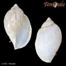
Harpovoluta charcoti is a species of sea snail, a marine gastropod mollusk in the family Volutidae, the volutes.[1]
The size of the shell varies between 25 mm and 75 mm.
Harpovoluta charcoti is widely found in and around Antarctica/sub-Antarctic or Southern Ocean.[2][3]
Harpovoluta charcoti is a species of sea snail, a marine gastropod mollusk in the family Volutidae, the volutes.