-
Ipoa fragila
-
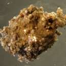
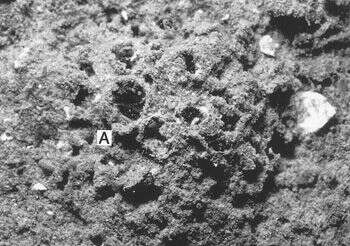
This image clearly shows the fine grains that make up the test surface. Inset: a closeup of the aperture. Image courtesy of Andrew J. Gooday, Southampton Oceanography Centre.
-
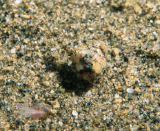
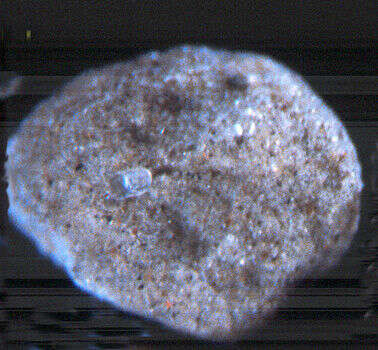
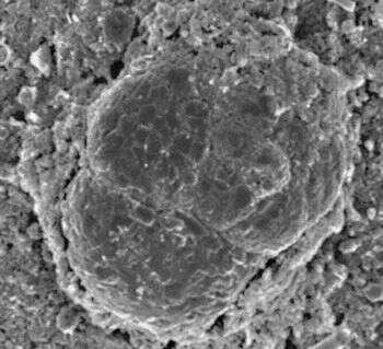
A live cell in its native environment. Notice that the foram has selected two discrete sizes of sand grains to make its test, and does not use the other sizes available to it. Photo courtesy of Robert Sanders. More information about this image is available at the
McMurdo Sound Underwater Field Guide.
-
Ipoa fragila specimen
-
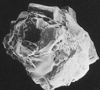
A closeup of the opening. Image courtesy of Elisabeth Alve, University of Oslo. Originally published in J. Foram. Res. 16: 261-284; used with permission.
-
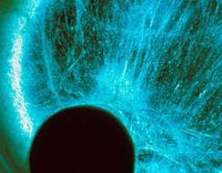
This darkfield image shows the reticulopodial network (blue fibers); the cell body is the dark circular mass at lower left. Image courtesy of Samuel S. Bowser, Wadsworth Center.
-
Reticulum sp.
-
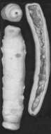
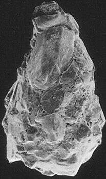
"Lagena" is a Latin word meaning "flask". This flask-shaped foram was found in the Oslofjord, Norway. Image courtesy of Elisabeth Alve, University of Oslo. Originally published in J. Foram. Res. 16: 261-284; used with permission.
-
An SEM of part of the reticulopodial network. A. rara reticulopods are unusually strong, capable of trapping and rending juvenile arthropods and echinoderms. Image courtesy of Samuel S. Bowser, Wadsworth Center.
-
This benthic species generally lives buried under 2-5 mm. of sediment. Image courtesy of Thomas Wilding, Southampton Oceanography Centre. This image first appeared in J. Foram. Res 32:358-363 and is used with permission.
-
This Antarctic allogromiid has a loosely-agglutinated but thick test made mostly of fine sand. The grains appear to be held together by reticulopodia, rather than by an organic cement. Image courtesy of Samuel S. Bowser, Wadsworth Center.
-
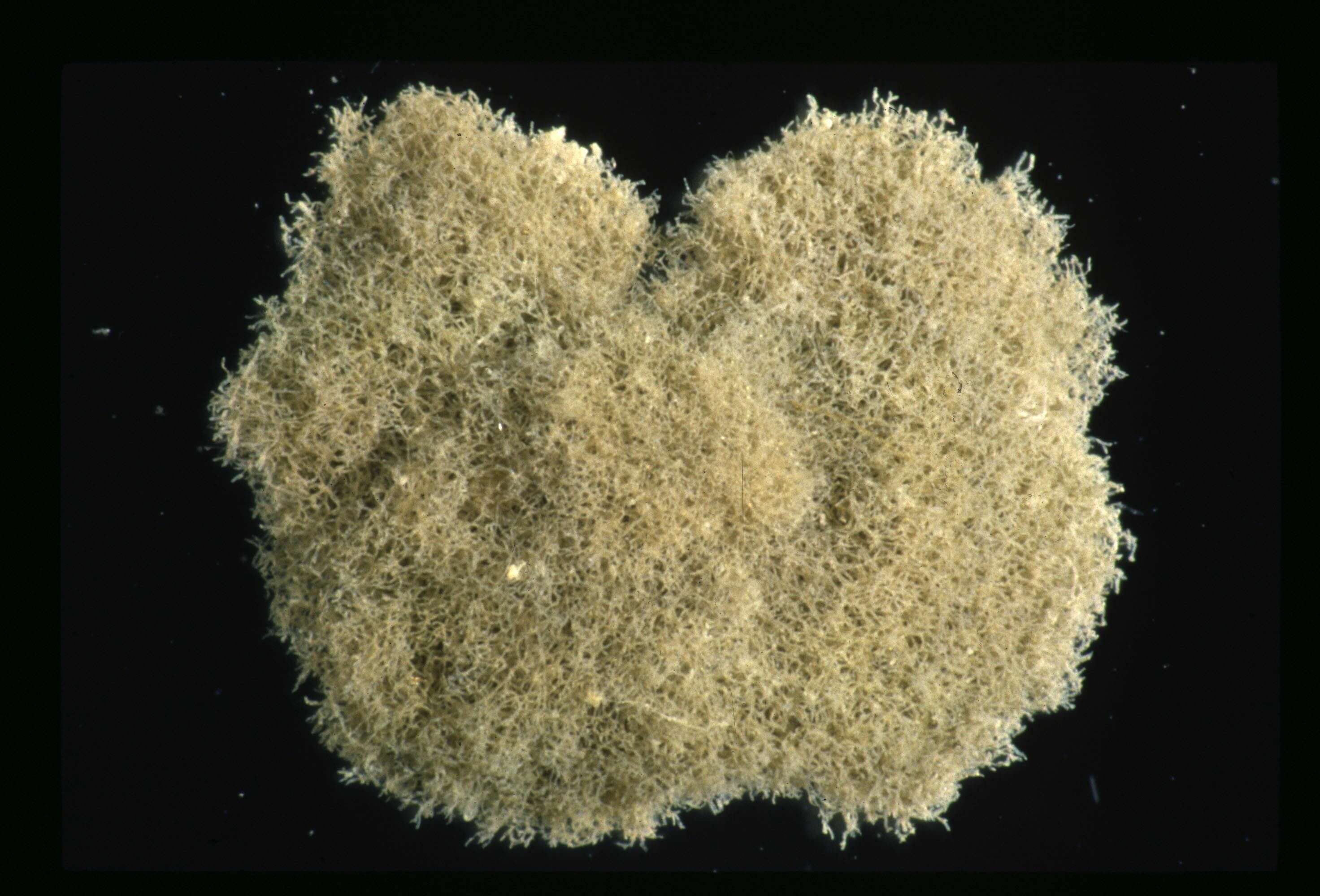
The opaque white color of the cytoplasm gives this species its name. Notice the loose structure of the cell mass. Image courtesy of Thomas Wilding, Southampton Oceanography Centre. This image first appeared in J. Foram. Res 32:358-363 and is used with permission.
-
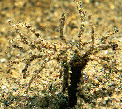
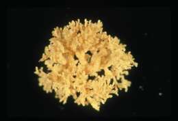
A live cell in its native environment. This species is found both in the "arborescent" morphology shown here and as a simple agglutinated sphere. Photo courtesy of Robert Sanders. More information about this image is available at the
McMurdo Sound Underwater Field Guide.
-
An individual that has contracted into a rounded shape after collection. Image courtesy of Thomas Wilding, Southampton Oceanography Centre. This image first appeared in J. Foram. Res 32:358-363 and is used with permission.
-
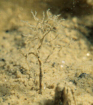
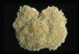
A live cell in its native environment. The tree-like structure allows the foram to lift its reticulopods a centimeter or more of the seafloor. Photo courtesy of Robert Sanders. More information about this image is available at the
McMurdo Sound Underwater Field Guide.
-
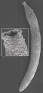
This foram was collected from fine carbonate muds off Little Duck Key, Florida. Image courtesy of Susan T. Goldstein, University of Georgia. This image first appeared in J. Foram Res. 32:375-383 and is used with permission.
-
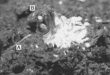
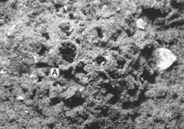
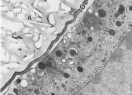
This species has a thick agglutinated test wall (A) underlain by a thin organic lining (IOL). Image courtesy of Susan T. Goldstein, University of Georgia. This image first appeared in J. Foram Res. 32:375-383 and is used with permission.
-
Image from Loeblich and Tappan's Foraminiferal Genera and their Classification. Originally appeared in Loeblich and Tappan 1964.
-
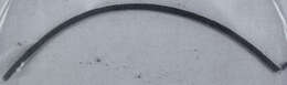
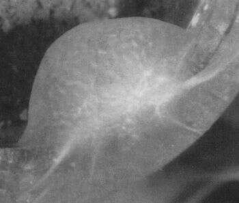
The foram in life position. The upper third protrudes from the seafloor, while the rest remains buried. Image courtesy of Andrew J. Gooday, Southampton Oceanography Centre. This image first appeared in J. Foram. Res. 22:129-146 (1992) and is used with permission.
-
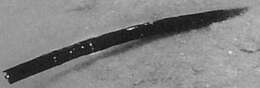
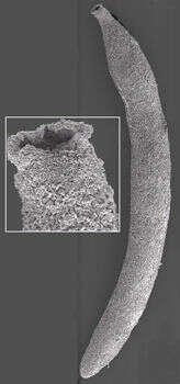
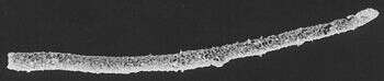
This approx. 10 cm. long individual was taken from the seafloor at 840 m. depth, 144 km. east of Cape Lookout, North Carolina. Image courtesy of Andrew J. Gooday, Southampton Oceanography Centre. This image first appeared in J. Foram. Res 22:129-146 (1992) and is used with permission.
-
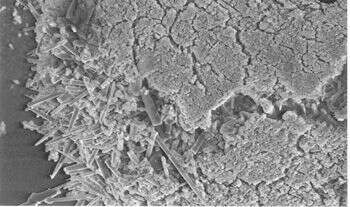
This SEM image shows the two distinct particle types that make up the foram's agglutinated wall. The outer surface is made of fine, black or brown particles, and is underlain by a much thicker layer of sponge spicules and small quartz particles. Image courtesy of Andrew J. Gooday, Southampton Oceanography Centre. This image first appeared in J. Foram. Res 22:129-146 (1992) and is used with permission.
-
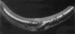
The white surface of this 6-cm-long foraminiferan is partially hidden by sediment trapped in mucus. The mucus was probably secreted by the polychaete worm Nicolea, which has been found on the test surface and is only about 20% as long as the foram is. Image courtesy of Andrew J. Gooday, Southampton Oceanography Centre. This image first appeared in J. Foram. Res 22:129-146 (1992) and is used with permission.
-
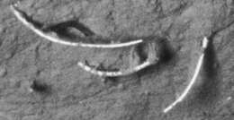
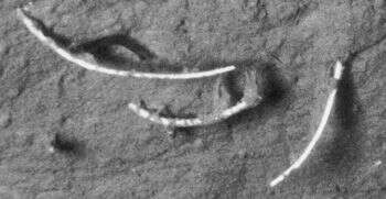
Three individuals photographed on the seafloor, at 850 m. depth, 72 km. NE of Cape Hatteras, North Carolina.Image courtesy of Andrew J. Gooday, Southampton Oceanography Centre. This image first appeared in J. Foram. Res 22:129-146 (1992) and is used with permission.
-
A closeup of a smaller foram (a member of the genus Trochammina) which is living on the surface of the Bathysiphon test. The Trochammina test is 100 microns in diameter. Image courtesy of Andrew J. Gooday, Southampton Oceanography Centre. This image first appeared in J. Foram. Res 22:129-146 (1992) and is used with permission.