-
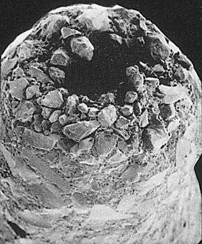
A closeup of the aperture. Image courtesy of Elisabeth Alve, University of Oslo. Originally published in J. Foram. Res. 16: 261-284; used with permission.
-
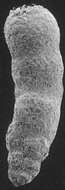
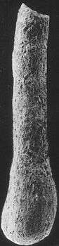
This genus is common in deep-water environments. Image courtesy of Elisabeth Alve, University of Oslo. Originally published in J. Foram. Res. 16: 261-284; used with permission.
-
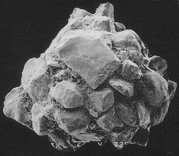
This species is common in the deep, marine ares of the Sandebukta, a branch of the Oslofjord. Image courtesy of Elisabeth Alve, University of Oslo. Originally published in J. Foram. Res. 16: 261-284; used with permission.
-
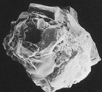
This specimen has two distinct apertures (one is quite visible at upper left). Image courtesy of Elisabeth Alve, University of Oslo. Originally published in J. Foram. Res. 16: 261-284; used with permission.
-
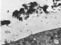
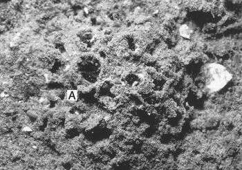
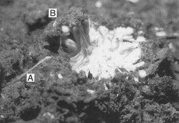
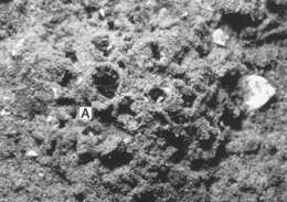
The wall of this foram includes a thick inner organic lining (IOL) underlying the agglutinated layer (A). The cell body is at lower right. Image courtesy of Susan T. Goldstein, University of Georgia. This image first appeared in J. Foram Res. 32:375-383 and is used with permission.
-
This specimen has no distinct apertures through the test, which is not uncommon in members of this genus. Image courtesy of Elisabeth Alve, University of Oslo. Originally published in J. Foram. Res. 16: 261-284; used with permission.
-
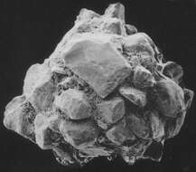
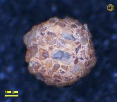
This foram was collected from a salt marsh on Sapelo Island, Georgia. The species name is due to this foram's habit of agglutinating fine, light-colored particls; it appears white under the light microscope. Image courtesy of Susan T. Goldstein, University of Georgia. This image first appeared in J. Foram Res. 32:375-383 and is used with permission.
-
Greenland Sea/Arctic, cruise of Polarstern found at 78.58N 07.37W at 192m depth 30.9.1995
-
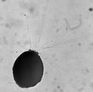
This image shows the foram's reticulopodia (the elaborate branching pseudopodia sticking out of the vase-like hole in the test). Reticulopods are the defining morphological characteristic of the Granuloreticulosea as a group. Image courtesy of Susan T. Goldstein, University of Georgia.
-
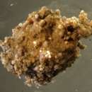
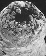
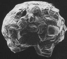
Tholosina species attach to surfaces and build an agglutinated dome over the cell body. The dome is more "inflated" looking than the ones produced by their apparent relatives, the genus Hemisphaerammina. Image courtesy of Elisabeth Alve, University of Oslo. Originally published in J. Foram. Res. 16: 261-284; used with permission.
-
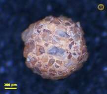
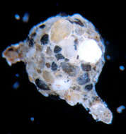
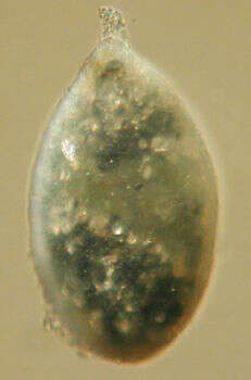
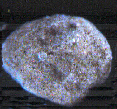
Psammophaga species are noted for taking sand grains into their bodies; the genus name means "sand eater" in Greek. You can see the coarse quartz sand through the translucent walls of the foram's test. Image courtesy of Susan T. Goldstein, University of Georgia.
-
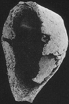
This view shows the inside of the dome-shaped test. Image courtesy of Elisabeth Alve, University of Oslo. Originally published in J. Foram. Res. 16: 261-284; used with permission.
-
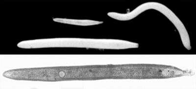
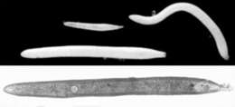
Top: Reflected-light image of three individuals of the species, showing size variation. The white color is caused by a very fine layer of sand particles (probably quartz) that the foram glues to its outer surface. Bottom: A slightly higher-magnification transmitted-light image, with the nucleus clearly visible. Length of this specimen: approximately 600 um. Image courtesy of Andrew J. Gooday, Southampton Oceanography Centre.
-
This giant Antarctic foraminiferan is often several millimeters across. Notice the two large projections (called stolons. In this species, the reticulopodia emerge from the ends of the stolons. Image courtesy of Samuel S. Bowser, Wadsworth Center.
-
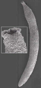
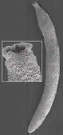
This image clearly shows the fine grains that make up the test surface. Inset: a closeup of the aperture. Image courtesy of Andrew J. Gooday, Southampton Oceanography Centre.
-
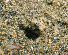
A live cell in its native environment. Notice that the foram has selected two discrete sizes of sand grains to make its test, and does not use the other sizes available to it. Photo courtesy of Robert Sanders. More information about this image is available at the
McMurdo Sound Underwater Field Guide.
-
A closeup of the opening. Image courtesy of Elisabeth Alve, University of Oslo. Originally published in J. Foram. Res. 16: 261-284; used with permission.
-
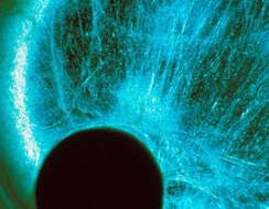
This darkfield image shows the reticulopodial network (blue fibers); the cell body is the dark circular mass at lower left. Image courtesy of Samuel S. Bowser, Wadsworth Center.
-
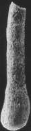
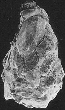
"Lagena" is a Latin word meaning "flask". This flask-shaped foram was found in the Oslofjord, Norway. Image courtesy of Elisabeth Alve, University of Oslo. Originally published in J. Foram. Res. 16: 261-284; used with permission.
-
An SEM of part of the reticulopodial network. A. rara reticulopods are unusually strong, capable of trapping and rending juvenile arthropods and echinoderms. Image courtesy of Samuel S. Bowser, Wadsworth Center.
-
This benthic species generally lives buried under 2-5 mm. of sediment. Image courtesy of Thomas Wilding, Southampton Oceanography Centre. This image first appeared in J. Foram. Res 32:358-363 and is used with permission.
-
This Antarctic allogromiid has a loosely-agglutinated but thick test made mostly of fine sand. The grains appear to be held together by reticulopodia, rather than by an organic cement. Image courtesy of Samuel S. Bowser, Wadsworth Center.
-
The opaque white color of the cytoplasm gives this species its name. Notice the loose structure of the cell mass. Image courtesy of Thomas Wilding, Southampton Oceanography Centre. This image first appeared in J. Foram. Res 32:358-363 and is used with permission.
-
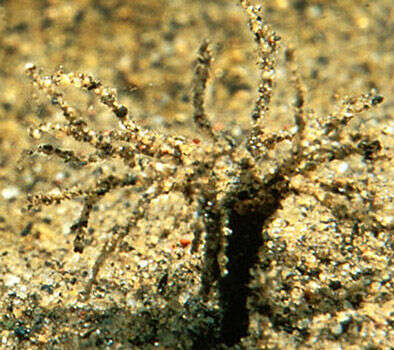
A live cell in its native environment. This species is found both in the "arborescent" morphology shown here and as a simple agglutinated sphere. Photo courtesy of Robert Sanders. More information about this image is available at the
McMurdo Sound Underwater Field Guide.