-
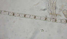
Chain forming diatom, which produces wing like extensions one valve end, which link adjacent cells. This is a cosmopolitan species.
-
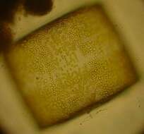
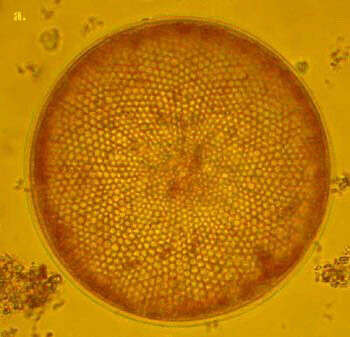
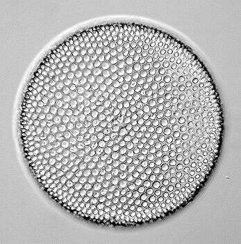
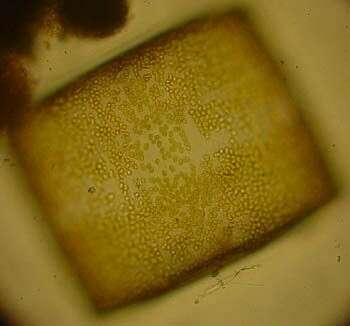
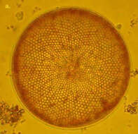
C. radiatus is one of the smaller Coscinodiscus species. It is box shaped in girdle view and the valves are very flat. The areolae form disctinct rows radiating from the valve centre. C. radiatus is a cosmopolitan species.
-
-
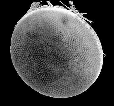
Scanning electron microscope image of valve. The organism is tentatively identified as C. radiatus. Sample taken from the water column off Martha's Vineyard, Massachusetts. Image by Charley O'Kelly and Shauna Murray.
-
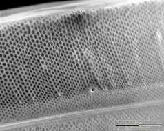
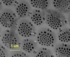
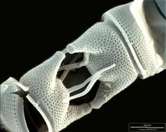
Scanning electron micrograph showing detail of the frustule of this diatom. The larger depressions are called areolae, and perforated region is called the cribrum, within which each perforation is referred to as a cribellum. The same term probably also refers to the perforations in the margins of the areolae. The species is probably C. radiatus. Sample from the water column off Martha's Vineyard. Images by Charley O'Kelly and Shauna Murray.
-
This species has a very fine aerolation. It can be distinguished from other species by the central hyaline area and the hyaline lines radiating from it between the areolae. It also has a distinctive shape in girdle view. The valve is very high (often higher than wide) and the valve margin appears to undulate slightly.
-
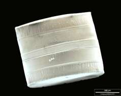
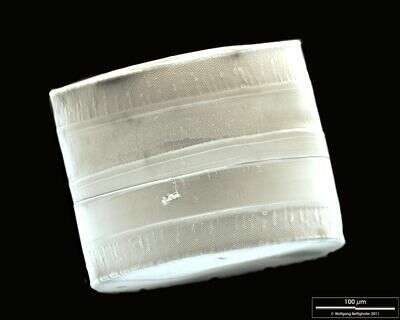
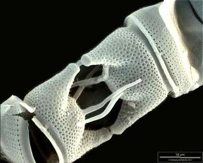
SEM of girdle view. The ligulae which fit in the open girdle bands are weakly visible. Scale bar indicates 100 µm. Sample from North Sea near Heligoland (spring diatom bloom). The image was built up using several photomicrographic frames with manual stacking technique. Use of SEM equipment courtesy of Lab Dr. Karl-Heinz Schäffner, Solingen, Germany.
-
Closeup of the lateral side of the valve. Scale bar indicates 25 µm. Sample from North Sea near Heligoland (spring diatom bloom). The image was built up using several photomicrographic frames with manual stacking technique. Use of SEM equipment courtesy of Lab Dr. Karl-Heinz Schäffner, Solingen, Germany.
-
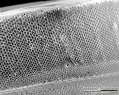
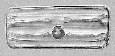
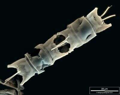
Valvar View. Scale bar indicates 100 µm. Sample from North Sea near Heligoland (spring diatom bloom). The image was built up using several photomicrographic frames with manual stacking technique. Use of SEM equipment courtesy of Lab Dr. Karl-Heinz Schäffner, Solingen, Germany.
-
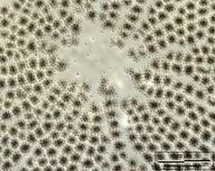
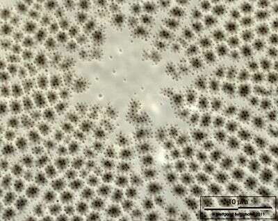
Closeup showing hyaline central area. Scale bar indicates 10 µm. Sample from North Sea near Heligoland (spring diatom bloom). Use of SEM equipment courtesy of Lab Dr. Karl-Heinz Schäffner, Solingen, Germany.
-
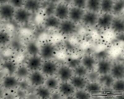
Closeup showing fine structure of valvar pores. Scale bar indicates 5 µm. Sample from North Sea near Heligoland (spring diatom bloom). Use of SEM equipment courtesy of Lab Dr. Karl-Heinz Schäffner, Solingen, Germany.
-
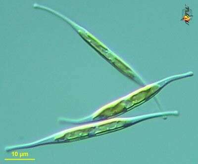
Cylindrotheca (sill-inn-dro-thee-ka) fusiformis, an elongate and slightly twisted pennate diatom (stramenopile), tends to move in a spiral motion, frustule not heavily silicifed and and can be deformed. Differential interference microscopy.
data on this strain.
-
-
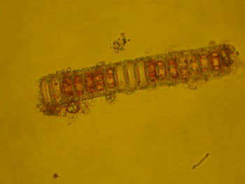
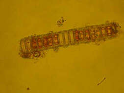
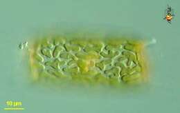
Thick walled, flat cells, which are united into tight chains, valve diameter 8-80 microns. P. sulcata is a benthic from but appears in the plankton in turbulent water
-
-
-
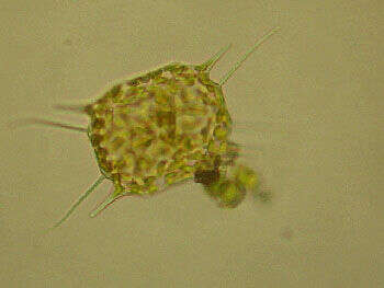
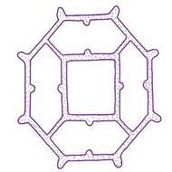
Distephanus octangulatus Wailes, 1931. Silicieous skeleton consisting of a frame forming a truncated cone, the basal ring octagonal with an external projection at each angle, from the centers of four sides arise supporting bars attached to the angles of the square apical ring, the side openings provided with small semicircular projections at the centers of the longer sides. Diameter of basal ring 35 microns
-
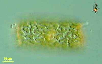
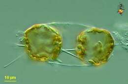
Odontella (owe-don-tell-a) mobiliensis, a centric diatom. The frustule or shell is formed of two valves joined by girdle bands. Many small peripheral chloroplasts and a central nucleus. Differential interference microscopy.
data on this strain.
-
Odontella (owe-don-tell-a) mobiliensis, a centric diatom. The frustule or shell is formed of two valves joined by girdle bands. With horns (spines) emerging from the apical margins of the valves and more spines (referred to as apical processes) arising closer to the centre of the valves. Many small peripheral chloroplasts and a central nucleus. Two daughter cells located within frustule of parental cell. Differential interference microscopy.
data on this strain.
-
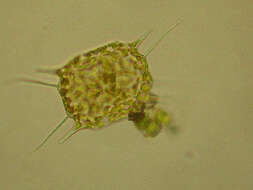
Cells are single or united into short chains by the long spines extending from the elevated central part of the valve face. The processes are slender and point diagonally outward. This species can be confused with O. regia.
-
Scale bar indicates 10 µm. Sample from North Sea near Heligoland (spring diatom bloom). Use of SEM equipment courtesy of Lab Dr. Karl-Heinz Schäffner, Solingen, Germany.
-
Scale bar indicates 25 µm. Sample from North Sea near Heligoland (spring diatom bloom). Use of SEM equipment courtesy of Lab Dr. Karl-Heinz Schäffner, Solingen, Germany.
-
-
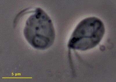
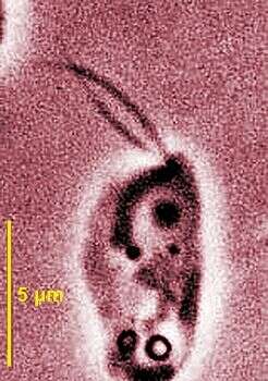
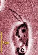
Phase contrast micrograph showing the two short anterior flagella, the nucleus and the mitochondrion close to the base of the flagella.