-
Cosmoeca ventricosa Thomsen in Thomsen and Boonruang, 1984. Cells solitary. Cell body when dried 6.7-7.5 x 2.5-4 microns, flagellum about 20 microns long. Lorica barrel-shaped, 23-31 microns high, composed of3 transverse costae and 9-12 longitudinal costae. The two almost equally sized anterior transverse costae (average widths 22.4 microns and 19.2 microns) are located at the anterior end of the lorica chamber and at the level of the joints between the second and the third longitudinal costal strip (counted from the anterior lorica end). The number of strips in both anterior transverse costae corresponds to the number of longitudinal costae. The third minute transverse costa is formed by 3-4 costal strips and located at the level of the joints between the third and the fourth longitudinal costal strip (counted from the anterior lorica end). A numerical reduction in the number of longitudinal costae occurs below the posterior transverse costa.
-
Syndetophyllum pulchellum (Leadbeater, 1974) Thomsen and Moestrup, 1983. Cell obovoid, 2 - 5 microns long x 2 - 4 microns wide, single flagellum approximately 15 microns long, funnel-shaped collar of approximately 20 tentacles 2 - 5 microns long. Lorica 6-8 microns high composed of two chambers. Anterior chamber consisting of 10 longitudinal costae and two transverse cos tae, one forming the anterior ring and one at the base of the chamber. Lower chamber formed by 10 longimdinal costae converging posteriorly. All costal strips forming the lorica broad with a thickened midrib with an elaborate pattern of perforations either side, costal strips of the anterior ring bearing an upright spine at one end.
-
Acanthoeca brevipoda Ellis, 1930. Lorica cup-shaped, 9.5-11 microns long, with spirally arranged costae. The strips towards the posterior are broad and flattened. Thirteen projections protrude anteriorly and are formed of more than a single costal strip basally.
-
Acanthoeca spectabilis Ellis, 1930. Cells are 3.5-5 microns in length, elongate with a pointed posterior, and fit closely to the main chamber of the lorica. Electron microscopy reveals that these cells are enclosed by fibrous material. The pseudopodial collar is shorter than the cell and the flagellum projects slightly from the top of the lorica. The main chamber of the lorica is conical and tapers posteriorly, giving rise to a pedicel. Costae in the main chamber give rise to longitudinal spines anteriorly. Fibres were present and arranged in the same pattern as costal strips in normal cells, except that, being less rigid, the spines are irregular and crumpled. The conical shape of the main chamber and the pedicel appears to be maintained by a membranous structure in the cells with a fibrous lorica.
-
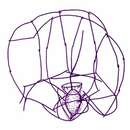
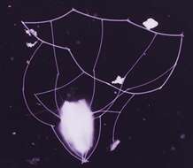
This image was made from samples taken during a scientific cruise in the Pacific. Water was filtered to concentrate the organisms that were present, then dried onto a thin sheet of plastic and then shadowed with a fine layer of metal to provide contrast. The preparation was then observed with an electron-microscope. This technique has been used to document the diversity of marine microbes, especially, protists in the oceans.
-
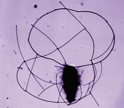
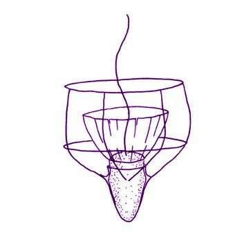
Crinolina isefiordensis Thomsen, 1981. Cell solitary, planktonic, living in skirt-shaped lorica, open anteriorly and posteriorly. Protoplast 8 microns long and 5 microns wide, without chloroplast. Single flagellum 2-3 times protoplast length, surrounded by a collar of tentacles. Height of collar approximately 6 microns, maximum diameter of collar 13 microns Lorica 25-30 microns long, diameter at base 20-30 microns, diameter at neck constriction 10-13 microns Longitudinal costae (12) 15-16, each composed of 6-7 costal strips. Two transverse costae. Longitudinal costae form spines at the anterior end of the lorica. Each spine is composed of two costal strips. Posteriorly the longitudinal costae project slightly beyond the transverse costal ring.
-

Parvicorbicula circularis Thomsen, 1976. Cell when dried approximately 3 x 5 microns The single flagellum, up to 15 microns long, protrudes above the lorica. The lorica is constructed of 4 longitudinal costae composed of 3 costal strips each and two transverse costae. One transverse costa (8 strips) delimits the lorica anteriorly. The bifurcated anterior tips of the longitudinal costae attach to the middle of every second transverse costal strip ( T -joints). The second transverse costa (6-8 strips) is located at the level of the joints between the first and the second longitudinal costal strip. In specimens with 8 costal strips in this costa, the longitudinal costae cross at every other joint between transverse costal strips. In specimens with 6-7 transverse costal strips, the crossings between longitudinal and transverse elements are less symmetric. The two posterior-most costal strips from each longitudinal costa overlap. The free ends of the penultimate longitudinal costal strips suspend a sheet which encompasses the cell.
-
Parvicorbicula manubriata Tong, 1997. Cells solitary or colonial. Cell 3.5-5.5 x 4.5-7 microns, flagellum 13-19 microns long surrounded by a collar of tentacles ~8 microns long. Lorica conical, ~8 microns long, consisting of a single transverse costa at the anterior of the lorica, and 8-10 longitudinal costae. The transverse costa consists of 10 or 11 slightly curved costal strips, one of which is duplicated. The longitudinal costae have no regular means of attachment to the transverse costa, joints may be in the middle or at the ends of the transverse costal strips. Longitudinal costae undergo numerical reduction, so that three to eight strips form the handle at the posterior of the lorica. All costal strips are narrow rods. Division is tectiform.
-
Parvicorbicula quadricostata Throndsen, 1970. The length of lorica is about 18 microns and its width is 24 microns
-
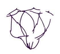
Parvicorbicula socialis (Meunier, 1910) Deflandre, 1960. The funnel-shaped lorica of this species is about 10 microns in diameter, with 10 longitudinal costae converging posteriorly, and two transverse costae. Each longitudinal costa consists of two costal strips. The anterior ring is made up of ten strips, each with a longitudinal costa attached to its middle part forming T-junctions. Drawing from a flattened lorica.
-
Parvicorbicula socialis socialis Deflandre, 1960. Lorica length 10 microns, lorica width 12 microns, longitudinal costae 10, transverse costae 2. Usually forms colonies.
-
This image was made from samples taken during a scientific cruise in the Pacific. Water was filtered to concentrate the organisms that were present, then dried onto a thin sheet of plastic and then shadowed with a fine layer of metal to provide contrast. The preparation was then observed with an electron-microscope. This technique has been used to document the diversity of marine microbes, especially, protists in the oceans.
-
Pleurasiga minima Throndsen, 1970. Lorica 9.5-16 microns long with 2 transverse costae and 7 longitudinal costae, all of which extend to the posterior of the lorica. The transverse costae are of about the same width, and the lorica narrows below the second costa to form a pointed base. Cell located at the posterior of the lorica, with flagellum protruding above it.
-
Pleurasiga minima minima Throndsen, 1970. The different varieties differ mainly in size. The lorica of P. minima minima is about 10 microns long and wide.
-
Pleurasiga reynoldsii Throndsen, 1970. The lorica is 23 microns long and 22 microns wide, respectively.
-
Pleurasiga echinocostata Espeland in Espeland and Throndsen, 1986. Cell 2.5-4.5 microns long, with single smooth flagellum surrounded basally by a pseudopodial collar. Lorica bell-shaped with 7 longitudinal costae, occasionally reduced to 6 or 5 in the posterior part, consisting of 3 costal strips, the 2 most anterior with a distal anterior dilatation. A single anterior transverse lorica ring consisting of 7 costal strips each with a terminal anteriorly pointing spine. Anterior longitudinal costa joining the middle of each coastal strip of the tranverse costa. Lorica length 7.5-10 microns, anterior diameter 5-8.3 microns Thje drawing is of a damaged lorica.
-
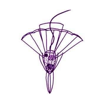
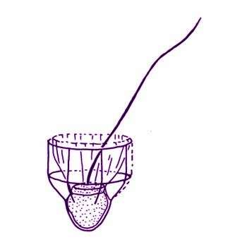
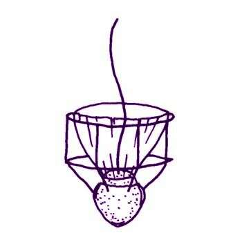
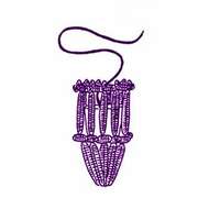
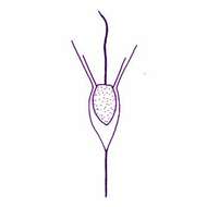
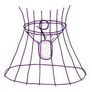
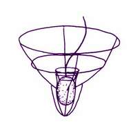
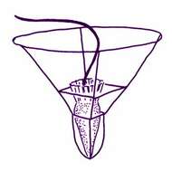
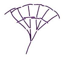
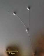
Polyoeca dichotoma Kent, 1880. Lorica of polythecium urceolate, pedicellate, tapering posterioly, slightly constricted at a distance of one-third of the total length from the anterior margin, and then widening out to their greatest diameter, pediceles of each separate lorica straight, slender, varying from the same to two or three times the length of the latter structure, contained animalcules ovate, occupying respectively about one-half of the cavities of the lorica, contour of polythecium subdichotomous, each zooid usually giving rise by trasverse fission to two new ones which attach themselves to opposite sides of the parent lorica. Length of separate lorica 10 microns
-

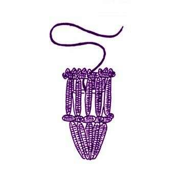
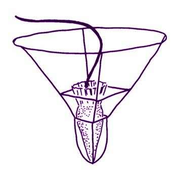
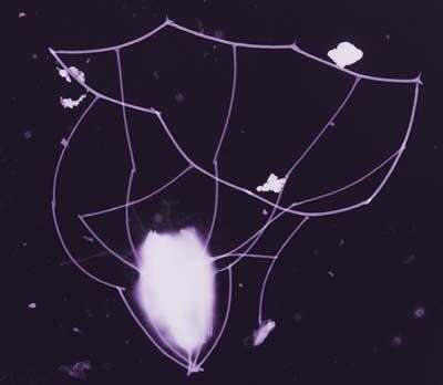
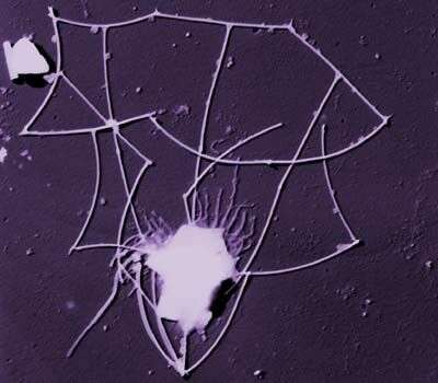
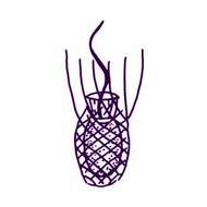
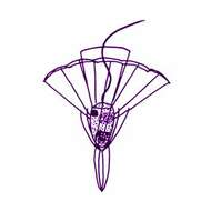
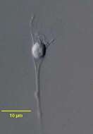
Portrait of the acanthoecid choanoflagellate, Polyoeca dichotoma (Kent, 1881). Cells are solitary or united to form linear or dendroid colonies which attach to the substrate. The lorica is funnel-shaped, constructed of numerous siliceous longitudinal costae, each made up of costal strips. The longitudinal costae terminate as anterior spines (11-17); 2 or more bands of equally spaced transverse costal strips (difficult to see in vivo) encircle the lorica chamber in which the protoplast (cell body) resides. At the apex of the funnel the longitudinal costae unite to form an aggregated pedicel. In dendroid colonies the pedicel of the daughter (anterior) cell attaches to the outside of the lorica of the parent cell. Details of lorica morphology are best seen with scanning electron microscopy. The cell body has one anterior flagellum (seen here) surrounded by a rhizopodial collar (not visible here). Division is nudiform (i.e. naked swarmers are formed which then form a lorica). Collected from a commercial saltwater aquarium in Boise, Idaho February 2004. DIC.
-
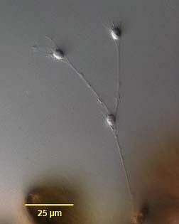
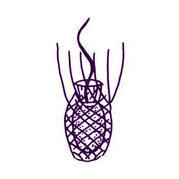
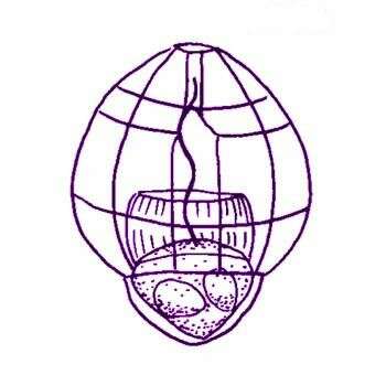
Savillea micropora (Norris, 1965) Leadbeater, 1975. Cells spherical to ovoid, with collar and flagellum. Cells located in base of lorica composed of longitudinal and transverse costae. Base of lorica with numerous closely spaced transverse costae, expanded anterior part of lorica composed of approximately 12 longitudinal costae traversed at regular or irregular intervals by 4-6 transverse costae. Terminal transverse costa forming pore with small diameter (approximately 1 microns diameter). Cell 2.5-3 microns diameter, base of lorica 1.6-3.5 microns long, 3 microns diameter, anterior part of lorica 5-6.3 microns long, 5-6.3 microns diameter.
-
Savillea parva (Ellis, 1930) Loeblich III, 1967. Cells are 2.5-4 microns (spherical), lorica 10-10.5 microns in length. The cell body is situated at the bottom of the lorica, the pseudopodial collar extends about two-thirds of the way up the lorica, and the flagellum extends to the top of the lorica. Unlike Savillea micropora, a flagellum is always present and active.
-
Acanthocorbis nana Thomsen et al, 1997. Cells solitary, attached to the substratum by means of single costal strips projecting from the posterior lorica end. Lorica funnel shaped, about 10 x 5 microns, and with anterior long spines (10-14) that continue downwards as longitudinal costae. Numerous, obliquely oriented transverse costal strips (>20) encircle the lorica at the level of or just below the joins between the anterior most longitudinal costal strips. The posterior part of the lorica encompasses numerous transverse costal strips that do not form distinct costae. Costal strips are rod-shaped, and 3-4 microns long. The cell body, about 4.5x3.5 microns, is posteriorly located within the lorica. The single flagellum is about 5 microns long, surrounded by a ring of tentacles (each about 2microns long), and with a conspicuous hairpoint. Division tectiform.