-
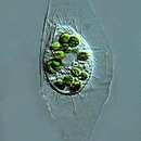
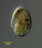
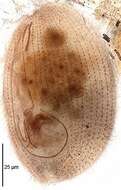
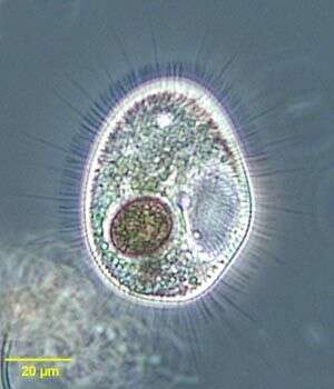
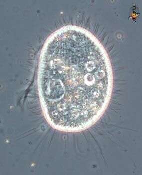
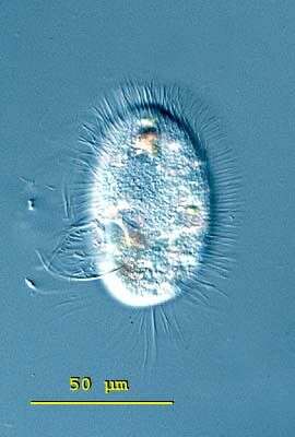
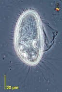
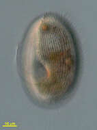
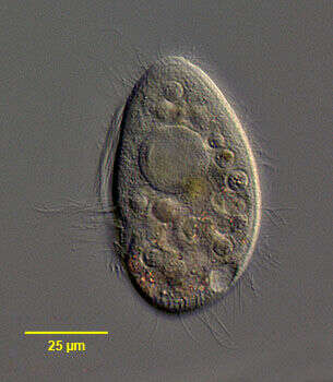
Histiobalantium is easy to distinguish from the similar genus Pleuronema by the stiff long cilia which intersperse the dense somatic ciliation. Histiobalantium have long cilia distributed over the whole cell whereas at Pleuronema this cilia are confined to the posterior region. The body of Histiobalantium natans is elliptical, with the right side slightly concave and anterior end a little narrower than the posterior. The peristome is deep, equipped with a well-developed undulating membrane. Several micronuclei are attached to the two round macronuclei. The contractile vacuole is located in the posterior half of the body. 55 - 105 microns long. From fresh water ponds and lakes. This free-swimming specimen of Histiobalantium natans was collected in freshwater ponds near Konstanz, Germany. The contractile vacuole is located in the posterior. 95 microns. Differential interference contrast.
-
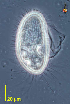
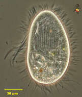
Histiobalantium is easy to distinguish from the similar genus Pleuronema by the stiff long cilia which intersperse the dense somatic ciliation. Histiobalantium have long cilia distributed over the whole cell whereas at Pleuronema this cilia are confined to the posterior region. The body of Histiobalantium natans is elliptical, with the right side slightly concave and anterior end a little narrower than the posterior. The peristome is deep, equipped with a well-developed undulating membrane. Several micronuclei are attached to the two round macronuclei. The contractile vacuole is located in the posterior half of the body. 55 - 105 microns long. From fresh water ponds and lakes. This specimen of Histiobalantium natans shows the stiff long cilia around the cell. 95 microns. From freshwater ponds near Konstanz, Germany. Differential interference contrast.
-
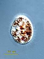
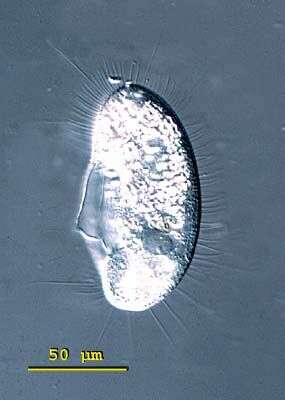
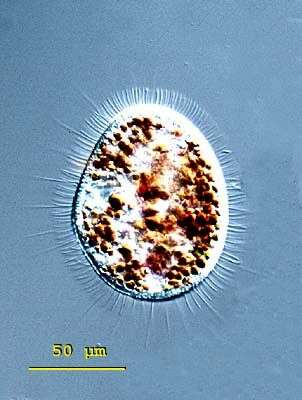
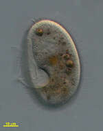
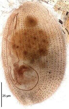
Histiobalantium is easy to distinguish from the similar genus Pleuronema by the stiff long cilia which intersperse the dense somatic ciliation. Histiobalantium have long cilia distributed over the whole cell whereas at Pleuronema this cilia are confined to the posterior region. The body of Histiobalantium natans is elliptical, with the right side slightly concave and anterior end a little narrower than the posterior. The peristome is deep, equipped with a well-developed undulating membrane. Several micronuclei are attached to the two round macronuclei. The contractile vacuole is located in the posterior half of the body. 55 - 105 microns long. From fresh water ponds and lakes. This well-fed specimen of Histiobalantium natans is from a freshwater pond near Konstanz, Germany. 88 microns. Differential interference contrast.
-
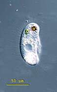
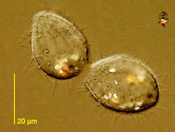
Histiobalantium natans(Claparede & Lachmann, 1858), a large scuticociliate - with long stiff cilia extending outwards from the body surface. This individual has ingested a Trachelomonas. Collected from a freshwater pond near Boise, Idaho.Phase contrast.
-
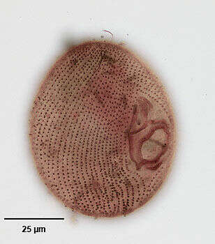
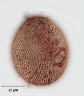
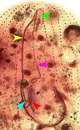
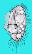
Ventral infraciliature of the hymenostome ciliate, Histiobalantium natans (Claparede & Lachmann, 1858). The cell is ovoid to reniform in outline with a broadly rounded posterior. The oval oral aperture is located in the mid-portion of the cell. There is an undulating membrane on the right margin of the peristome curving around the posteriorly located cytostome to form a cup or pouch. There are three adoral membranelles on the left side of the peristome. M1 and M2 are parallel and oriented obliquely to the undulating membrane while membranelle M3 is inclined slightly posteriorly between the posterior ends of M1 and M2 and the undulating membrane, forming a triangle. The somatic ciliature is composed of longitudinal kineties. Longer bristle-like cilia are interspersed with more numerous short cilia. Both pre- and post-oral sutures are present. A long caudal cilium absent. There are three contractile vacuoles. Macronucleus with an irregular shape, usually divided into two parts with several micronuclei. Similar in appearance to Pleuronema which may have long caudal cilia but whose other somatic cilia are of equal length. Stained by the silver carbonate technique (see Foissner, W. Europ. J. Protistol., 27:313-330;1991). Collected from a freshwater pond near Boise, Idaho, february 2005. Brightfield.
-
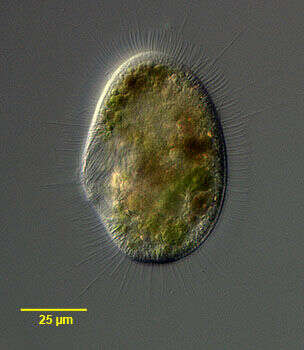
Portraitof the hymenostome ciliate, Histiobalantium natans (Claparede & Lachmann, 1858). The cell is ovoid to reniform in outline with a broadly rounded posterior. The oval oral aperture is located in the mid-portion of the cell. There is an undulating membrane on the right margin of the peristome curving around the posteriorly located cytostome to form a cup or pouch. There are three adoral membranelles on the left side of the peristome. M1 and M2 are parallel and oriented obliquely to the undulating membrane while membranelle M3 is inclined slightly posteriorly between the posterior ends of M1 and M2 and the undulating membrane, forming a triangle. The somatic ciliature is composed of longitudinal kineties. Longer bristle-like cilia are interspersed with more numerous short cilia. Both pre- and post-oral sutures are present. A long caudal cilium absent. There are three contractile vacuoles. Macronucleus with an irregular shape, usually divided into two parts with several micronuclei. Similar in appearance to Pleuronema which may have long caudal cilia but whose other somatic cilia are of equal length. Stained by the silver carbonate technique (see Foissner, W. Europ. J. Protistol., 27:313-330;1991). Collected from a freshwater pond near Boise, Idaho, february 2005.DIC.
-
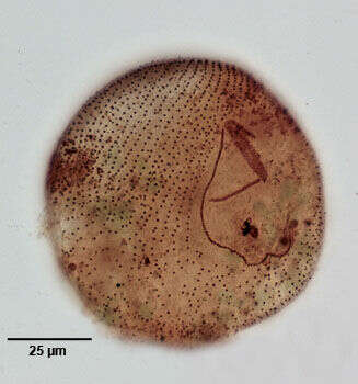
Oral infraciliature of the hymenostome ciliate, Histiobalantium natans (Claparede & Lachmann, 1858). The cell is ovoid to reniform in outline with a broadly rounded posterior. The oval oral aperture is located in the mid-portion of the cell. There is an undulating membrane on the right margin of the peristome curving around the posteriorly located cytostome to form a cup or pouch. There are three adoral membranelles on the left side of the peristome. M1 and M2 are parallel and oriented obliquely to the undulating membrane while membranelle M3 is inclined slightly posteriorly between the posterior ends of M1 and M2 and the undulating membrane, forming a triangle. The somatic ciliature is composed of longitudinal kineties. Longer bristle-like cilia are interspersed with more numerous short cilia. Both pre- and post-oral sutures are present. A long caudal cilium absent. There are three contractile vacuoles. Macronucleus with an irregular shape, usually divided into two parts with several micronuclei. Similar in appearance to Pleuronema which may have long caudal cilia but whose other somatic cilia are of equal length. Stained by the silver carbonate technique (see Foissner, W. Europ. J. Protistol., 27:313-330;1991). Collected from a freshwater pond near Boise, Idaho, february 2005. Brightfield.
-

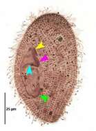
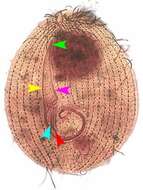
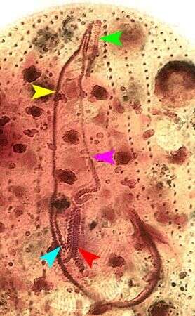
Oral infraciliature of the hymenostome ciliate, Histiobalantium natans (Claparede & Lachmann, 1858). The cell is ovoid to reniform in outline with a broadly rounded posterior. The oval oral aperture is located in the mid-portion of the cell. There is an undulating membrane on the right margin of the peristome curving around the posteriorly located cytostome to form a cup or pouch (green arrowhead). There are three adoral membranelles on the left side of the peristome. M1 and M2 are parallel and oriented obliquely to the undulating membrane (yellow and pink arrowheads respectively) while membranelle M3 is inclined slightly posteriorly between the posterior ends of M1 and M2 and the undulating membrane, forming a triangle (light blue arrowhead). The somatic ciliature is composed of longitudinal kineties. Longer bristle-like cilia are interspersed with more numerous short cilia. Both pre- and post-oral sutures are present. A long caudal cilium absent. There are three contractile vacuoles. Macronucleus with an irregular shape, usually divided into two parts with several micronuclei. Similar in appearance to Pleuronema which may have long caudal cilia but whose other somatic cilia are of equal length. Stained by the silver carbonate technique (see Foissner, W. Europ. J. Protistol., 27:313-330;1991). Collected from a freshwater pond near Boise, Idaho, february 2005. Brightfield.
-
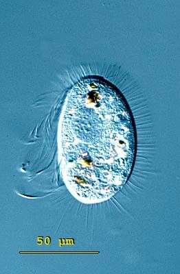
Pleuronema (ploo-row-knee-ma) is a scuticociliate - a group that distinguished by a well developed undulating membrane associated with the mouth. The scuticus refers to the hook -shape of the line of cilia associated with the mouth. The scuticus is used to help capture suspended bacteria which are ingested as food. In this individual. Mostly marine. Phase contrast.
-
Pleuronema (ploo-row-knee-ma) is a scuticociliate - a group that distinguished by a well developed undulating membrane associated with the mouth. The scuticus refers to the hook -shape of the line of cilia associated with the mouth, and the curve of this hook can be seen as the light curving line to the left near the equator of the cell. The scuticus is used to help capture suspended bacteria which are ingested as food. In this individual, the membrane (or sail) is not extended. Mostly marine. Phase contrast.
-
Pleuronema (ploo-row-knee-ma) is a scuticociliate - a group that distinguished by a well developed undulating membrane associated with the mouth. The scuticus refers to the hook -shape of the line of cilia associated with the mouth. The scuticus is used to help capture suspended bacteria which are ingested as food. In this individual. Mostly marine. Phase contrast.
-
-
Cedar Swamp, Woods Hole, Massachusetts, USA. Photoed by Hwan Su Yoon.
-
Cedar Swamp, Woods Hole, Massachusetts, USA. Photoed by Hwan Su Yoon.
-
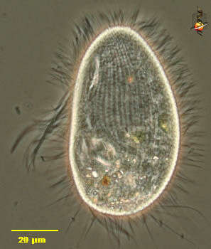
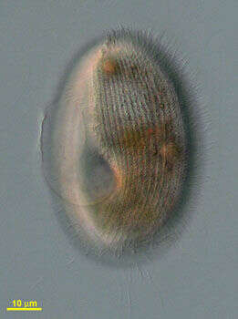
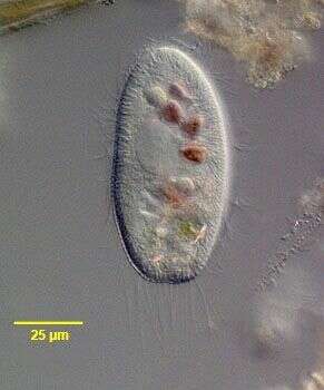
Pleuronema (ploo-row-knee-ma); a genus of very fast swimming ovoid ciliates. When feeding, the ciliates rest motionless and can be observed with care. Cells of Pleuronema coronatum, this species, measure 60 - 90 microns. There is a large prominent undulating membrane marking off the right edge of the peristome. The macronucleus is spherical and more or less centrally located. The posterior end of the cell has several long caudal cilia. The contractile vacuole is located in the posterior third of the cell. This species is found in fresh water ponds and lakes. This specimen is a resting specimen with a round macronucleus and an extended undulating membrane. 76 microns. Differential interference contrast.
-
Pleuronema (ploo-row-knee-ma); a genus of very fast swimming ovoid ciliates. When feeding, the ciliates rest motionless and can be observed with care. Cells of Pleuronema coronatum, this species, measure 60 - 90 microns. There is a large prominent undulating membrane marking off the right edge of the peristome. The macronucleus is spherical and more or less centrally located. The posterior end of the cell has several long caudal cilia. The contractile vacuole is located in the posterior third of the cell. This species is found in fresh water ponds and lakes. This mage emphasizes the caudal cilia. 76 microns. Differential interference contrast.
-
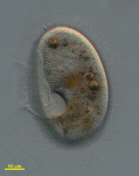
Left lateral view of the hymenostome ciliate, Pleuronema coronatum (Kent, 1881). The cell is ovoid to fusiform and slightly dorsoventrally compressed. The teardrop shaped peristome begins subapically and extends about 2/3 the cell length. There is a prominent undulating membrane on its right border. This curves around the posterior margin of the peristome forming a distinct pocket. The cytostome is in the center of this pocket. Fibrils radiate from the margin of this pocket to the cytostome. There are three adoral membranelles. . There are many longer caudal cilia. The otherwise very similar P. crassum has no tuft of longer caudal cilia. There is a fring of fusoform subcortical extrusomes (visible here at the posterior end).The spherical macronucleus is central and has 1 to 8 adjacent micronuclei. The single posterior contractile vacuole empties through a dorsal excretory pore. P. coronatum, like Histiobalantium, swims very rapidly, intermittently coming to rest. Members of the genus Pleuronema can be distinguished in vivo from the similar genus, Histiobalantium, by the absence of longer, stiff bristle-like cilia interspersed with shorter somatic cilia seen in the latter genus. Collected from a freshwater pond near Boise, Idaho March 2005. DIC.
-
Left lateral view of the hymenostome ciliate, Pleuronema coronatum (Kent, 1881). The cell is ovoid to fusiform and slightly dorsoventrally compressed. The teardrop shaped peristome begins subapically and extends about 2/3 the cell length. There is a prominent undulating membrane on its right border. This curves around the posterior margin of the peristome forming a distinct pocket. The cytostome is in the center of this pocket. Fibrils radiate from the margin of this pocket to the cytostome. There are three adoral membranelles. . There are many longer caudal cilia. there is a fringe of fusiform subcortical extrusomes (seen well here). The otherwise very similar P. crassum has no tuft of longer caudal cilia. The spherical macronucleus is central and has 1 to 8 adjacent micronuclei. The single posterior contractile vacuole empties through a dorsal excretory pore. P. coronatum, like Histiobalantium, swims very rapidly, intermittently coming to rest. Members of the genus Pleuronema can be distinguished in vivo from the similar genus, Histiobalantium, by the absence of longer, stiff bristle-like cilia interspersed with shorter somatic cilia seen in the latter genus. Collected from a freshwater pond near Boise, Idaho March 2005. DIC.
-

Ventral infraciliature of the hymenostome ciliate, Pleuronema coronatum (Kent, 1881). The cell is ovoid to fusiform and slightly dorsoventrally compressed. The teardrop shaped peristome begins subapically and extends about 2/3 the cell length. There is a prominent undulating membrane on its right border. This curves around the posterior margin of the peristome forming a distinct pocket. The cytostome is in the depths of this pocket. Fibrils radiate from the margin of this pocket to the cytostome. There are three adoral membranelles. M1 begins at the anterior end of the undulating membrane and forms an acute angle with it as it extends posteriorly for a short distance. M2 is bipartite. The long anterior portion begins in the angle between M1 and the undulating membrane and extends posteriorly to the termination of the undulating membrane. At the posterior end of this part there is a sharp indentation rightward toward the peristome. This feature of M2 differentiates the "coronatum"� type oral apparatus from the "marinum" type in this genus. There is a separate V-shaped part of M2 on the right side of the peristomal pocket. Some workers describe this as M3 but Foissner includes it as part of M2 and designates the two divergent kineties to its left as M3 (Foissner W., Berger H and Kohmann F. Taxonomische und ökologische Revision der Ciliaten des Saprobiensystems- Band III: Hymenostomata, Prostomatida, Nassulida. Informationsberichte Bayer. Landesamtes für Wasserwirtschaft. 1/94: 278-284, 1994). There are approximate 35 somatic kineties with 2 to 6 preoral kineties and no postoral kineties. There is a short preoral suture and a broad postoral suture in the center of which the cytopyge is located. The anterior apex of the cell is unciliated. There are many longer caudal cilia. The otherwise very similar P. crassum has no tuft of longer caudal cilia. The spherical macronucleus is central and has 1 to 8 adjacent micronuclei. The single posterior contractile vacuole empties through a dorsal excretory pore. Stained by the silver carbonate technic (see Foissner, W. Europ. J. Protistol., 27:313-330;1991). Collected from a freshwater pond near Boise, Idaho March 2005. Brightfield.
-
Detail of oral infraciliature of the hymenostome ciliate, Pleuronema coronatum (Kent, 1881). The cell is ovoid to fusiform and slightly dorsoventrally compressed. The teardrop shaped peristome begins subapically and extends about 2/3 the cell length. There is a prominent undulating membrane on its right border. This curves around the posterior margin of the peristome forming a distinct pocket. The cytostome is in the depths of this pocket. Fibrils radiate from the margin of this pocket to the cytostome. There are three adoral membranelles. M1 begins at the anterior end of the undulating membrane and forms an acute angle with it as it extends posteriorly for a short distance. M2 is bipartite. The long anterior portion begins in the angle between M1 and the undulating membrane and extends posteriorly to the termination of the undulating membrane. At the posterior end of this part there is a sharp indentation rightward toward the peristome. This feature of M2 differentiates the "coronatum"� type oral apparatus from the "marinum" type in this genus. There is a separate V-shaped part of M2 on the right side of the peristomal pocket. Some workers describe this as M3 but Foissner includes it as part of M2 and designates the two divergent kineties to its left as M3 (Foissner W., Berger H and Kohmann F. Taxonomische und ökologische Revision der Ciliaten des Saprobiensystems- Band III: Hymenostomata, Prostomatida, Nassulida. Informationsberichte Bayer. Landesamtes für Wasserwirtschaft. 1/94: 278-284, 1994). There are approximate 35 somatic kineties with 2 to 6 preoral kineties and no postoral kineties. There is a short preoral suture and a broad postoral suture in the center of which the cytopyge is located. The anterior apex of the cell is unciliated. There are many longer caudal cilia. The otherwise very similar P. crassum has no tuft of longer caudal cilia. The spherical macronucleus is central and has 1 to 8 adjacent micronuclei. The single posterior contractile vacuole empties through a dorsal excretory pore. Stained by the silver carbonate technic (see Foissner, W. Europ. J. Protistol., 27:313-330;1991). Collected from a freshwater pond near Boise, Idaho March 2005.
-
Oral infraciliature of the hymenostome ciliate, Pleuronema coronatum (Kent, 1881). The cell is ovoid to fusiform and slightly dorsoventrally compressed. The teardrop shaped peristome begins subapically and extends about 2/3 the cell length. There is a prominent undulating membrane on its right border. This curves around the posterior margin of the peristome forming a distinct pocket. The cytostome is in the depths of this pocket. Fibrils radiate from the margin of this pocket to the cytostome. There are three adoral membranelles. M1 begins at the anterior end of the undulating membrane and forms an acute angle with it as it extends posteriorly for a short distance. M2 is bipartite. The long anterior portion begins in the angle between M1 and the undulating membrane and extends posteriorly to the termination of the undulating membrane. At the posterior end of this part there is a sharp indentation rightward toward the peristome. This feature of M2 differentiates the "coronatum"� type oral apparatus from the "marinum" type in this genus. There is a separate V-shaped part of M2 on the right side of the peristomal pocket. Some workers describe this as M3 but Foissner includes it as part of M2 and designates the two divergent kineties to its left as M3 (Foissner W., Berger H and Kohmann F. Taxonomische und ökologische Revision der Ciliaten des Saprobiensystems- Band III: Hymenostomata, Prostomatida, Nassulida. Informationsberichte Bayer. Landesamtes für Wasserwirtschaft. 1/94: 278-284, 1994). There are approximate 35 somatic kineties with 2 to 6 preoral kineties and no postoral kineties. There is a short preoral suture and a broad postoral suture in the center of which the cytopyge is located. The anterior apex of the cell is unciliated. There are many longer caudal cilia. The otherwise very similar P. crassum has no tuft of longer caudal cilia. The spherical macronucleus is central and has 1 to 8 adjacent micronuclei. The single posterior contractile vacuole empties through a dorsal excretory pore. Stained by the Protargol technic (Wilbert modification) (see Foissner, W. Europ. J. Protistol., 27:313-330;1991). Collected from a freshwater pond near Boise, Idaho.Brightfield
-
Originally described by Ehrenberg under the name Paramecium chrysalis.
-
Originally described by Ehrenberg under the name Paramecium chrysalis.
-
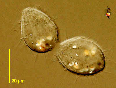
Cyclidium (sigh-clid-ee-um) is a small scuticociliate, and is here seen in late division. Ciliates multiply by dividing in two. Ciliates divide across the kineties (i.e. along a line that is parallel to the axis of the flagellar bases), and flagellates divide longitudinally (but for them that also means that the vision is in parallel to the axis of the flagella). The two cells shown here are joined by the tiniest strand of umbilical cytoplasm. Phase contrast.