in vivo
Description:
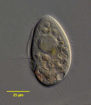
Left lateral view of the hymenostome ciliate, Pleuronema coronatum (Kent, 1881). The cell is ovoid to fusiform and slightly dorsoventrally compressed. The teardrop shaped peristome begins subapically and extends about 2/3 the cell length. There is a prominent undulating membrane on its right border. This curves around the posterior margin of the peristome forming a distinct pocket. The cytostome is in the center of this pocket. Fibrils radiate from the margin of this pocket to the cytostome. There are three adoral membranelles. . There are many longer caudal cilia. The otherwise very similar P. crassum has no tuft of longer caudal cilia. There is a fring of fusoform subcortical extrusomes (visible here at the posterior end).The spherical macronucleus is central and has 1 to 8 adjacent micronuclei. The single posterior contractile vacuole empties through a dorsal excretory pore. P. coronatum, like Histiobalantium, swims very rapidly, intermittently coming to rest. Members of the genus Pleuronema can be distinguished in vivo from the similar genus, Histiobalantium, by the absence of longer, stiff bristle-like cilia interspersed with shorter somatic cilia seen in the latter genus. Collected from a freshwater pond near Boise, Idaho March 2005. DIC.
Included On The Following Pages:
- Life (creatures)
- Cellular (cellular organisms)
- Eukaryota (eukaryotes)
- SAR (Stramenopiles, Alveolates, Rhizaria)
- Alveolata (alveolates)
- Ciliophora (ciliates)
- Intramacronucleata
- Oligohymenophorea
- Scuticociliatia
- Pleuronematida
- Pleuronematidae
- Pleuronema
- Pleuronema coronatum
This image is not featured in any collections.
Source Information
- license
- cc-by-nc
- author
- William Bourland
- provider
- micro*scope
- original
- original media file
- visit source
- partner site
- micro*scope
- ID