-
-
-
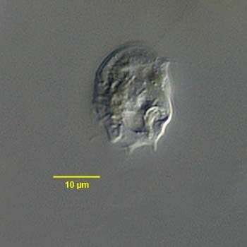
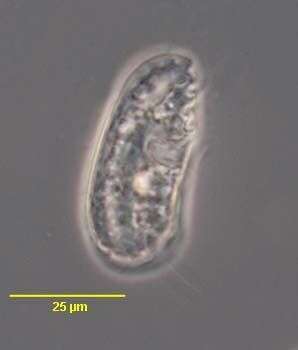
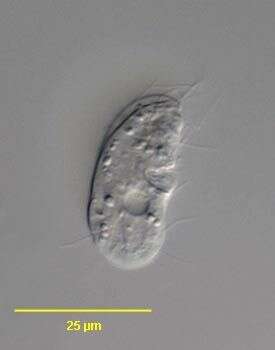
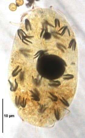
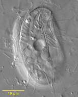
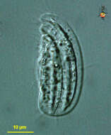
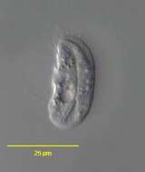
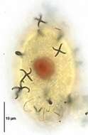
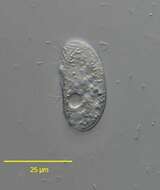
Trochiliopsis opaca (Pennard 1922), a colorless microthoracid ciliate synonymous with Trichopelma opaca (Kahl 1931). This genus contains one other species, T. australis. The rigid body of T. opaca is ovoid and strongly laterally compressed, the anterior curving ventrally into a small beak (seen in accompanying images). The right and left surfaces are ribbed longitudinally. This image is of the sparsely ciliated left surface (sometimes designated as dorsal). Two posterior cilia are seen. There is a kinety curving toward the ventral surface and a cluster of cilia near the oral aperture. Two of the large obliquely oriented extrusomes are seen in the central anterior half of the cell. T. opaca is bactivorous. From putrefying freshwater specimen containing decomposing filamentous algae collected near Boise, Idaho. Differential interference contrast.
-

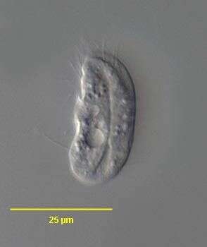
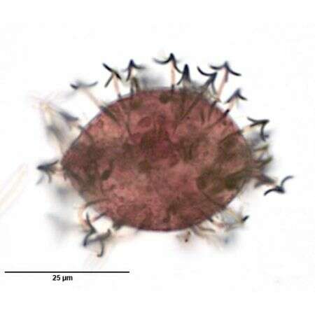
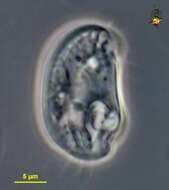
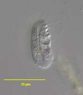
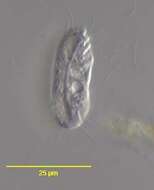
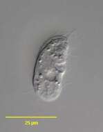
Trochiliopsis opaca (Pennard 1922), a colorless microthoracid ciliate synonymous with Trichopelma opaca (Kahl 1931). This genus contains one other species, T. australis. The rigid body of T. opaca is ovoid and strongly laterally compressed, the anterior curving ventrally into a small beak (seen in here). The right and left surfaces are ribbed longitudinally. This image is of the right surface (sometimes designated as ventral) which bears most of the somatic and oral ciliature. The left anterior oral aperture is seen here. There is a cyrtos which is visible only with silver impregnation techniques. The cyrtos of T. australis is visible in vivo. The central contractile vacuole has a prominent duct connecting it with a pellicular pore through which it empties at the posterior end of the paraoral kinety (the duct is seen in this image extending from the left lower quadrant of the contractile vacuole). The round macronucleus is central and very inconspicuous. T. Opaca is bactivorous. From putrefying freshwater specimen containing decomposing filamentous algae collected near Boise, Idaho. Differential interference contrast.
-

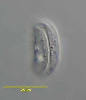
Trochiliopsis opaca (Pennard 1922), a colorless microthoracid ciliate synonymous with Trichopelma opaca (Kahl 1931). This genus contains one other species, T. australis. The rigid body of T. opaca is ovoid and strongly laterally compressed, the anterior curving ventrally into a small beak (seen in here). The right and left surfaces are ribbed longitudinally. This image is of the right surface (sometimes designated as ventral) which bears most of the somatic and oral ciliature (seen in this image). The left anterior oral aperture is seen here. Characteristic extrusomes are seen along the anterior curved cell margin. There is a cyrtos which is visible only with silver impregnation techniques. The cyrtos of T. australis is visible in vivo. The contractile vacuole is central. The round macronucleus is central and very inconspicuous. T. Opaca is bactivorous. From putrefying freshwater specimen containing decomposing filamentous algae collected near Boise, Idaho. Differential interference contrast.
-
Microthorax (mike-row-though-racks), small ciliate, stiff body, sparsely ciliates, with cytostome (posterior right) located in rear part of the cell Phase contrast.
-
Microthorax - a ciliate. With a mouth structure that is supported internally by stiff rods, consumes detritus and bacteria which are manipulated into the mouth by the rods From Lake Donghu, China. Differential interference contrast micrograph.
-
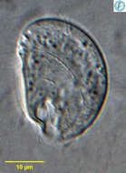
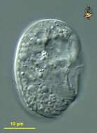
Portrait of Microthorax tridentatus (Penard,1922), a small nassophorean ciliate. The body is strongly dorsoventrally flattened. The right side is gently rounded and the left is straight. The body has delicately ridged armor, which bears three short posterior spines in this species. The cytostome is in a notch in the left posterior body (seen here). The spheroid macronucleus is central. One or two contractile vacuoles are usually adjacent to the oral aperture. Collected from a slow moving organically enriched freshwater stream near Boise, Idaho. DIC optics.
-
Portrait of Microthorax tridentatus (Penard,1922), a small nassophorean ciliate. The body is strongly dorsoventrally flattened. The right side is gently rounded and the left is straight. The body has delicately ridged armor, which bears three short posterior spines in this species. The cytostome is in a notch in the left posterior body. The spheroid macronucleus is central. One or two contractile vacuoles are usually adjacent to the oral aperture. Collected from a slow moving organically enriched freshwater stream near Boise, Idaho. DIC optics.
-
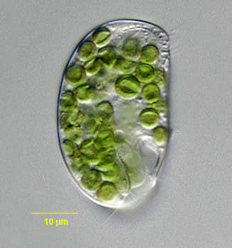
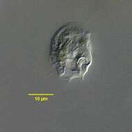
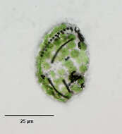
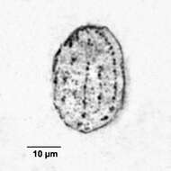
Portrait of Microthorax viridis (Penard,1922), a small nassophorean ciliate. The body is strongly dorsoventrally flattened. The right side is gently rounded and the left is straight. This genus has delicately ridged armor. The cytostome is in a notch in the left posterior body (seen here). The spheroid macronucleus is central. One or two contractile vacuoles are usually adjacent to the oral aperture. M. viridis has endosymbiotic algae (probably Chlorella). Collected from a eutrophic freshwater dredge pit near Idaho City, Idaho September 2003. DIC optics.
-
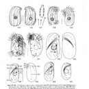
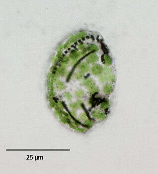
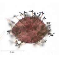
Infraciliature (right side) of Microthorax viridis (Penard,1922), a small nassophorean ciliate. The body is strongly dorsoventrally flattened. The right side is gently rounded and the left is straight. Somatic ciliature is reduced to three interrupted kineties on the right side. There is a short undulating membrane on the right margin of the cytostome and a small adoral membranelle in the depths of the oral depression. There are a few small clusters of preoral cilia on the left side anteriorly. This genus has delicately ridged armor. The cytostome is in a notch in the left posterior body (seen here). The spheroid macronucleus is central. One or two contractile vacuoles are usually adjacent to the oral aperture. M. viridis has endosymbiotic algae (probably Chlorella). Stained by the dry silver nitrate technic (see Foissner, W.Europ. J. Protistol.27,313-330;1991). Collected from a eutrophic freshwater dredge pit near Idaho City, Idaho September 2003.Brightfield.
-
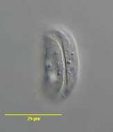
Drepanomonas (dree-pan-owe-moan-ass) is an odontostome ciliate. This is a small group of small flattened and sparsely ciliated ciliates which are most usually found in anoxic habitats. Kineties are associated with the surface ridges, contractile vacuole near the middle of the cell. Differential interference contrast with closed condenser iris.
-
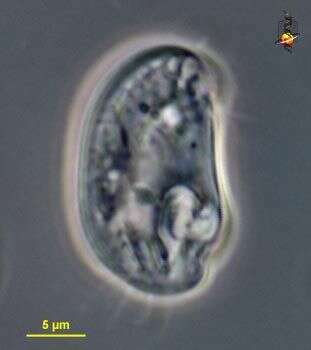
Drepanomonas revoluta PENARD, 1922. The deep furrow of the left side is visible throught the transparent pellicle in this optical section. DIC.
-
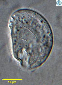
Drepanomonas revoluta PENARD,1922.DIC
-
Drepanomonas revoluta PENARD,1922. Phase contrast.
-
Drepanomonas revoluta PENARD,1922.DIC.
-
Drepanomonas revoluta PENARD,1922.DIC.
-
Drepanomonas revoluta PENARD,1922.DIC.
-
Drepanomonas revoluta PENARD, 1922. Partially exploded extrusomes.Stained by the silver carbonate technique (see Foissner, W. Europ. J. Protistol., 27:313-330;1991).Brightfield.
-
Drepanomonas revoluta PENARD, 1922. Completely exploded extrusomes.Stained by the silver carbonate technique (see Foissner, W. Europ. J. Protistol., 27:313-330;1991).Brightfield.
-
Drepanomonas revoluta PENARD, 1922. Completely exploded extrusomes.Stained by the silver carbonate technique (see Foissner, W. Europ. J. Protistol., 27:313-330;1991).Brightfield.
-
Upside down cell (anterior to bottom) - identified by Bill Bourland. Differential interference contrast. Material from Nymph Creek and Nymph Lake, thermal sites within Yellowstone National Park, photograph by Kathy Sheehan and David Patterson.
-
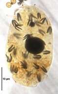
Silver nitrate impregnated (left side) microthoracid ciliate, Leptopharynx costatus (Mermod, 1914). Cell shape is lenticular in outline and strongly laterally flattened. The rigid pellicle is colorless with shallow curved concentric ridges on the right and left sides. There are four short oblique grooves on the ventral surface anteriorly. The somatic ciliature is reduced to 9 kineties. The four curved right-sided kineties have more densely packed kinetids (seen here) than the four left sided kineties. The 9th kinety is on the posterior half of the ventral surface (seen here). There is a small adoral zone of membranelles just to the left of the cytostome (not seen here). The cytostome is in the anterior 1/3 of the body. There are four short oblique preoral kineties. The transversely oriented cytopharynx is supported by fine trichites (not seen in this image). The round macronucleus and adjacent micronucleus are central (not seen here). The single contractile vacuole is ventral, posterior to the cytostome. The cytopyge is on the right surface posterior to the cytostome (not seen here). Extrusomes are spindleform (not seen in this image). Feeds on bacteria and small algae. Collected from freshwater in roadside ditch near Boise, Idaho June 2004. Brightfield optics.
-

Portrait (left side) of microthoracid ciliate, Leptopharynx costatus (Mermod, 1914). Cell shape is lenticular in outline and strongly laterally flattened. The rigid pellicle is colorless with shallow curved concentric ridges on the right and left sides. There are four short oblique grooves on the ventral surface anteriorly. The somatic ciliature is reduced to 9 kineties. The four curved right-sided kineties have more densely packed kinetids then the four left sided kineties. The 9th kinety is on the posterior half of the ventral surface. There is a small adoral zone of membranelles just to the left of the cytostome. The cytostome is in the anterior 1/3 of the body (not seen here). There are four short oblique preoral kineties. The transversely oriented cytopharynx is supported by fine trichites (visible in this image). The round macronucleus and adjacent micronucleus are central (not seen here). The single contractile vacuole is ventral, posterior to the cytostome. The cytopyge is on the right surface posterior to the cytostome. Extrusomes are spindleform. Feeds on bacteria and small algae. Collected from freshwater in roadside ditch near Boise, Idaho June 2004. DIC optics.