-
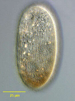
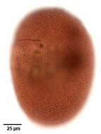
Nassula microstoma.
-

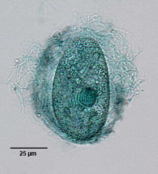
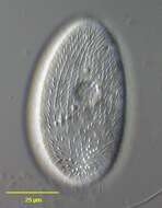
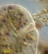
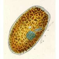
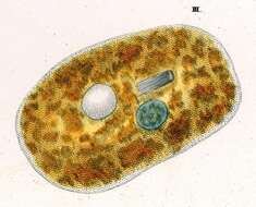
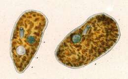
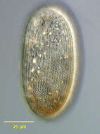
Ventral view of the nassulid ciliate, Furgasonia trichocystis (Stokes, 1894) Jankowski, 1964. Synonym: Cyclogramma. The cell shape is a slightly dorsoventrally flattened ellipsoid. The left side is flattened and the right side slightly convex. The cytostome is in the anterior 1/5 of the cell in a shallow depression. It is supported by a prominent basket of obliquely oriented cytopharyngeal trichites. The somatic ciliature consists of about 32 to 26 longitudinal kineties. On the ventral surface the right kineties arch to the left anterior to the cytostome to terminate on a short but wide preoral suture. The straight left kineties terminate on this suture to the left of the cytostome. There is a short curved right paraoral membrane. There are three approximately rectangular paroral polykineties. The most anterior (M1) is also least conspicuous. It is obliquely oriented in the preoral suture. The middle membrane (M2) is to the left of the cytostome and almost perpendicular to the long axis of the cell. The most posterior membranelle (M3) is posterior to the cytostome (often obscured by the trichites in silver carbonate preparations) almost parallel to the long axis of the cell. These three distinctive small polykineties distinguish Furgasonia from other nassulid genera. The spherical macronucleus and adjacent micronucleus are slightly posterior to the equator. The single contractile vacuole (visible here posterior to the cytopharyngeal basket) is located in the cell center with an excretory pore on its ventral aspect. There is a prominent layer of fusiform subpellicular extrusomes (mucocysts). The cytoplasm is colorless in these bactivorous individuals. It is unclear whether this species is synonymous with F. rubens which is orange to blue colored due to ingested cyanobacteria. Morphologically the two species are quite similar aside from this coloration (see Faur�-Fremiet, E. Le Genre Cyclogramma, Perty, 1852. J. Protozool. 14: 456-464, 1967.) Collected from a temporary rainwater pool with abundant decaying grass near Boise, Idaho. March, 2005. DIC.
-
Dorsal view of the nassulid ciliate, Furgasonia trichocystis (Stokes, 1894) Jankowski, 1964. Synonym: Cyclogramma. The cell shape is a slightly dorsoventrally flattened ellipsoid. The left side is flattened and the right side slightly convex. There is a prominent layer of fusiform subpellicular extrusomes (mucocysts). The cytoplasm is colorless in these bactivorous individuals. It is unclear whether this species is synonymous with F. rubens which is orange to blue colored due to ingested cyanobacteria. Morphologically the two species are quite similar aside from this coloration (see Faur�-Fremiet, E. Le Genre Cyclogramma, Perty, 1852. J. Protozool. 14: 456-464, 1967.) Collected from a temporary rainwater pool with abundant decaying grass near Boise, Idaho. March, 2005. DIC.
-
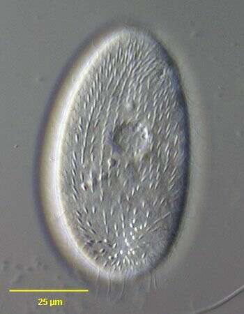
Ventral anterior view of the nassulid ciliate, Furgasonia trichocystis (Stokes, 1894) Jankowski, 1964. Synonym: Cyclogramma. The cell shape is a slightly dorsoventrally flattened ellipsoid. The left side is flattened and the right side slightly convex. The cytostome is in the anterior 1/5 of the cell in a shallow depression. It is supported by a prominent basket of obliquely oriented cytopharyngeal trichites. The somatic ciliature consists of about 32 to 26 longitudinal kineties. On the ventral surface the right kineties arch to the left anterior to the cytostome to terminate on a short but wide preoral suture. The straight left kineties terminate on this suture to the left of the cytostome. There is a short curved right paraoral membrane. There are three approximately rectangular paroral polykineties. The most anterior (M1) is obliquely oriented in the preoral suture. The middle membrane (M2) is to the left of the cytostome and almost perpendicular to the long axis of the cell. The most posterior membranelle (M3) is posterior to the cytostome (often obscured by the trichites in silver carbonate preparations) almost parallel to the long axis of the cell. These three distinctive small polykineties distinguish Furgasonia from other nassulid genera. The spherical macronucleus and adjacent micronucleus are slightly posterior to the equator. The single contractile vacuole (visible here posterior to the cytopharyngeal basket) is located in the cell center with an excretory pore on its ventral aspect. There is a prominent layer of fusiform subpellicular extrusomes (mucocysts). The cytoplasm is colorless in these bactivorous individuals. It is unclear whether this species is synonymous with F. rubens which is orange to blue colored due to ingested cyanobacteria. Morphologically the two species are quite similar aside from this coloration (see Faurè-Fremiet, E. Le Genre Cyclogramma, Perty, 1852. J. Protozool. 14: 456-464, 1967). Collected from a temporary rainwater pool with abundant decaying grass near Boise, Idaho. March, 2005. Silver carbonate stain (see Foissner, W. Europ. J. Protistol., 27:313-330;1991). Brightfield.
-
Ventral infraciliature of the nassulid ciliate, Furgasonia trichocystis (Stokes, 1894) Jankowski, 1964. Synonym: Cyclogramma. The cell shape is a slightly dorsoventrally flattened ellipsoid. The left side is flattened and the right side slightly convex. The cytostome is in the anterior 1/5 of the cell in a shallow depression. It is supported by a prominent basket of obliquely oriented cytopharyngeal trichites. The somatic ciliature consists of about 32 to 26 longitudinal kineties. On the ventral surface the right kineties arch to the left anterior to the cytostome to terminate on a short but wide preoral suture. The straight left kineties terminate on this suture to the left of the cytostome. There is a short curved right paraoral membrane. There are three approximately rectangular paroral polykineties. The most anterior (M1) is obliquely oriented in the preoral suture. The middle membrane (M2) is to the left of the cytostome and almost perpendicular to the long axis of the cell. The most posterior membranelle (M3) is posterior to the cytostome (often obscured by the trichites in silver carbonate preparations) almost parallel to the long axis of the cell. These three distinctive small polykineties distinguish Furgasonia from other nassulid genera. The spherical macronucleus and adjacent micronucleus are slightly posterior to the equator. The single contractile vacuole (visible here posterior to the cytopharyngeal basket) is located in the cell center with an excretory pore on its ventral aspect. There is a prominent layer of fusiform subpellicular extrusomes (mucocysts). The cytoplasm is colorless in these bactivorous individuals. It is unclear whether this species is synonymous with F. rubens which is orange to blue colored due to ingested cyanobacteria. Morphologically the two species are quite similar aside from this coloration (see Faurè-Fremiet, E. Le Genre Cyclogramma, Perty, 1852. J. Protozool. 14: 456-464, 1967). Collected from a temporary rainwater pool with abundant decaying grass near Boise, Idaho. March, 2005. Silver carbonate stain (see Foissner, W. Europ. J. Protistol., 27:313-330;1991). Brightfield.
-
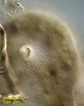
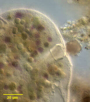
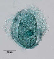
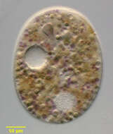
Discharged extrusomes (mucocysts) of the nassulid ciliate, Furgasonia trichocystis (Stokes, 1894) Jankowski, 1964. Synonym: Cyclogramma. The spherical macronucleus (stained dark green here)and adjacent micronucleus are slightly posterior to the equator. There is a prominent layer of fusiform subpelicular extrusomes (mucocysts). This preparation demonstartes the discharged extrusomes and mucus surrounding the cell. Collected from a temporary rainwater pool with abundant decaying grass near Boise, Idaho. March, 2005. Methyl green Pyronin-Y stain (see Foissner, W. Europ. J. Protistol., 27:313-330;1991). Brightfield.
-
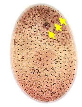
Ventral infraciliature of the nassulid ciliate, Furgasonia trichocystis (Stokes, 1894) Jankowski, 1964. Synonym: Cyclogramma. The cell shape is a slightly dorsoventrally flattened ellipsoid. The left side is flattened and the right side slightly convex. The cytostome is in the anterior 1/5 of the cell in a shallow depression. It is supported by a prominent basket of obliquely oriented cytopharyngeal trichites. The somatic ciliature consists of about 32 to 26 longitudinal kineties. On the ventral surface the right kineties arch to the left anterior to the cytostome to terminate on a short but wide preoral suture. The straight left kineties terminate on this suture to the left of the cytostome. There is a short curved right paraoral membrane. There are three approximately rectangular paroral polykineties (nassulid organelles). The most anterior (M1,long arrow) is obliquely oriented in the preoral suture. The middle membrane (M2, medium arrow) is to the left of the cytostome and almost perpendicular to the long axis of the cell. The most posterior membranelle (M3, short arrow) is posterior to the cytostome, almost parallel to the long axis of the cell. These three distinctive small polykineties distinguish Furgasonia from other nassulid genera. The spherical macronucleus and adjacent micronucleus are slightly posterior to the equator. The single contractile vacuole (visible here posterior to the cytopharyngeal basket) is located in the cell center with an excretory pore on its ventral aspect. There is a prominent layer of fusiform subpellicular extrusomes (mucocysts). The cytoplasm is colorless in these bactivorous individuals. It is unclear whether this species is synonymous with F. rubens which is orange to blue colored due to ingested cyanobacteria. Morphologically the two species are quite similar aside from this coloration (see Fauré-Fremiet, E. Le Genre Cyclogramma, Perty, 1852. J. Protozool. 14: 456-464, 1967). Collected from a temporary rainwater pool with abundant decaying grass near Boise, Idaho. March, 2005. Silver carbonate (see Foissner, W. Europ. J. Protistol., 27:313-330;1991). Brightfield. This image was taken by William Bourland. He now uses a Zeiss Axioskop 2 with a Flex camera (Diagnostic Instruments).
-
Dorsal view of the nassulid ciliate, Furgasonia trichocystis (Stokes, 1894) Jankowski, 1964. Synonym: Cyclogramma. The cell shape is a slightly dorsoventrally flattened ellipsoid. The left side is flattened and the right side slightly convex. There is a prominent layer of fusiform subpellicular extrusomes (mucocysts). The cytoplasm is colorless in these bactivorous individuals. It is unclear whether this species is synonymous with F. rubens which is orange to blue colored due to ingested cyanobacteria. Morphologically the two species are quite similar aside from this coloration (see Faur�-Fremiet, E. Le Genre Cyclogramma, Perty, 1852. J. Protozool. 14: 456-464, 1967.) Collected from a temporary rainwater pool with abundant decaying grass near Boise, Idaho. March, 2005. DIC.
-

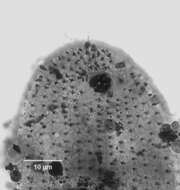
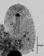
Right dorsolateral view of the infraciliature of the large nassulid ciliate Obertrumia aurea (Ehrenberg, 1833; Foissner 1987). Synonym of Nassula aurea. Obertrumia is distinguished by it's bipartite "hypostomial frange", linear arrays of ciliary tufts. A sigmoid ventrolateral frange runs anteriorly and to the left. The dorsal termination of this part is seen as a vertical arrangement of rectangular collections of kinetids on the viewer's left in this image. It terminates at the left end of the horizontal line of ciliary tufts on the dorsal surface.The uniform longitudinal somatic kineties are well seen here. In vivo the cell appears brightly colored (orange, green or violet) due to multiple food vacuoles containing ingested cyanobacteria. Numerous small mucocysts give the cortex a roughly granular appearance. O. aurea feeds mainly on cyanobacteria. Silver carbonate stain (see Foissner, W.Europ. J. Protistol.27,313-330;1991). Collected from a freshwater aquaculture pond near Boise,Idaho November 2004. Brightfield.
-
Ventral infraciliature of the large nassulid ciliate Obertrumia aurea (Ehrenberg, 1833; Foissner 1987). Synonym of Nassula aurea. Obertrumia is distinguished by it's bipartite "hypostomial frange",linear arrays of ciliary tufts. A sigmoid ventrolateral frange begins posterior to the cytostome running anteriorly and to the left (viewer's right) around to the dorsal surface. The dorsal termination is not seen in this image. The ventrolateral part of the frange terminates at the left end of the horizontal line of ciliary tufts on the dorsal surface. The uniform longitudinal somatic kineties, interrupted by the hypostomial frange, are well seen here. The multiple darkly stained micronuclei are seen overlying the spherical macronucleus in this image.The small circular structure to the viewer's left just posterior to the macronucleus is the excretory pore of the contractile vacuole. The thin dark longitudinal line posterior to this is the cytopyge (cell anus).In vivo the cell appears brightly colored (orange, green or violet) due to multiple food vacuoles containing ingested cyanobacteria. Numerous small mucocysts give the cortex a roughly granular appearance. O. aurea feeds mainly on cyanobacteria. Silver carbonate stain (see Foissner, W.Europ. J. Protistol.27,313-330;1991). Collected from a freshwater aquaculture pond near Boise, Idaho,Idaho November 2004. Brightfield optics.
-
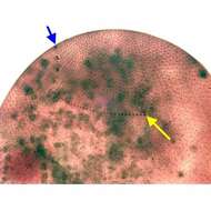
Dorsal infraciliature of the large nassulid ciliate Obertrumia aurea (Ehrenberg, 1833; Foissner 1987). Synonym of Nassula aurea. Obertrumia is distinguished by it's bipartite "hypostomial frange", linear arrays of ciliary tufts. A sigmoid ventrolateral frange runs anteriorly and to the left. The dorsal termination of this part is seen as a vertical arrangement of rectangular collections of kinetids (blue arrow). It terminates at the left end of the horizontal line of ciliary tufts on the dorsal surface (yellow arrow). The uniform longitudinal somatic kineties are well seen here. In vivo the cell appears brightly colored (orange, green or violet) due to multiple food vacuoles containing ingested cyanobacteria. Numerous small mucocysts give the cortex a roughly granular appearance. O. aurea feeds mainly on cyanobacteria. Silver carbonate stain (see Foissner, W.Europ. J. Protistol.27,313-330;1991). Collected from a freshwater pond near Idaho City, Idaho Septmeber 2004. Brightfield optics.
-
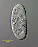
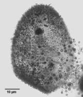
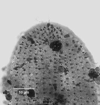
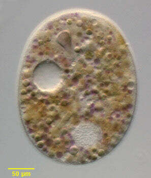
Portrait of Obertrumia aurea(Ehrenberg, 1833; Foissner 1987). Widely distributed nassulid ciliate with well developed cytopharyngeal basket or cyrtos (viewer's upper left). Fed organisms may contain brightly colored orange, blue and purple food vacuoles. O. aurea has a bipartite hypostomial frange (not seen here). From a freshwater pond near Boise, Idaho. Oblique illumination This image was taken by William Bourland. He now uses a Zeiss Axioskop 2 with a Spot Insight CCD camera (Diagnostic Instruments).
-
Detail of the large Nnassulid ciliate Obertrumia aurea (Ehrenberg, 1833; Foissner 1987). Synonym of Nassula aurea. Obertrumia is distinguished by it's bipartite "hypostomial frange", linear arrays of ciliary tufts. A sigmoid ventrolateral frange (seen most clearly posterior to the cytostome in this image) runs anteriorly and to the left and is separated from a horizontal line of ciliary tufts on the dorsal surface. The cell appears brightly colored (orange, green or violet) due to multiple food vacuoles containing ingested cyanobacteria. Numerous small mucocysts give the cortex a roughly granular appearance. O. aurea feeds mainly on cyanobacteria. Collected from a freshwater pond near Boise, Idaho November 2003. DIC optics.
-
Detail of the large nassulid ciliate Obertrumia aurea (Ehrenberg, 1833; Foissner 1987). Synonym of Nassula aurea. Obertrumia is distinguished by it's bipartite "hypostomial frange", linear arrays of ciliary tufts. A sigmoid ventrolateral frange runs anteriorly and to the left (not seen in this image) and is separated from a horizontal line of ciliary tufts on the dorsal surface (seen here). The cell appears brightly colored (orange, green or violet) due to multiple food vacuoles containing ingested cyanobacteria. Numerous small mucocysts give the cortex a roughly granular appearance. O. aurea feeds mainly on cyanobacteria. Collected from a freshwater pond near Boise, Idaho November 2003. DIC optics.
-
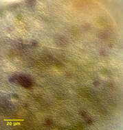
Detail of the large nassulid ciliate Obertrumia aurea (Ehrenberg, 1833; Foissner 1987). Synonym of Nassula aurea. Obertrumia is distinguished by it's bipartite "hypostomial frange", linear arrays of ciliary tufts. There is a prominent cytopharyngeal basket (nasse) composed of stout nematodesmata at the base of a shallow oral introitus in the anterior 1/3 of the cell. Focal swellings of the nematodesmata give the appearance of a ring (annulus) near the outer end of the nasse. Numerous small mucocysts give the cortex a roughly granular appearance. O. aurea feeds mainly on cyanobacteria. Collected from a freshwater pond near Boise, Idaho November 2003. DIC optics.
-
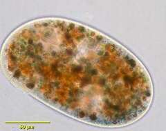
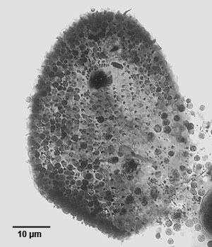
Portrait of the large nassulid ciliate Obertrumia aurea (Ehrenberg, 1833; Foissner 1987). Synonym of Nassula aurea. Obertrumia is distinguished by it's bipartite "hypostomial frange", linear arrays of ciliary tufts. A sigmoid ventrolateral frange runs anteriorly and to the left and is separated from a horizontal line of ciliary tufts that lies on the dorsal surface. This specimen is slightly compressed, the cell outline normally appearing as a more elongate ellipsoid. The cell appears brightly colored (orange, green or violet) due to multiple food vacuoles containing ingested cyanobacteria. The single spherical macronucleus is seen posteriorly. There is one large lateral contractile vacuole, which fills from smaller surrounding vacuoles rather than collecting canals (seen well here). There is a prominent cytopharyngeal basket (nasse) composed of stout nematodesmata at the base of a shallow oral introitus in the anterior 1/3 of the cell. Focal swellings of the nematodesmata give the appearance of a ring near the outer end of the nasse. Numerous small mucocysts give the cortex a roughly granular appearance. O. aurea feeds mainly on cyanobacteria. Collected from a freshwater pond near Boise, Idaho November 2003. DIC optics.
-

Right dorsolateral view of the infraciliature of the large nassulid ciliate Obertrumia aurea (Ehrenberg, 1833; Foissner 1987). Synonym of Nassula aurea. Obertrumia is distinguished by it's bipartite "hypostomial frange", linear arrays of ciliary tufts. A sigmoid ventrolateral frange runs anteriorly and to the left. The dorsal termination of this part is seen as a vertical arrangement of rectangular collections of kinetids on the viewer's left in this image. It terminates at the left end of the horizontal line of ciliary tufts on the dorsal surface.The uniform longitudinal somatic kineties are well seen here. In vivo the cell appears brightly colored (orange, green or violet) due to multiple food vacuoles containing ingested cyanobacteria. Numerous small mucocysts give the cortex a roughly granular appearance. O. aurea feeds mainly on cyanobacteria. Silver carbonate stain (see Foissner, W.Europ. J. Protistol.27,313-330;1991). Collected from a freshwater aquaculture pond near Boise,Idaho February 2006. Brightfield.
-
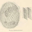
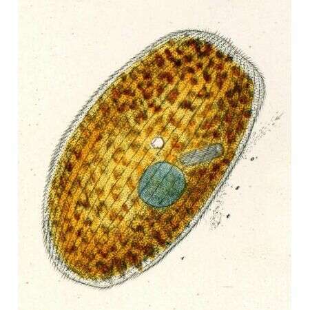
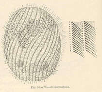
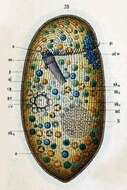
Originally described by Schewiakoff under the name Nassula aurea. Ventral view, slightly rotated. a--Anus al--Pellicular alveoli ad. w--Adoral ciliated zone (hypostomial frange) cp--Cortical plasma cv--Contractile vacuole g--Gelatinous layer h--Ectoplasm of a homogenous appearance oe--Throat N--Macronucleus ncl -- Micronucleus nk1-nk4 -- Food particles in various stages of digestion o -- Mouth oe -- Throat pe -- Excretory pore pi -- Pigmented spot
-
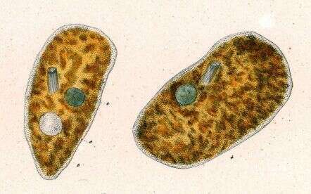
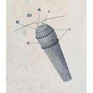
Originally described by Schewiakoff under the name Nassula aurea. Detail of cytopharyngeal ""eel-basket."" Key to abbreviations: kr--Plasmatic collar of the rod system o--Mouth r--Plasmatic ring encircling the cyrtos (basket) st--Cytopharyngeal basket vh--Cavity before mouth opening
-
Originally described by Ehrenberg under the name Nassula aurea.
-
Originally described by Ehrenberg under the name Nassula elegans.
-
Originally described by Ehrenberg under the name Nassula elegans.
-
Portrait of the marine nassulid ciliate, Paranassula brunnea (Fabre-Domerge,1885) Fauré-Fremiet,1963. The cell body is an elongate cylinder rounded anteriorly and posteriorly. Size varies from medium to large (100-300 µm). The genus is distinguished by the unique oral apparatus, which lies in the anterior 1/4 of the body. There is an atrial depression within which two short polykinetids are located. This atrium leads to a stout cytopharyngeal basket. The somatic ciliature is composed of closely spaced uniform longitudinal kineties. There is a short straight preoral suture. There is a long centrally located vermiform macronucleus. A single contractile vacuole is located in the mid-body. Collected from a commercial saltwater aquarium in Boise, Idaho. DIC.