-
Centers for Disease Control/Division of Parasitic Diseases and Malaria
EOL staff
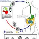
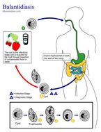
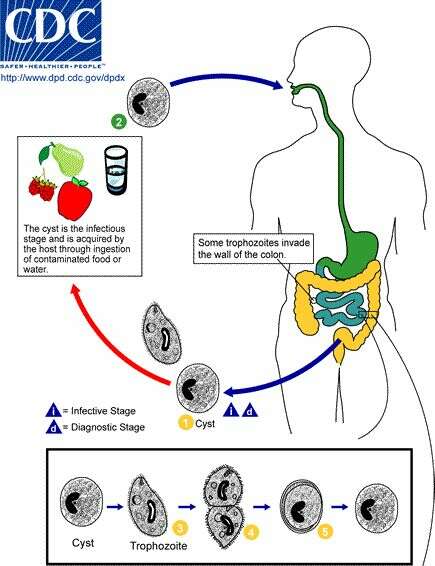
Life cycle of Balantidium coli, the cause of human balantidiasis The cyst stage (1) of the B. coli life cycle is responsible for transmission.The host most often acquires the cyst through ingestion of contaminated food or water (2). Following ingestion, excystation occurs in the small intestine and the trophozoites colonize the large intestine (3). The trophozoites reside in the lumen of the large intestine of humans and other animals, where they reproduce by binary fission, during which conjugation may occur (4). Trophozoites undergo encystation to produce infective cysts (5). Some trophozoites invade the wall of the colon and multiply. Some return to the lumen and disintegrate. Mature cysts are passed with feces (1).From
Centers for Disease Control Parasites and Health website
-
This illustration depicts the life cycle of Balantidium coli, the causal agent of Balantidiasis.Created: 2002
-
Roland Yao Wa Kouassi, Scott William McGraw, Patrick Kouassi Yao, Ahmed Abou-Bacar, Julie Brunet, Bernard Pesson, Bassirou Bonfoh, Eliezer Kouakou N’goran and Ermanno Candolfi
Wikimedia Commons
Description: English:
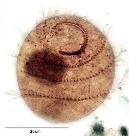
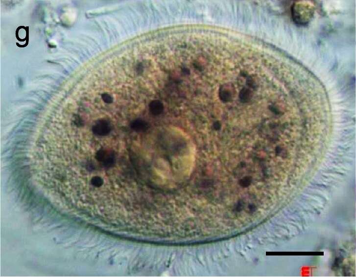
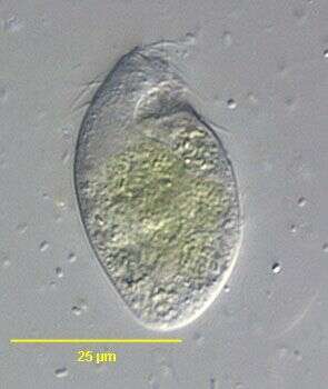
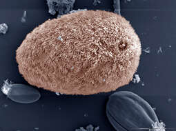
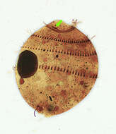
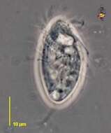
Balantidium coli, found in a primate of the Taï National Park. Scale bar: 5 μm. Date: 13 January 2020. Source:
File:Parasite140080-fig2 Gastrointestinal parasites in seven primates of the Taï National Park - Protozoa.png, Original: Fig. 2 at
doi:10.1051/parasite/2015001 Diversity and prevalence of gastrointestinal parasites in seven non-human primates of the Taï National Park, Côte d’Ivoire. Parasite, 2015, 22, 1. Cropped: Fig. 2g. Author: Roland Yao Wa Kouassi, Scott William McGraw, Patrick Kouassi Yao, Ahmed Abou-Bacar, Julie Brunet, Bernard Pesson, Bassirou Bonfoh, Eliezer Kouakou N’goran and Ermanno Candolfi. Permission(
Reusing this file): "This is an Open Access article distributed under the terms of the Creative Commons Attribution License (
http://creativecommons.org/licenses/by/4.0)".
-
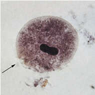
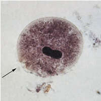
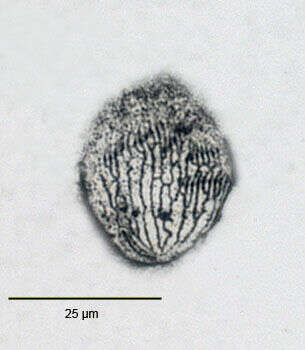
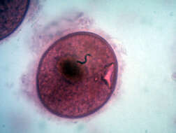
Description: English: Balantidium coli trophozoit العربية: أتروفة القربية القولونية. Date: 24 April 2012, 11:53:25. Source: Own work. Author:
Sara Nabih.
-
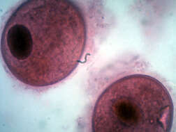
Description: English: Balantidium coli trophozoit العربية: أتروفة القربية القولونية. Date: 29 April 2012, 11:51:53. Source: Own work. Author:
Sara Nabih.
-
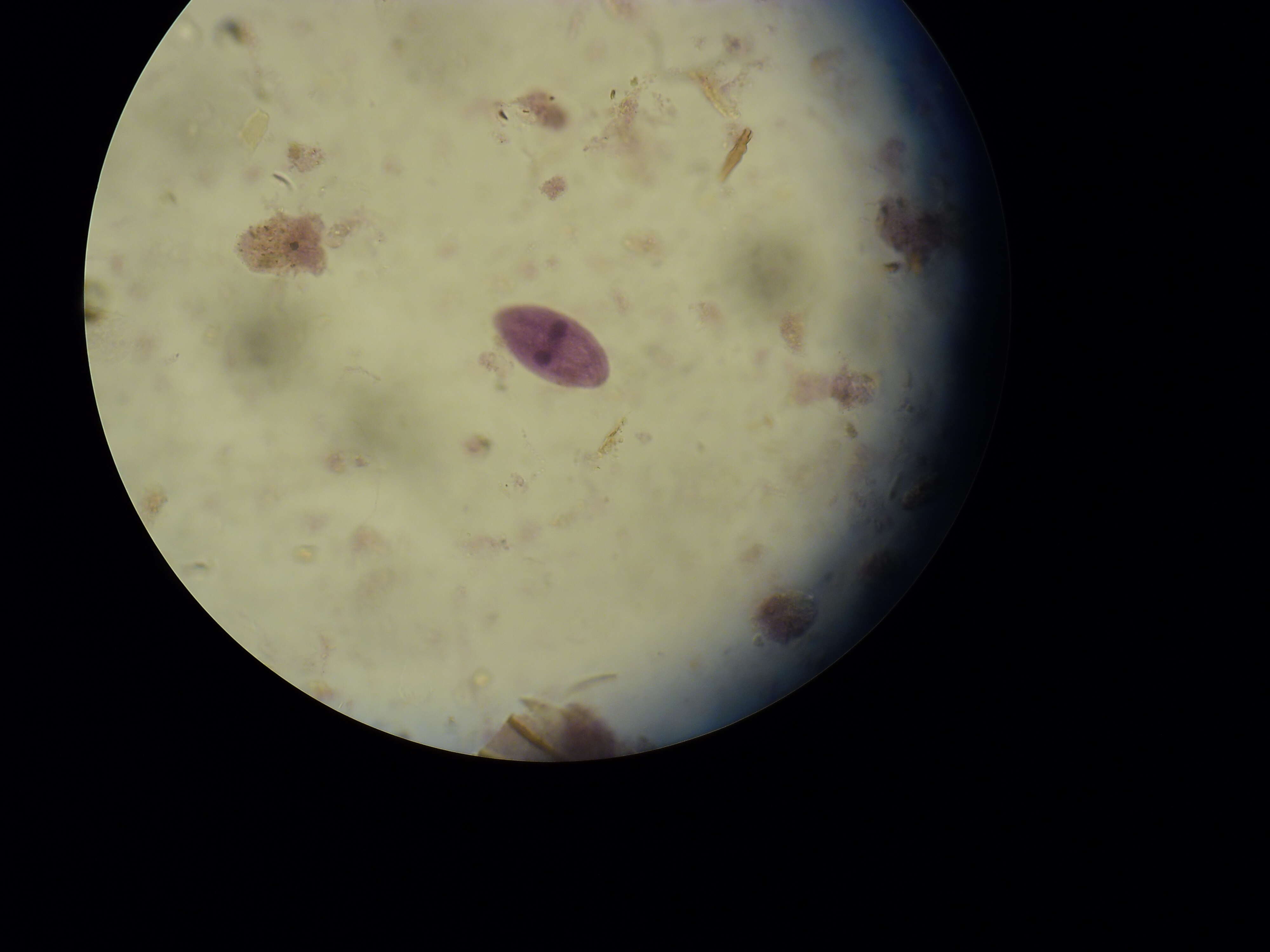
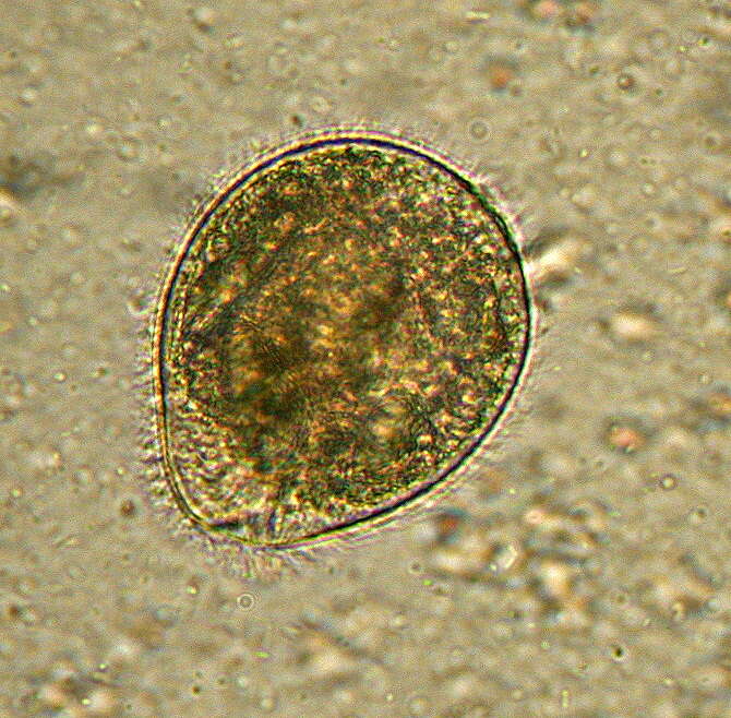
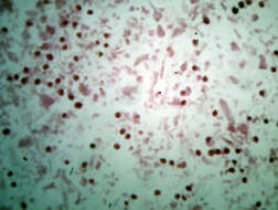
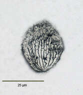
Description: English: Balantidium coli 1000x oil. from a fecal sample (prepared slide) (The worm-like line issue to the camera and does not represent an organelle.). Date: 7 September 2022, 05:35:33. Source: Own work. Author:
The Other 95%.
-
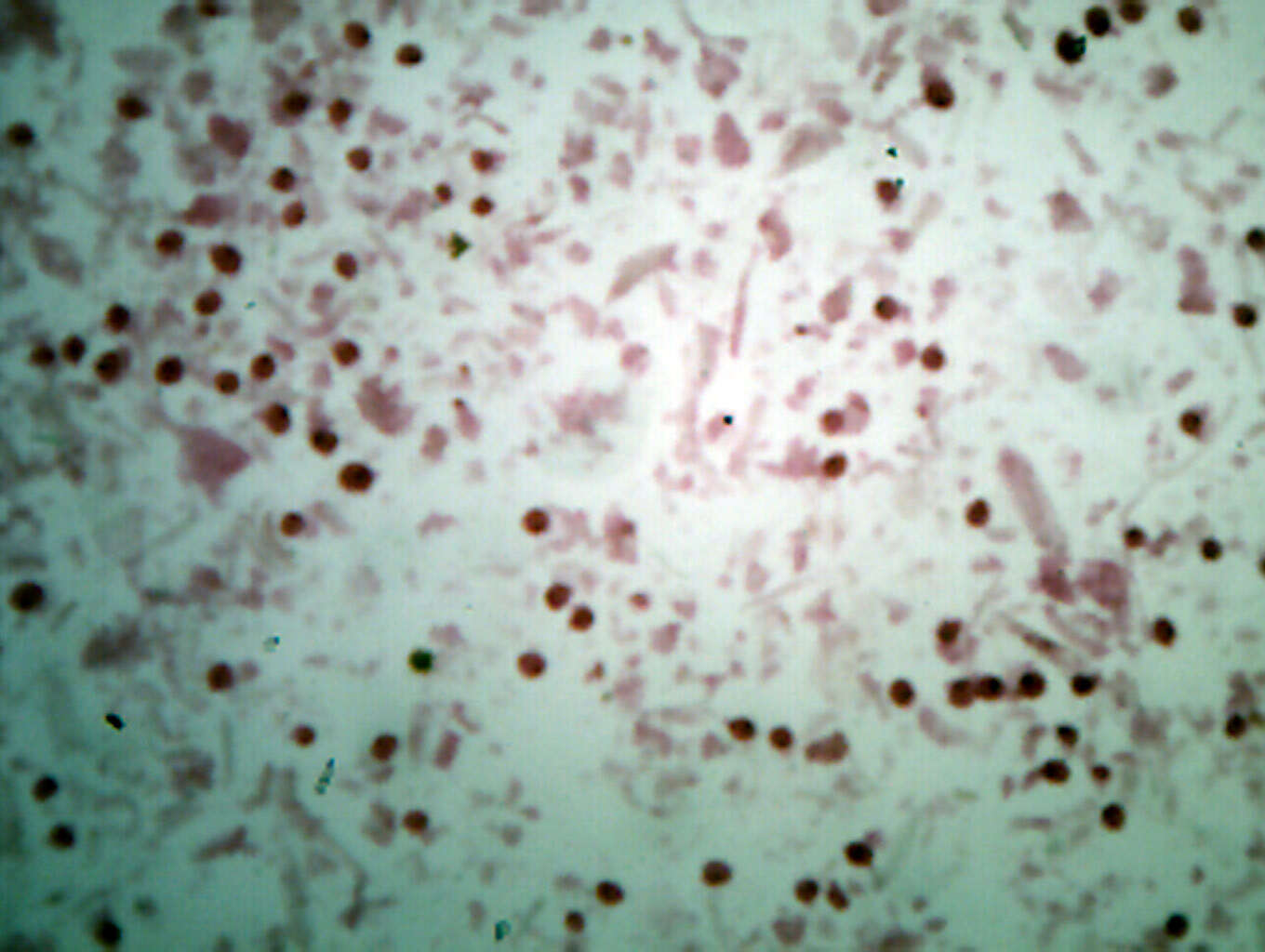
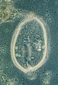
Description: English: Balantidium coli from a fecal sample (prepared slide) (The worm-like line issue to the camera and does not represent an organelle.). Date: 7 September 2022, 05:29:32. Source: Own work. Author:
The Other 95%.
-
-
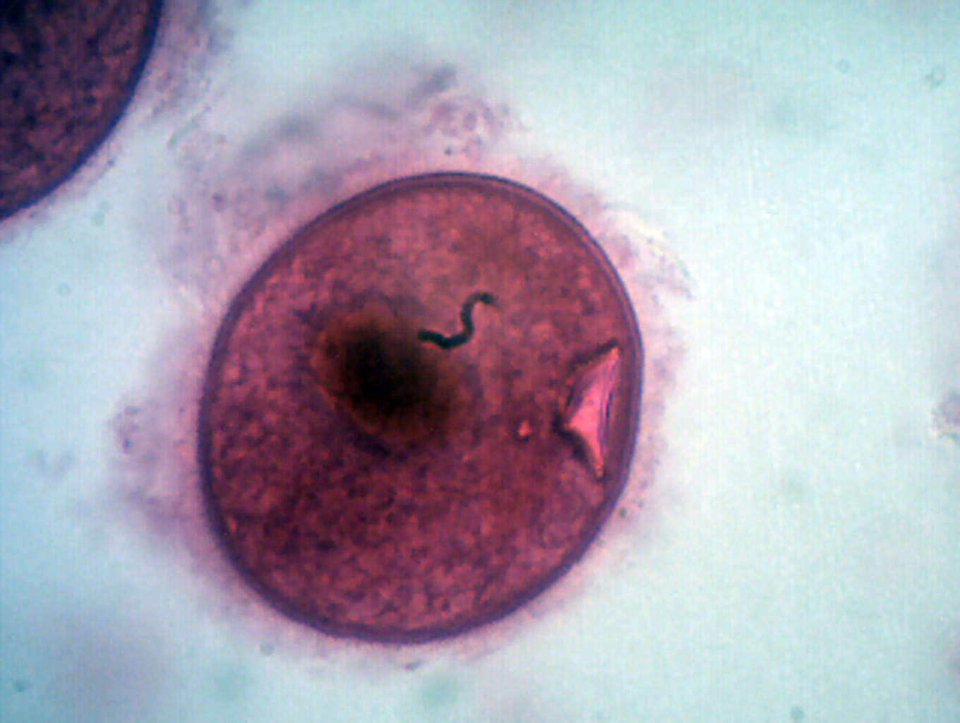
Description: English: Balantidium coli 1000x oil. from a fecal sample (prepared slide) (The worm-like line issue to the camera and does not represent an organelle.). Date: 7 September 2022, 05:36:20. Source: Own work. Author:
The Other 95%.
-
-
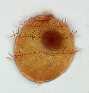
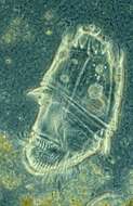
Description: English: Isotricha intestinalis, a protozoan distinguished by its mouth position. This protozoan can be up to 200 micrometers long, making it the largest in the rumen of sheep. Date: February 2006. Source:
Image Number D383-3. Author: Photo by Sharon Franklin. Colorization by Stephen Ausmus.
-
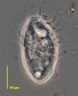
Right lateral view of the colorles ciliate Trimyema compressum (Lackey,1925).The cell is fusiform with bluntly rounded anterior and posterior ends. The funnel-shaped anterior oral aperture is subapical (seen here). 50-60 somatic kineties are reduced to three cilated basal bodies in each row.The arrangement of the somatic kineties gives the appearance of three discontinuous slightly spiral rows of cilia when viewed from ventral aspect. There is a single long caudal cilium (not seen here).Two long C- shaped kineties border the oral aperture. There is a short 3rd innermost kinety at the posterior end of the oral aperture. Near the anterior end of the oral kineties is a smal group of dikinetids representing the "adoral membranelles". There is a spherical central macronucleus.A single lateral contractile vacuole is located in the posterior 1/2 of the cell.From polysaprobic sediments of a freshwater rain barrel near Boise, Idaho.December 2005.DIC.
-
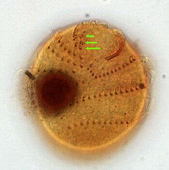
Right lateral view of the colorles ciliate Trimyema compressum (Lackey,1925).The cell is fusiform with bluntly rounded anterior and posterior ends. The funnel-shaped anterior oral aperture is subapical (seen here). 50-60 somatic kineties are reduced to three cilated basal bodies in each row.The arrangement of the somatic kineties gives the appearance of three discontinuous slightly spiral rows of cilia when viewed from ventral aspect. There is a single long caudal cilium (not seen here).Two long C- shaped kineties border the oral aperture. There is a short 3rd innermost kinety at the posterior end of the oral aperture. Near the anterior end of the oral kineties is a smal group of dikinetids representing the "adoral membranelles" (green arrows). There is a spherical central macronucleus.A single lateral contractile vacuole is located in the posterior 1/2 of the cell.From polysaprobic sediments of a freshwater rain barrel near Boise, Idaho.December 2005.Stained by the silver carbonate technique (see Foissner, W. Europ. J. Protistol., 27:313-330;1991).Brightfield.
-

Dorsal infraciliature of the colorles ciliate Trimyema compressum (Lackey,1925).The cell is fusiform with bluntly rounded anterior and posterior ends. The funnel-shaped anterior oral aperture is subapical. 50-60 somatic kineties are reduced to three cilated basal bodies in each row.The arrangement of the somatic kineties gives the appearance of three discontinuous slightly spiral rows of cilia when viewed from ventral aspect. There is a single long caudal cilium (not seen here).Two long C- shaped kineties border the oral aperture. There is a short 3rd innermost kinety at the posterior end of the oral aperture. Near the anterior end of the oral kineties is a smal group of dikinetids representing the "adoral membranelles". There is a spherical central macronucleus.A single lateral contractile vacuole is located in the posterior 1/2 of the cell.From polysaprobic sediments of a freshwater rain barrel near Boise, Idaho.December 2005.Stained by the silver carbonate technique (see Foissner, W. Europ. J. Protistol., 27:313-330;1991).Brightfield.
-
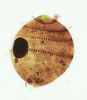
Left lateral view of infraciliature of the colorles ciliate Trimyema compressum (Lackey,1925).The cell is fusiform with bluntly rounded anterior and posterior ends. The funnel-shaped anterior oral aperture is subapical. 50-60 somatic kineties are reduced to three cilated basal bodies in each row.The arrangement of the somatic kineties gives the appearance of three discontinuous slightly spiral rows of cilia when viewed from ventral aspect. There is a single long caudal cilium (only a short remnant of it is seen here).Two long C- shaped kineties border the oral aperture. There is a short 3rd innermost kinety at the posterior end of the oral aperture (green arrow). Near the anterior end of the oral kineties is a smal group of dikinetids representing the "adoral membranelles". There is a spherical central macronucleus.A single lateral contractile vacuole is located in the posterior 1/2 of the cell.From polysaprobic sediments of a freshwater rain barrel near Boise, Idaho.December 2005.Stained by the silver carbonate technique (see Foissner, W. Europ. J. Protistol., 27:313-330;1991).Brightfield.
-
Ventral view of the silverline system of Trimyema compressum (Lackey,1925).Stained by the dry silver nitrate technique (see Foissner, W. Europ. J. Protistol., 27:313-330;1991).Brightfield.
-
Right lateral view of the colorles ciliate Trimyema compressum (LACKEY,1925).The cell is fusiform with bluntly rounded anterior and posterior ends. The funnel-shaped anterior oral aperture is subapical. 50-60 somatic kineties are reduced to three cilated basal bodies in each row.The arrangement of the somatic kineties gives the appearance of three discontinuous slightly spiral rows of cilia when viewed from ventral aspect. There is a single long caudal cilium (not seen here).Two long C- shaped kineties border the oral aperture. There is a short 3rd innermost kinety at the posterior end of the oral aperture. Near the anterior end of the oral kineties is a smal group of dikinetids representing the "adoral membranelles". There is a spherical central macronucleus.A single lateral contractile vacuole is located in the posterior 1/2 of the cell.From polysaprobic sediments of a freshwater rain barrel near Boise, Idaho.December 2005.Stained by the silver carbonate technique (see Foissner, W. Europ. J. Protistol., 27:313-330;1991).Brightfield.
-
Isotricha, from the rumen of domestic cattle. Phase contrast micrograph.
-
Entodiniomorph ciliate from the rumen of domestic cattle. Species such as this are predators, and they reveal the emergence of a multilayered microbial ecosystem within this habitat that - in evolutionary terms - only became available relatively recently. Phase contrast micrograph.
-
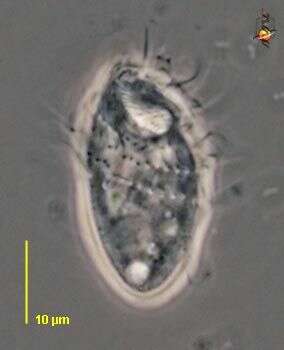
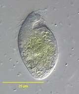
Trimyema (try-my-ee-ma) is a scuticociliate, with an anterior mouth region, and cilia arranged in sparse spiral kineties. With caudal cilium. Phase contrast.
-
Trimyema (try-my-ee-ma) is a scuticociliate, with an anterior mouth region, and cilia arranged in sparse spiral kineties. With caudal cilium. Phase contrast.


