-
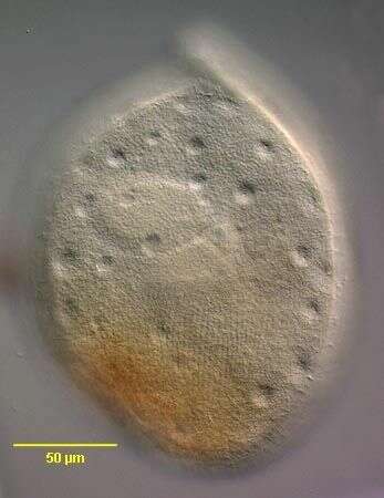
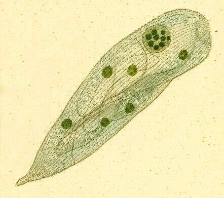
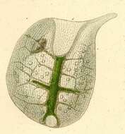
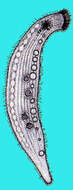
Portrait of Trachelius ovum, (EHRENBERG, 1831) EHRENBERG,1838 a haptorid ciliate with rounded body and short curved proboscis. Cytostome is located at the convexity at the base of the proboscis. Cytoplasm is extensively vacuolated. Coarsely granular bipartite macronucleus. From freshwater pond near Boise, Idaho.Oblique illumination.
-
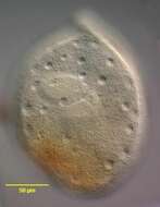
Portrait of Trachelius ovum (EHRENBERG,1831) EHRENBERG,1838 , a haptorid ciliate with rounded body and short curved proboscis. Cytostome is located at the convexity at the base of the proboscis. Cytoplasm is extensively vacuolated. Coarsely granular bipartite macronucleus. From freshwater pond near Boise, Idaho. Phase contrast.
-
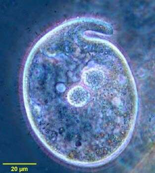
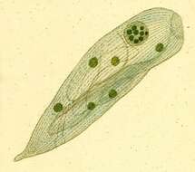
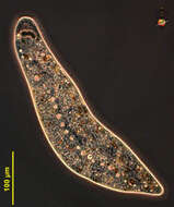
Trachelius ovum (EHRENBERG,1831)EHRENBERG,1838.Dorsolateral view showing numerous scattered small contractile vacuoles,each with a solitary excretory pore.DIC.
-
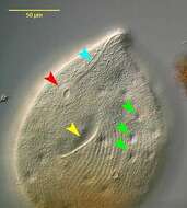
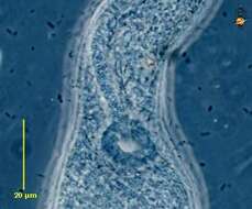
Trachelius ovum (EHRENBERG,1831)EHRENBERG,1838.Left ventrolateral view showing the pellicular groove or fossa (yellow arrowhead). The function of this structure is unknown but some speculate that it may be a site of cell adherence to the substratum. The circular cytostome (red arrowhead) is located at the base of the anterior snout. The circumoral and perioral kineties (light blue arrowhead) encircle the cytostome and converge to parallel each other along the ventral surface of the snout terminating at its tip.Several of the numerous small contractile vacuoles, each with a solitary excretory pore are indicated by the green arrowheads.DIC.
-
-
-
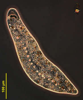
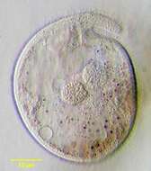
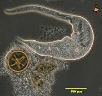
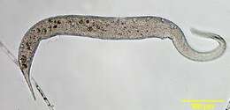
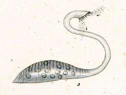
Dileptus (die-leapt-us) is a predatory ciliate. There is a body and a neck. The body is elliptical, tapering posteriorly, but extending anteriorly to form the neck. The neck sweeps around to try to find other protozoa or other prey. It is armed with extrusomes which kill the prey and which is then manipulated into food vacuoles by the mouth which is located at the bottom of the neck. The cells have multiple contractile vacuoles (clear structures) and an elongate macronucleus (dark in this image). This image includes an occupied and a vacated cyst. The cysts do not protect against desiccation, but probably are used when there is no food. Phase contrast.
-
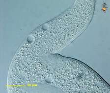
Dileptus (die-leapt-us) is a predatory ciliate. There is a body and a neck. The body is elliptical, tapering posteriorly, but extending anteriorly to form the neck. The neck sweeps around to try to find other protozoa or other prey. It is armed with extrusomes which kill the prey and which is then manipulated into food vacuoles by the mouth which is located at the bottom of the neck, and is evident here as a slightly stiffer region just below the fold. This is the circular structure that is visible in this micrograph. The cells have multiple contractile vacuoles (clear structures). Phase contrast.
-
Dileptus (die-leapt-us) is a predatory ciliate. There is a body and a neck. The body is elliptical, tapering posteriorly, but extending anteriorly to form the neck. The neck sweeps around to try to find other protozoa or other prey. The flexibility of the cells is illustrated here. It is armed with extrusomes which kill the prey and which is then manipulated into food vacuoles by the mouth which is located at the bottom of the neck. The cells have multiple contractile vacuoles (clear structures) and an elongate macronucleus (dark in this image). Phase contrast.
-
Dileptus (die-leapt-us) is a predatory ciliate. There is a body and a neck. The body is elliptical, tapering posteriorly, but extending anteriorly to form the neck. The neck sweeps around to try to find other protozoa or other prey. It is armed with extrusomes which kill the prey and which is then manipulated into food vacuoles by the mouth which is located at the bottom of the neck. The cells have multiple contractile vacuoles (clear structures) and an elongate macronucleus (dark in this image). Phase contrast.
-
Dileptus (die-leapt-us) is a predatory ciliate. There is a body and a neck. The body is elliptical, tapering posteriorly, but extending anteriorly to form the neck. The neck sweeps around to try to find other protozoa or other prey. It is armed with extrusomes which kill the prey and which is then manipulated into food vacuoles by the mouth which is located at the bottom of the neck. This is the circular structure that is visible in this micrograph. Phase contrast.
-
-
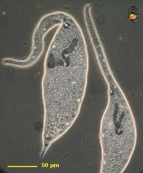
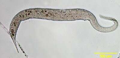
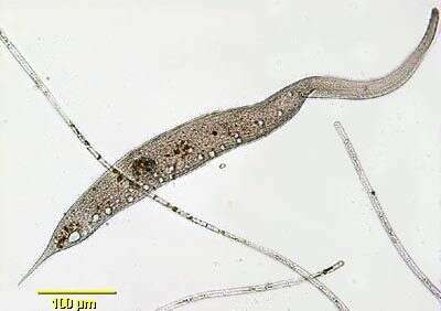
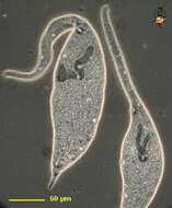
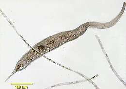
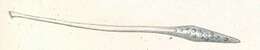
Portrait of Dileptus, large haptorid ciliate with long very flexible proboscis - directed to the right, which the organism casts about as part of its feeding behaviour. The oval cytostome is located at the junction of body and proboscis. Densely packed toxicysts line the ventral surface of the proboscis. The cell is pointed posteriorly. Multiple small contractile vacuoles along dorsum. From aquaculture pond at koi farm near Boise, Idaho. Brightfield.
-
Portrait of Dileptus, large haptorid ciliate with long very flexible proboscis - directed to the right, which the organism casts about as part of its feeding behaviour. The oval cytostome is located at the junction of body and proboscis. Densely packed toxicysts line the ventral surface of the proboscis. The cell is pointed posteriorly. Multiple small contractile vacuoles along dorsum. From aquaculture pond at koi farm near Boise, Idaho. Brightfield.n
-
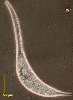
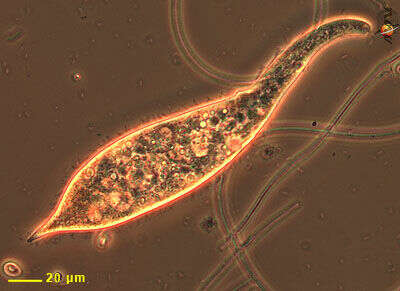
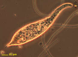
Predatory ciliate, the snout (to the right) sweeps around and if prey are encountered toxic extrusomes are discharged. The mouth is at the base of the snout. The body is evenly ciliates. Phase contrast microscopy.
-
-
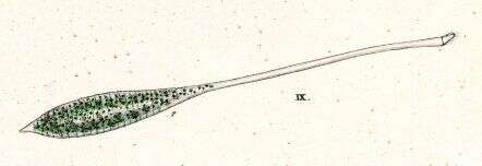
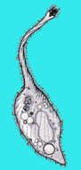
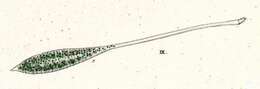
Originally described by Ehrenberg under the name Trachelocerca viridis.
-
Originally described by Ehrenberg under the name Trachelocerca viridis.
-
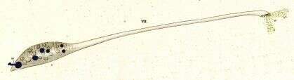
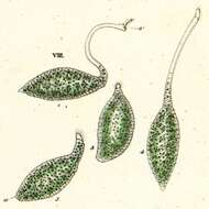
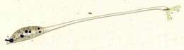
Originally described by Ehrenberg under the name Trachelocerca olor.
-
Originally described by Ehrenberg under the name Trachelocerca olor.
-
Originally described by Ehrenberg under the name Trachelocerca olor.
-
-
Homalozoon, a elongate ribbon-like predatory ciliate. The body is truncated (cut) off at the front end where the mouth is located, and pointed posteriorly. It has rows of cilia mostly on the ventral side, it glides over the substrate, sometimes contracting. Feeds on detritus and other protists. This image shows the line of contractile vacuoles which extends along the body. Flattened. Phase contrast micrograph.
-
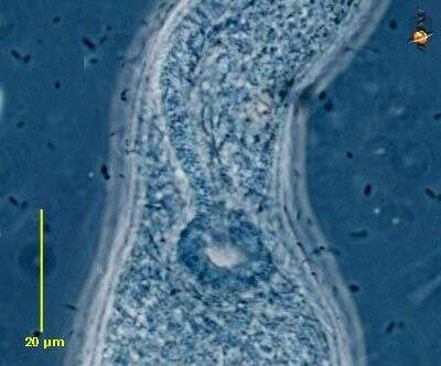
Homalozoon, a elongate-ribbon like predatory ciliate. The body is truncated (cut) off at the front end where the mouth is located, and pointed posteriorly. It has rows of cilia mostly on the ventral side, it glides over the substrate, sometimes contracting. Feeds on detritus and other protists. This image shows the macronucleus, which takes to form of a row of ellip[tical beads attached end-to-end. The micronuclei are small round dark structures adjacent to the macronucleus. Phase contrast micrograph.