-
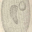
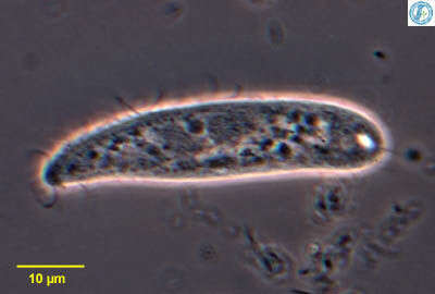
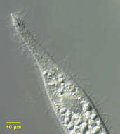
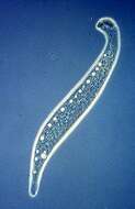
Detail of the anterior end of the haptorid ciliate, Chaenea teres (Dujardin,1841). Probably synonymous with C. stricta (Dujardin 1841, Foissner, 1995) which is also found in freshwater habitats. The cell is elongate,ovoid in cross section,flexible but only slightly contractile. There is a short anterior snout with an inconspicuous apical cytostome. Small trichites support the cytopharynx. Longitudinal somatic kineties are widely spaced. The short snout has densely packed slightly spiral kineties with longer cilia (seen well here). The cytoplasm of this individual contains highly refractile crystalline inclusions.This specimen was collected from a commercial saltwater aquarium in Boise, Idaho May 2004. DIC optics.
-
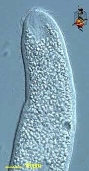
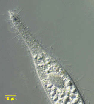
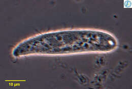
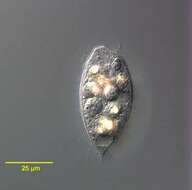
Enchelyodon (ench-elly-owe-don), a cylindrical predatory ciliate, body fairly flexible, mouth is a slit zone at anterior end, underlain by a number of extrusomes. Differential interference contrast.
-
-
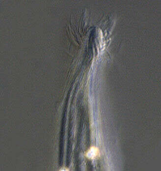
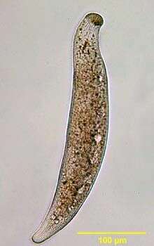
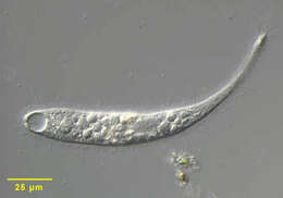
Trachelophyllum, small predatory ciliate, with a wreath of flagella projecting from the front of the cell. This cell has two large macronuclei, one on each side of the small micronucleus. From Lake Donghu, China. Phase contrast micrograph.
-
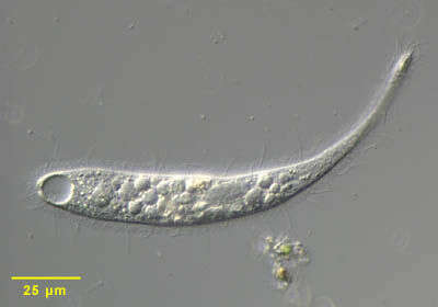
Trachelophyllum, a predatory haptorid ciliate. The mouth is located at the anterior pole. Extrusomes lie internal to the mouth. Contractile vacuole located at posterior end. Differential interference contrast optics.
-
Trachelophyllum, a predatory haptorid ciliate. The mouth is located at the anterior pole. Extrusomes lie internal to the mouth. Contractile vacuole located at posterior end.
-
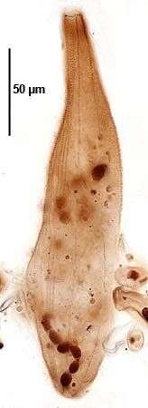
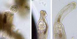
Portrait of large haptorid ciliate Homalozoon vermiculare (STOKES,1887)STOKES,1890. Laterally flattened. Bluntly rounded anteriorly and tapered posteriorly. Contractile. Slit-like oral aperture with prominent oral bulge extrusomes. Multiple small contractile vacuoles along lateral margin. Macronucleus moniliform. Brightfield. From standing freshwater with abundant decomposing leaves near Boise, Idaho.
-
Detail of anterior of Homalozoon vermiculare (STOKES,1887) STOKES,1890 showing dense collection of oral bulge extrusomes. The characteristic dense anterior aggregate of pharyngeal granules is well seen. The function of these is unclear. Brightfield. From freshwater pond near Boise, Idaho.
-
Homalozoon vermiculare (STOKES,1887) STOKES,1890 seen here preying on a peritrich ciliate. The characteristic dense aggregate of granules can be seen. This is displaced by the ingested prey and then disperses as the food vacuole proceeds distally. From freshwater pond near Boise, Idaho. Brightfield
-
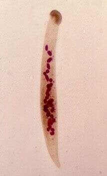
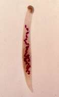
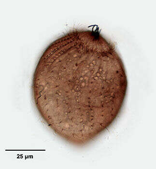
This cell has been killed and then stained with Feulgen stain which shows up the nuclei. As with all ciliates, there are two kinds of nuclei, a large macronucleus which takes the form of a string of beads, and smaller micronuclei which in this species are numerous small structures located near the macronucleus.
-
Phase contrast micrograph of a living cell. The line of contractile vacuoles, lines of the kineties and the band of extrusomes just under the mouth are visible.
-
Infraciliature (right side) of Homalozoon vermiculare (STOKES,1887) STOKES,1890.Collected from a eutrophic freshwater pond in Boise,Idaho June 2008.Stained by the Protargol A technique (see Foissner, W. Europ. J. Protistol., 27:313-330;1991).Brightfield.
-
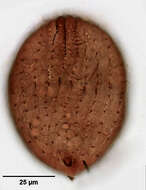
Infraciliature (ventral side) of Homalozoon vermiculare (STOKES,1887) STOKES,1890. the posterior portion of the moniliform macronucleus and one of the numerous micronuclei are seen here.Collected from a eutrophic freshwater pond in Boise,Idaho June 2008.Stained by the Protargol A technique (see Foissner, W. Europ. J. Protistol., 27:313-330;1991).Brightfield.
-
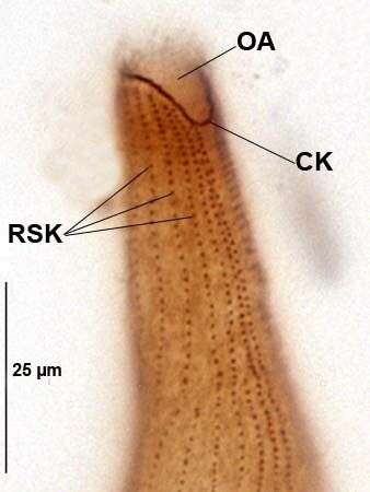
Infraciliature (right side) of Homalozoon vermiculare (STOKES,1887) STOKES,1890. CK=circumoral kinety.OA=oral aperture.RSK=right somatic kineties.Collected from a eutrophic freshwater pond in Boise,Idaho June 2008.Stained by the Protargol A technique (see Foissner, W. Europ. J. Protistol., 27:313-330;1991).Brightfield.
-
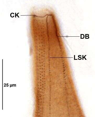
Infraciliature (ventral side) of Homalozoon vermiculare (STOKES,1887) STOKES,1890. CK=circumoral kinety.DB=dorsal brush.LSK=left somatic kineties.Collected from a eutrophic freshwater pond in Boise,Idaho June 2008.Stained by the Protargol A technique (see Foissner, W. Europ. J. Protistol., 27:313-330;1991).Brightfield.
-

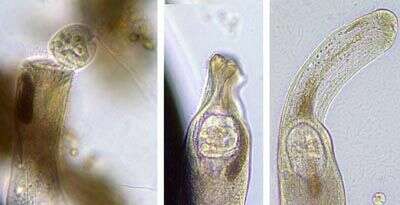
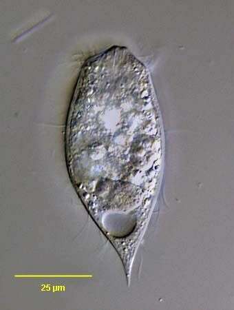
Right lateral view of the haptorid ciliate, Acropisthium mutabile (Perty, 1852). The cell body is ovoid to cylindrical. The posterior tapers to a short point. The fixation and staining process swells the cells. The anterior end forms a blunt snout with an apical cytostome. Short trichites support the cytopharynx (not seen here). There is a girdle of longer cilia just posterior to the bare anterior snout. There are 22 widely spaced uniform longitudinal somatic kineties. This individual is in the middle stage of division. The equatorial band of closely spaced kintosomes will form the circumoral ciliary girdle of the posterior daughter cell (opisthe). The anterior halves of three dorsal kineties are made up of clavate (short club-shaped) cilia forming a dorsal brush (seen well in this view). The dorsal brush of the opisthe is seen well here. Collected from freshwater pond near Boise, Idaho August 2004. This specimen is stained by a silver carbonate technique (see Foissner, W.Europ. J. Protistol.27,313-330;1991). Brightfield optics.
-
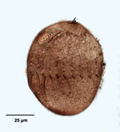
Ventral view of the haptorid ciliate, Acropisthium mutabile (Perty, 1852). The cell body is ovoid to cylindrical. The posterior tapers to a short point. The fixation and staining process swells the cells. The anterior end forms a blunt snout with an apical cytostome. Short trichites support the cytopharynx (not seen here). There is wreath of longer cilia just posterior to the bare anterior snout. There are 22 widely spaced uniform longitudinal somatic kineties. The anterior halves of three dorsal kineties are made up of clavate (short club-shaped) cilia forming a dorsal brush (not seen in this view).Collected from freshwater pond near Boise, Idaho August 2004. This specimen is stained by a silver carbonate technique (see Foissner, W.Europ. J. Protistol.27,313-330;1991). Brightfield optics.
-
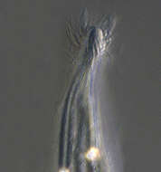
Right lateral view of the haptorid ciliate, Acropisthium mutabile (Perty, 1852). The cell body is ovoid to cylindrical. The posterior tapers to a short point. The fixation and staining process swells the cells. The anterior end forms a blunt snout with an apical cytostome in the center of a bare area. Short trichites support the cytopharynx. A cluster of extrusomes (stained black here) protrudes from the cytostome. There is a girdle of longer cilia just posterior to the bare anterior snout. The closely packed kinetosomes of this circumoral ciliary girdle are angled obliquely to the long axis (seen well here). There are 22 widely spaced uniform longitudinal somatic kineties. The anterior halves of three dorsal kineties are made up of clavate (short club-shaped) cilia forming a dorsal brush (seen well in this view). Collected from freshwater pond near Boise, Idaho August 2004. This specimen is stained by a silver carbonate technique (see Foissner, W.Europ. J. Protistol.27,313-330;1991). Brightfield optics.
-

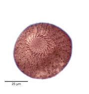
Anterior apical view of the haptorid ciliate, Acropisthium mutabile (Perty, 1852). The cell body is ovoid to cylindrical. The posterior tapers to a short point. The anterior end forms a blunt snout with an apical cytostome. Short trichites support the cytopharynx. There is a girdle of longer cilia just posterior to the bare anterior snout. There is a girdle of longer cilia just posterior to the bare anterior snout. The closely packed kinetosomes of this circumoral ciliary girdle are angled obliquely to the long axis (seen well here). Radiating fibrils can be seen between the circumoral kineties and the cytostome. There are 22 widely spaced uniform longitudinal somatic kineties. The anterior halves of three dorsal kineties are made up of clavate (short club-shaped) cilia forming a dorsal brush (seen well in this view at 12 o'clock). Stained by the silver carbonate technic (see Foissner, W. Europ. J. Protistol.27, 313-330; 1991). Collected from a freshwater pond near Boise, Idaho. Brightfield.
-
Dorsal view of the haptorid ciliate, Acropisthium mutabile (Perty, 1852). The cell body is ovoid to cylindrical. The posterior tapers to a short point. The fixation and staining process swells the cells. The anterior end forms a blunt snout with an apical cytostome. Short trichites support the cytopharynx (not seen here). There is a girdle of longer cilia just posterior to the bare anterior snout. There are 22 widely spaced uniform longitudinal somatic kineties. This individual is in the early stage of division. The equatorial band of closely spaced kintosomes will form the circumoral ciliary girdle of the posterior daughter cell (opisthe). The anterior halves of three dorsal kineties are made up of clavate (short club-shaped) cilia forming a dorsal brush (seen well in this view). Collected from freshwater pond near Boise, Idaho August 2004. This specimen is stained by a silver carbonate technique (see Foissner, W.Europ. J. Protistol.27,313-330;1991). Brightfield optics.
-
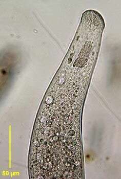
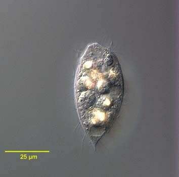
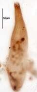
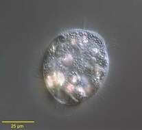
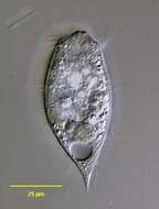
Portrait of the haptorid ciliate, Acropisthium mutabile (Perty, 1852). The cell body is ovoid. The posterior tapers to a short point. The anterior end forms a blunt snout with an apical cytostome. Short trichites support the cytopharynx. There is wreath of longer cilia just posterior to the bare anterior snout. The uniform longitudinal somatic kineties are are widely spaced. Three anterior rows of clavate cilia form a dorsal brush (seen here on viewer's left anteriorly). The cytoplasm contains highly refractile crystaline inclusions. The spherical macronucleus is posterior. There is a single posterior terminal contractile vacuole. Collected from freshwater pond near Boise, Idaho May 2004. DIC optics.
-
Portrait of the haptorid ciliate, Acropisthium mutabile (Perty, 1852). The cell body is ovoid. The posterior tapers to a short point. The anterior end forms a blunt snout with an apical cytostome. Short trichites support the cytopharynx. There is wreath of longer cilia just posterior to the bare anterior snout. The uniform longitudinal somatic kineties are are widely spaced. Three anterior rows of clavate cilia form a dorsal brush. The cytoplasm contains highly refractile crystaline inclusions. The spherical macronucleus is posterior. There is a single posterior terminal contractile vacuole. Collected from freshwater pond near Boise, Idaho May 2004. DIC optics.
-
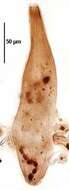
Portrait of the haptorid ciliate, Acropisthium mutabile (Perty, 1852). The cell body is ovoid. The posterior tapers to a short point. The anterior end forms a blunt snout with an apical cytostome. Short trichites support the cytopharynx. There is wreath of longer cilia just posterior to the bare anterior snout. The uniform longitudinal somatic kineties are are widely spaced. Three anterior rows of clavate cilia form a dorsal brush. The cytoplasm contains highly refractile crystaline inclusions. The ellipsoid macronucleus is seen just anterior to the contractile vacuole. There is a single posterior terminal contractile vacuole. Collected from freshwater pond near Boise, Idaho February 2005. DIC optics.
-
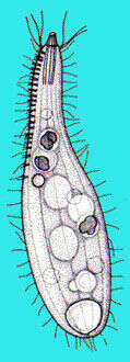
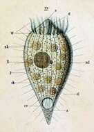
Originally described as Dinophrya liberkuhnii (Butschli) a -- Anus cl -- Cilia cv -- Contractile vacuole ek -- Ectoplasm N -- Macronucleus ncl -- Micronucleus nk -- Food particle o -- Mouth nk -- Food particle p -- Pellicle st -- Cytopharyngeal basket W -- Ciliated ring