-
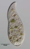
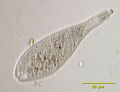
Luporinophrys micelae (FOISSNER,2005). Collected from an ephemeral puddle on a flood-irrigated grass lawn in Boise, Idaho, 2007.Phase contrast.
-
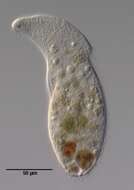
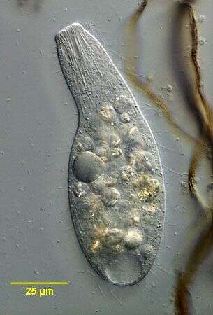
Enchelyodon armatus (KAHL,1926) KAHL,1930 from a freshwater pond near Boise, Idaho. Oblique illumination.
-
Enchelyodon armatus (KAHL, 1926) KAHL, 1930.DIC.
-
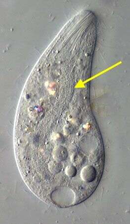
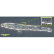
In vivo portrait of Enchelyodon armatus (KAHL,1926),KAHL,1930 demonstrating the band-form macronucleus.
-
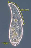
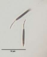
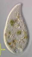
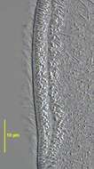
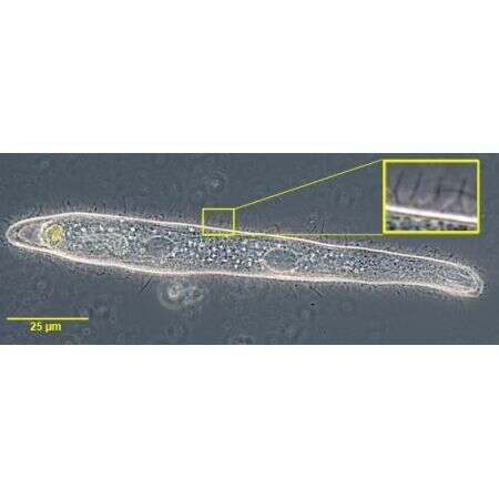
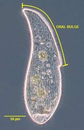
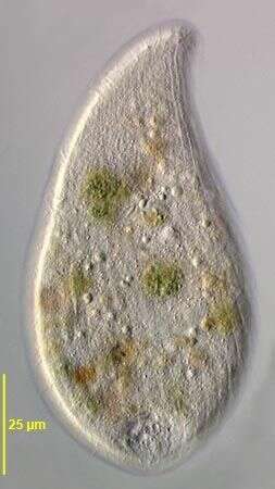
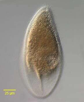
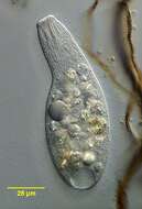
The long oral bulge (~50% of cell length) is one of the main distinguishing features of this subspecies of A. cultriforme. This specimen is somewhat stouter than the cells described by Foissner (Protistology 4 (1), 5-55 (2005) probably due to contraction after transfer from the culture dish to the slide. When observed undisturbed under the dissecting microscope the cells appear more slender.Phase contrast.
-
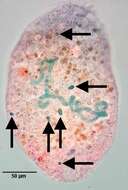
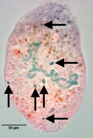
Six of the multiple (7-18) micronuclei are in the focal plane of this image. Stained by the methylgreen-pyroninY technique (see Foissner, W.Europ. J. Protistol.27:313-330;1991).Brightfield.
-
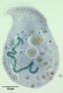
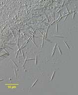
Band-form macronucleus of Arcuospathidum cultriforme scalpriforme (KAHL,1930) FOISSNER,2003.Stained by the methylgreen-pyroninY technique (see Foissner, W.Europ. J. Protistol.27:313-330;1991).Brightfield.
-
-
-
Stained by the silver carbonate technique (see Foissner, W.Europ. J. Protistol.27:313-330;1991).Brightfield.
-
-
2 of the three dorsal brush rows are seen in this image.DIC.
-
-
-
-
-
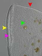
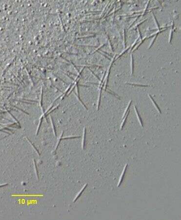
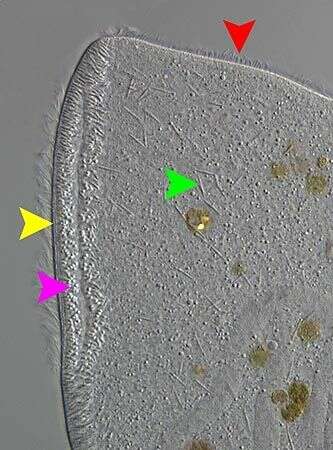
Complete circumoral kinety separate from somatic kineties (yellow). Extrusomes scattered randomly on each half of oral bulge (pink). extrusome (green). Clavate cilia of one of the dorsal brush rows (red).DIC.
-
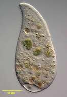
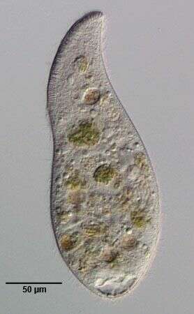
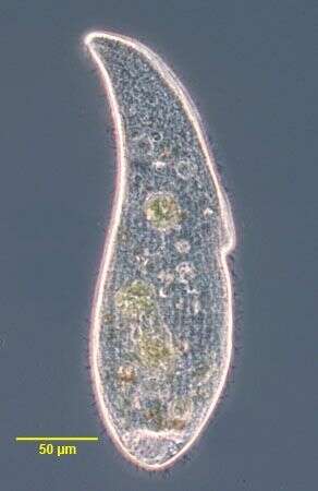
Arcuospathidium cultriforme scalpriforme (KAHL,1930) FOISSNER,2003. Phase contrast
-
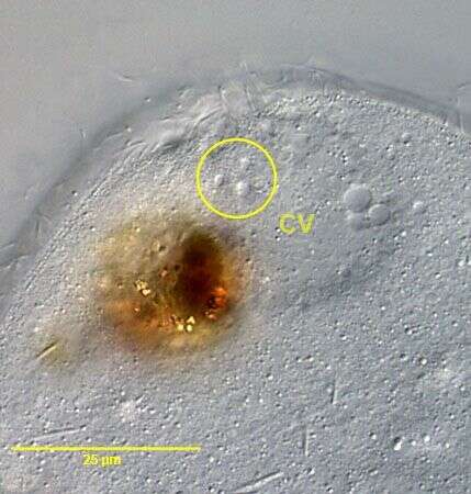
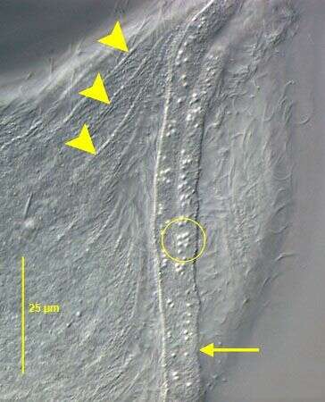
The posterior terminal contractile vacuole empties through multiple pores three of which are indicated within the yellow circle. DIC.
-
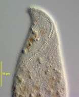
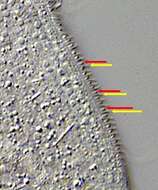
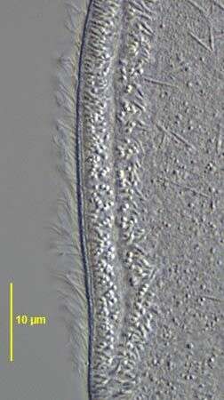
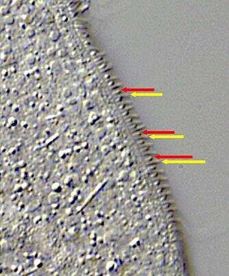
The dorsal brush rows have alternating longer and shorter cilia. In brush rows 1 and 2 the anterior cilium of each dikinetid is longer but in the 3rd dorsal brush row (seen here) the posterior cilium (yellow arrow) is longer than the anterior one (red arrow). DIC.
-
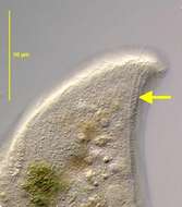
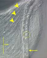
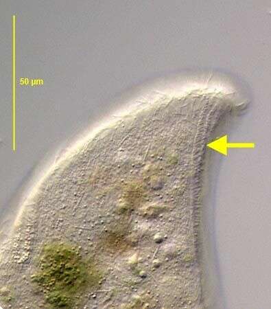
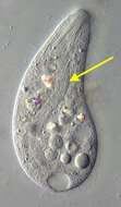
The oral bulge is indicated by the yellow arrow.The ends of the oral bulge extrusomes appear as bright spots here (yellow circle). Several right somatic kineties are indicated by the yellow arrowheads.The slit-like oral aperture is between the two halves of the oral bulge. DIC.
-
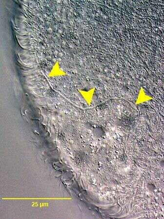
Highly compressed specimen showing part of the circumoral kinety (yellow arrowheads)which is separate from the anterior ends of the dorsally curving somatic kineties. DIC.
-
Left side of the pleurostome ciliate,Opisthodon niemeccense (Stein,1859).The flexible cell is lanceolate in outline and laterally compressed.The somatic kineties of the right side meet at an anterior suture.A single file dorsal brush terminates in a small anterior depression (seen here).Thre are two perioral kineties.The oral aperture extends from the anterior end to the mid-body (on viewer's left). The macronucleus is bipartite. There is one contractile vacuole dorsally(on viewer's right here). Collected from sapropelic bottom sediments of a freshwater pond near Boise, Idaho;43°19'07.45"N 115°27'31.99"W, elev.4712 ft.October 2005.DIC.
-
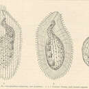
a -- Anus cv -- contractile vacuole ek -- Ectoplasm N -- Macronucleus nk -- food particle o -- Mouth oe -- Throat tr -- Trichocysts st -- Cytopharyngeal basket