-
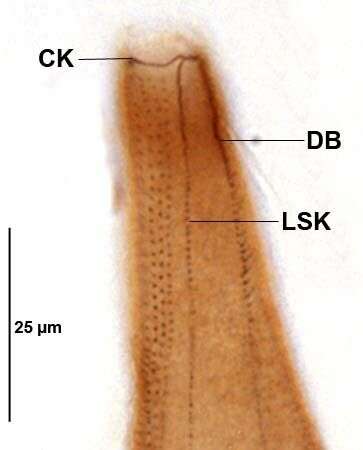
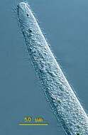
Infraciliature (ventral side) of Homalozoon vermiculare (STOKES,1887) STOKES,1890. CK=circumoral kinety.DB=dorsal brush.LSK=left somatic kineties.Collected from a eutrophic freshwater pond in Boise,Idaho June 2008.Stained by the Protargol A technique (see Foissner, W. Europ. J. Protistol., 27:313-330;1991).Brightfield.
-

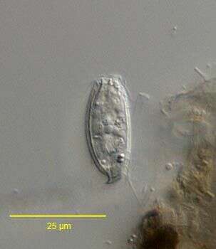
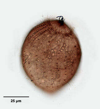
Right lateral view of the haptorid ciliate, Acropisthium mutabile (Perty, 1852). The cell body is ovoid to cylindrical. The posterior tapers to a short point. The fixation and staining process swells the cells. The anterior end forms a blunt snout with an apical cytostome. Short trichites support the cytopharynx (not seen here). There is a girdle of longer cilia just posterior to the bare anterior snout. There are 22 widely spaced uniform longitudinal somatic kineties. This individual is in the middle stage of division. The equatorial band of closely spaced kintosomes will form the circumoral ciliary girdle of the posterior daughter cell (opisthe). The anterior halves of three dorsal kineties are made up of clavate (short club-shaped) cilia forming a dorsal brush (seen well in this view). The dorsal brush of the opisthe is seen well here. Collected from freshwater pond near Boise, Idaho August 2004. This specimen is stained by a silver carbonate technique (see Foissner, W.Europ. J. Protistol.27,313-330;1991). Brightfield optics.
-
Ventral view of the haptorid ciliate, Acropisthium mutabile (Perty, 1852). The cell body is ovoid to cylindrical. The posterior tapers to a short point. The fixation and staining process swells the cells. The anterior end forms a blunt snout with an apical cytostome. Short trichites support the cytopharynx (not seen here). There is wreath of longer cilia just posterior to the bare anterior snout. There are 22 widely spaced uniform longitudinal somatic kineties. The anterior halves of three dorsal kineties are made up of clavate (short club-shaped) cilia forming a dorsal brush (not seen in this view).Collected from freshwater pond near Boise, Idaho August 2004. This specimen is stained by a silver carbonate technique (see Foissner, W.Europ. J. Protistol.27,313-330;1991). Brightfield optics.
-
Right lateral view of the haptorid ciliate, Acropisthium mutabile (Perty, 1852). The cell body is ovoid to cylindrical. The posterior tapers to a short point. The fixation and staining process swells the cells. The anterior end forms a blunt snout with an apical cytostome in the center of a bare area. Short trichites support the cytopharynx. A cluster of extrusomes (stained black here) protrudes from the cytostome. There is a girdle of longer cilia just posterior to the bare anterior snout. The closely packed kinetosomes of this circumoral ciliary girdle are angled obliquely to the long axis (seen well here). There are 22 widely spaced uniform longitudinal somatic kineties. The anterior halves of three dorsal kineties are made up of clavate (short club-shaped) cilia forming a dorsal brush (seen well in this view). Collected from freshwater pond near Boise, Idaho August 2004. This specimen is stained by a silver carbonate technique (see Foissner, W.Europ. J. Protistol.27,313-330;1991). Brightfield optics.
-

Anterior apical view of the haptorid ciliate, Acropisthium mutabile (Perty, 1852). The cell body is ovoid to cylindrical. The posterior tapers to a short point. The anterior end forms a blunt snout with an apical cytostome. Short trichites support the cytopharynx. There is a girdle of longer cilia just posterior to the bare anterior snout. There is a girdle of longer cilia just posterior to the bare anterior snout. The closely packed kinetosomes of this circumoral ciliary girdle are angled obliquely to the long axis (seen well here). Radiating fibrils can be seen between the circumoral kineties and the cytostome. There are 22 widely spaced uniform longitudinal somatic kineties. The anterior halves of three dorsal kineties are made up of clavate (short club-shaped) cilia forming a dorsal brush (seen well in this view at 12 o'clock). Stained by the silver carbonate technic (see Foissner, W. Europ. J. Protistol.27, 313-330; 1991). Collected from a freshwater pond near Boise, Idaho. Brightfield.
-
Dorsal view of the haptorid ciliate, Acropisthium mutabile (Perty, 1852). The cell body is ovoid to cylindrical. The posterior tapers to a short point. The fixation and staining process swells the cells. The anterior end forms a blunt snout with an apical cytostome. Short trichites support the cytopharynx (not seen here). There is a girdle of longer cilia just posterior to the bare anterior snout. There are 22 widely spaced uniform longitudinal somatic kineties. This individual is in the early stage of division. The equatorial band of closely spaced kintosomes will form the circumoral ciliary girdle of the posterior daughter cell (opisthe). The anterior halves of three dorsal kineties are made up of clavate (short club-shaped) cilia forming a dorsal brush (seen well in this view). Collected from freshwater pond near Boise, Idaho August 2004. This specimen is stained by a silver carbonate technique (see Foissner, W.Europ. J. Protistol.27,313-330;1991). Brightfield optics.
-
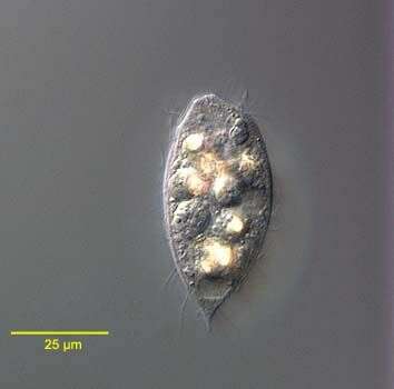
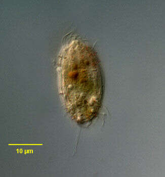
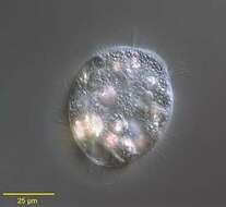
Portrait of the haptorid ciliate, Acropisthium mutabile (Perty, 1852). The cell body is ovoid. The posterior tapers to a short point. The anterior end forms a blunt snout with an apical cytostome. Short trichites support the cytopharynx. There is wreath of longer cilia just posterior to the bare anterior snout. The uniform longitudinal somatic kineties are are widely spaced. Three anterior rows of clavate cilia form a dorsal brush (seen here on viewer's left anteriorly). The cytoplasm contains highly refractile crystaline inclusions. The spherical macronucleus is posterior. There is a single posterior terminal contractile vacuole. Collected from freshwater pond near Boise, Idaho May 2004. DIC optics.
-
Portrait of the haptorid ciliate, Acropisthium mutabile (Perty, 1852). The cell body is ovoid. The posterior tapers to a short point. The anterior end forms a blunt snout with an apical cytostome. Short trichites support the cytopharynx. There is wreath of longer cilia just posterior to the bare anterior snout. The uniform longitudinal somatic kineties are are widely spaced. Three anterior rows of clavate cilia form a dorsal brush. The cytoplasm contains highly refractile crystaline inclusions. The spherical macronucleus is posterior. There is a single posterior terminal contractile vacuole. Collected from freshwater pond near Boise, Idaho May 2004. DIC optics.
-
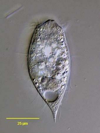
Portrait of the haptorid ciliate, Acropisthium mutabile (Perty, 1852). The cell body is ovoid. The posterior tapers to a short point. The anterior end forms a blunt snout with an apical cytostome. Short trichites support the cytopharynx. There is wreath of longer cilia just posterior to the bare anterior snout. The uniform longitudinal somatic kineties are are widely spaced. Three anterior rows of clavate cilia form a dorsal brush. The cytoplasm contains highly refractile crystaline inclusions. The ellipsoid macronucleus is seen just anterior to the contractile vacuole. There is a single posterior terminal contractile vacuole. Collected from freshwater pond near Boise, Idaho February 2005. DIC optics.
-
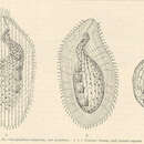
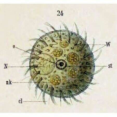
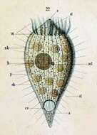
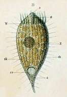
Originally described as Dinophrya liberkuhnii (Butschli) a -- Anus cl -- Cilia cv -- Contractile vacuole ek -- Ectoplasm N -- Macronucleus ncl -- Micronucleus nk -- Food particle o -- Mouth nk -- Food particle p -- Pellicle st -- Cytopharyngeal basket W -- Ciliated ring
-
Originally described as Dinophrya lieberkuhnii (Butschli) Shown with the posterior extended to a tail-like appendage. a -- Anus cv -- Contractile vacuole ek -- Ectoplasm N -- Macronucleus nk -- Food particle st -- Cytopharyngeal basket W -- Ciliated ring
-
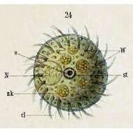
Originally described as Dinophrya lieberkuhnii (Butschli). Oral view. cl -- Cilia N -- Macronucleus nk -- Fppd particle 0 -- Mouth st -- Cytopharyngeal basket W -- Ciliated ring
-
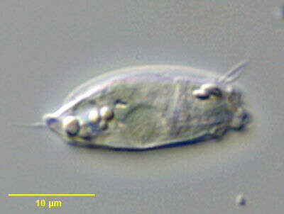
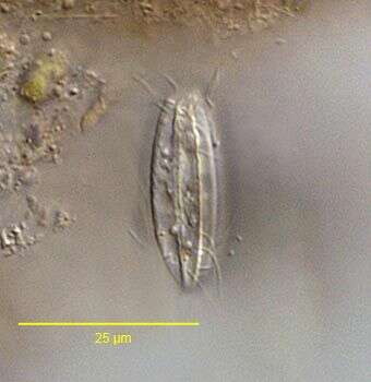
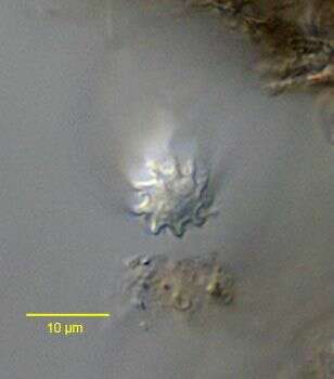
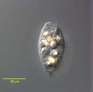
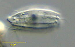
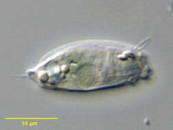
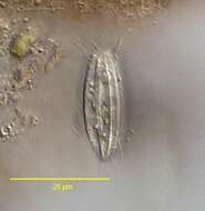
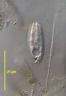
Surface view of Pithothorax processus a small haptorid ciliate found in polysaprobic habitats. The body is a slightly flattened cylinder. The pellicle is rigid with longitudinal ribbing. The oral aperture is at the anterior apex surrounded by projections of the pellicular ridges. There is a curved funnel-shaped posterior process from which a long caudal cilium protrudes (seen here). The round macronucleus is anterior and the contractile vacuole is located in the posterior 1/3 at the periphery (seen here). The somatic ciliature is confined to the anterior and posterior quarters of the body. From stagnant organically enriched freshwater pond near Boise, Idaho. DIC optics.
-
Saggital optical section of Pithothorax processus (Kahl, 1926), a small haptorid ciliate found in polysaprobic habitats. The body is a slightly flattened cylinder. The pellicle is rigid with longitudinal ribbing. The oral aperture is at the anterior apex surrounded by projections of the pellicular ridges. There is a curved funnel-shaped posterior process from which a long caudal cilium protrudes (seen here). The round macronucleus is anterior and the contractile vacuole is located in the posterior 1/3 at the periphery (seen here). The somatic ciliature is confined to the anterior and posterior quarters of the body. From stagnant organically enriched freshwater pond near Boise, Idaho. DIC optics.
-
Surface view of Pithothorax processus (Kahl, 1926) a small haptorid ciliate found in polysaprobic habitats. The body is a slightly flattened cylinder. The pellicle is rigid with longitudinal ribbing. The oral aperture is at the anterior apex surrounded by projections of the pellicular ridges. There is a curved funnel-shaped posterior process from which a long caudal cilium protrudes (seen here). The round macronucleus is anterior and the contractile vacuole is located in the posterior 1/3 at the periphery (seen here). The somatic ciliature is confined to the anterior and posterior quarters of the body. From stagnant organically enriched freshwater pond near Boise, Idaho. DIC optics.
-
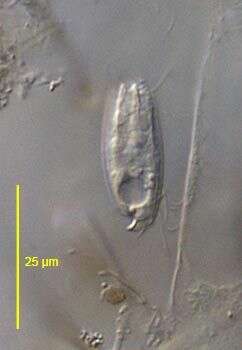
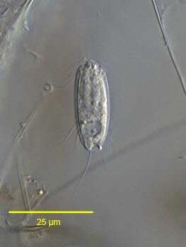
In vivo portrait of Pithothorax processus (Kahl, 1926), a small haptorid ciliate found in polysaprobic habitats. The body is a slightly flattened cylinder. The pellicle is rigid with longitudinal ribbing. The oral aperture is at the anterior apex surrounded by projections of the pellicular ridges. There is a curved funnel-shaped posterior process from which a long caudal cilium protrudes (seen here). The round macronucleus is anterior and the contractile vacuole is located in the posterior 1/3 at the periphery (seen here). The somatic ciliature is confined to the anterior and posterior quarters of the body. From stagnant organically enriched freshwater pond near Boise, Idaho. June 2005. DIC optics.
-
Portrait of Pithothorax processus (Kahl, 1926), a small haptorid ciliate found in polysaprobic habitats. The body is a slightly flattened cylinder. The pellicle is rigid with longitudinal ribbing. The oral aperture is at the anterior apex surrounded by pointed projections of the pellicular ridges. There is a funnel shaped posterior process from which a long caudal cilium protrudes (visible here). The round macronucleus is anterior. The somatic ciliature is confined to the anterior and posterior quarters of the body. Although this specimen was stained by the silvercarbonate technic (see Foissner, W. Europ. J. Protistol., 27:313-330;1991, the somatic kinetids did not impregnate. Short rod shaped partially discharged extrusomes (stained brown here) are visible anteriorly. From stagnant organically enriched freshwater pond near Boise, Idaho. Brighfield.
-
In vivo portrait of Pithothorax processus (Kahl, 1926), a small haptorid ciliate found in polysaprobic habitats. The body is a slightly flattened cylinder. The pellicle is rigid with longitudinal ribbing. The oral aperture is at the anterior apex surrounded by projections of the pellicular ridges. There is a curved funnel-shaped posterior process from which a long caudal cilium protrudes (seen here). The round macronucleus is anterior and the contractile vacuole is located in the posterior 1/3 at the periphery (seen here). The somatic ciliature is confined to the anterior and posterior quarters of the body. From stagnant organically enriched freshwater pond near Boise, Idaho. June 2005. DIC optics.
-
Cross-sectional view of Pithothorax processus (Kahl, 1926) a small haptorid ciliate found in polysaprobic habitats. The body is a slightly flattened cylinder. The pellicle is rigid with longitudinal ribbing giving it a fluted appearance. From stagnant organically enriched freshwater pond near Boise, Idaho.June 2005. DIC.
-
Lasteral view of Pithothorax processus (Kahl, 1926), a small haptorid ciliate found in polysaprobic habitats. The body is a slightly flattened cylinder. The pellicle is rigid with longitudinal ribbing. The oral aperture is at the anterior apex surrounded by projections of the pellicular ridges. There is a curved funnel-shaped posterior process from which a long caudal cilium protrudes. The round macronucleus is anterior and the contractile vacuole is located in the posterior 1/3 at the periphery. The somatic ciliature is confined to the anterior and posterior quarters of the body. From stagnant organically enriched freshwater pond near Boise, Idaho. June 2005. DIC optics.
-
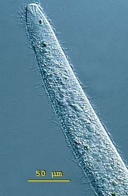
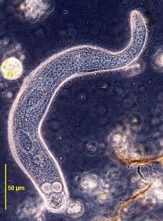
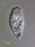
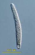
Spathidium (spa-thid-ee-um) moniliforme, the body is elongate, the posterior end is bluntly pointed or rounded but the anterior end is distinctively swollen - often fan-shaped and obliquely truncated. There is an ciliated apical ridge which is lined by toxicysts. The oral aperture is a slit that lies along the length of this ridge. The cilia are uniformly distributed in longitudinal parallel rows on both lateral surfaces. The macronucleus is highly variable, often elongate, ribbon-like or moniliform. The contractile vacuole is single and at the end of the cell. Spathidium feeds on other ciliates. It lives in fresh water ponds and lakes. This specimen was collected in a freshwater pond near Konstanz, Germany. This swimming cell is 250 microns long. Differential interference contrast.
-
Spathidium (spa-thid-ee-um) moniliforme, the body is elongate, the posterior end is bluntly pointed or rounded but the anterior end is distinctively swollen - often fan-shaped and obliquely truncated. There is an ciliated apical ridge which is lined by toxicysts. The oral aperture is a slit that lies along the length of this ridge. The cilia are uniformly distributed in longitudinal parallel rows on both lateral surfaces. The macronucleus is highly variable, often elongate, ribbon-like or moniliform. The contractile vacuole is single and at the end of the cell. Spathidium feeds on other ciliates. It lives in fresh water ponds and lakes. This cell is squashed allowing ribbon-like macronucleus and the fan-like arrangement of toxicysts at the front of the cell to be seen. Differential interference contrast.
-
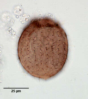
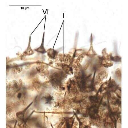
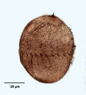
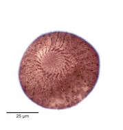
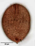
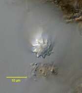
Two of the three types of lepidosomes of Luporinophrys micelae (FOISSNER,2005). The type II are not seen in this image.Stained by the silver carbonate technique (see Foissner, W. Europ. J. Protistol., 27:313-330;1991).Brightfield.
-
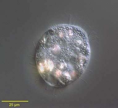
Luporinophrys micelae (FOISSNER,2005). Collected from an ephemeral puddle on a flood-irrigated grass lawn in Boise, Idaho, 2007.Phase contrast.