-
Centers for Disease Control/Division of Parasitic Diseases and Malaria
EOL staff
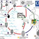
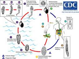
Life cycle of the trematode parasites Fasciola hepatica and F. giganticaImmature eggs are discharged in the biliary ducts and in the stool (1). Eggs become embryonated in water (2) and release
miracidia (3), which invade a suitable snail intermediate host (4), including snails in the genera Galba, Fossaria, and Pseudosuccinea. In the snail, the parasites pass through several developmental stages (
sporocyst (4a),
redia (4b), and
cercaria (4c)). The
cercariae are released from the snail (5) and encyst as
metacercariae on aquatic vegetation or other surfaces. Mammals acquire the infection by eating vegetation containing
metacercariae. Humans can become infected by ingesting metacercariae-containing freshwater plants, especially watercress (6). After ingestion, the
metacercariae excyst in the duodenum (7) and migrate through the intestinal wall, the peritoneal cavity, and the liver parenchyma into the biliary ducts, where they develop into adults (8). In humans, maturation from
metacercariae into adult flukes takes approximately 3 to 4 months. The adult flukes (Fasciola hepatica: up to 30 mm by 13 mm; F. gigantica: up to 75 mm) reside in the large biliary ducts of the mammalian host. Fasciola hepatica infect a variety of mammals, but mostly herbivores.From
Centers for Disease Control Parasites and Health website.
-
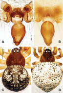
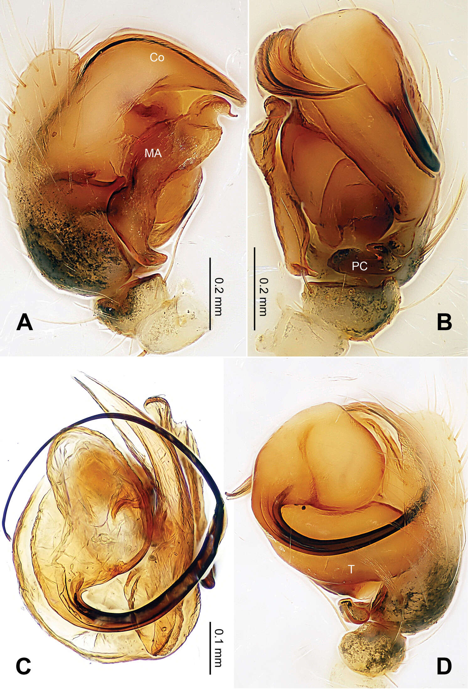
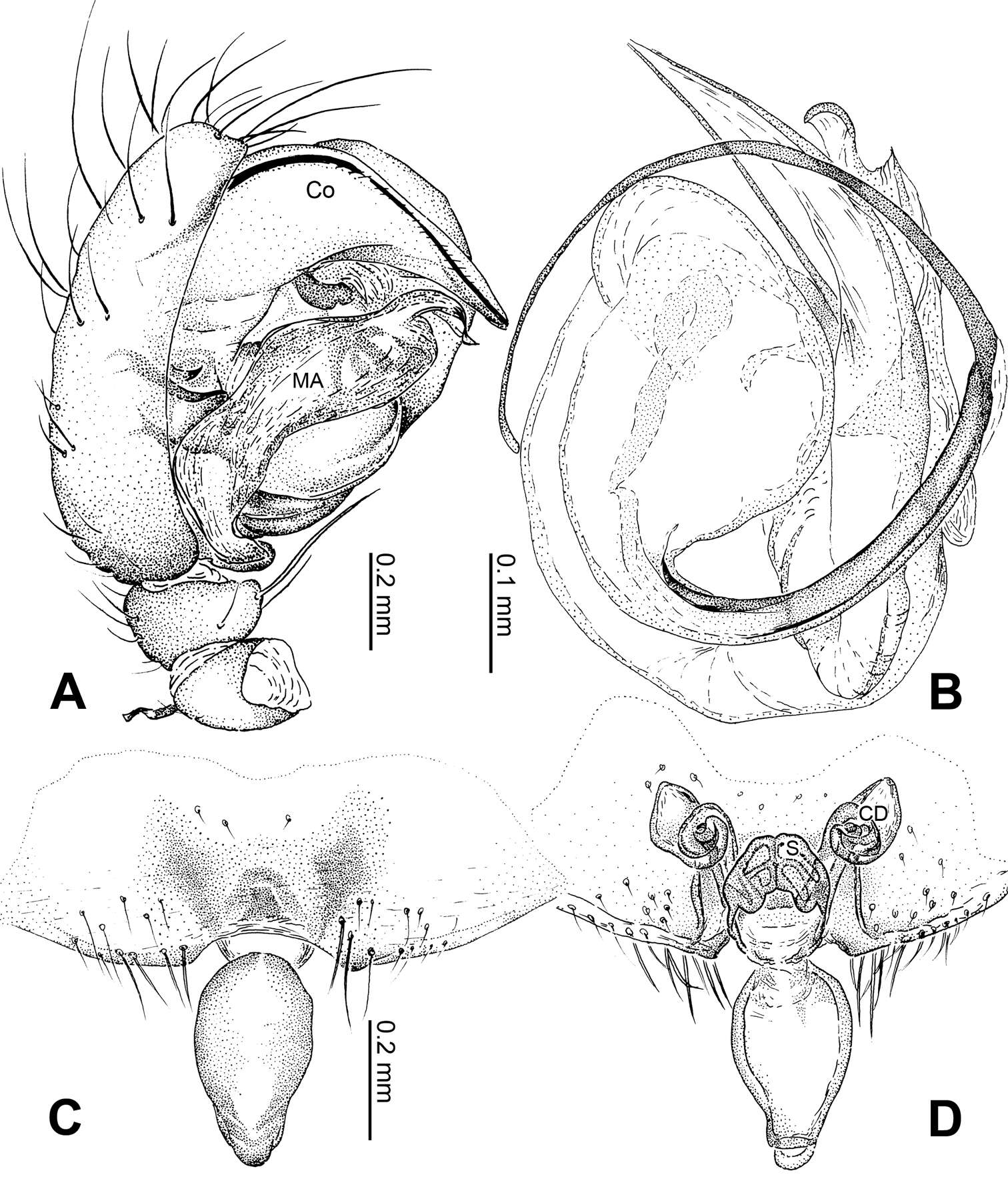
Figure 1.Alaria chengguanensis sp. n., male holotype. A Pedipalp, prolateral view B Pedipalp, ventral view C Embolic division, dorsal view D Pedipalp, retrolateral view. Co conductor; MA median apophysis; PC paracymbium; T tegulum. Scale bars: D as A.
-
Centers for Disease Control/Division of Parasitic Diseases and Malaria
EOL staff
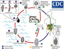
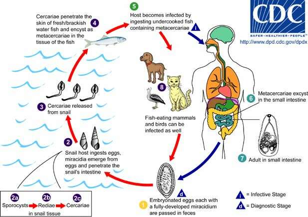
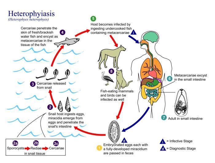
Life cycle of the trematode parasite Heterophyes heterophyesAdults release embryonated eggs, each with a fully-developed
miracidium, and eggs are passed in the host's feces (1). After ingestion by a suitable snail (first intermediate host), the eggs hatch and release
miracidia which penetrate the snail’s intestine (2). Genera Cerithidia and Pironella are important snail hosts in Asia and the Middle East, respectively. The
miracidia pass through several developmental stages in the snail:
sporocysts (2a),
rediae (2b), and
cercariae (2c). Many
cercariae are produced from each
redia. The
cercariae are released from the snail (3) and encyst as
metacercariae in the tissues of a suitable fresh/brackish water fish (second intermediate host) (4). The definitive host becomes infected by ingesting undercooked or salted fish containing
metacercariae (5). After ingestion, the
metacercariae excyst, attach to the mucosa of the small intestine (6) and mature into adults (measuring 1.0 to 1.7 mm by 0.3 to 0.4 mm) (7). In addition to humans, various fish-eating mammals (e.g., cats and dogs) and birds can be infected by H. heterophyes.From
Centers for Disease Control Parasites and Health website.
-
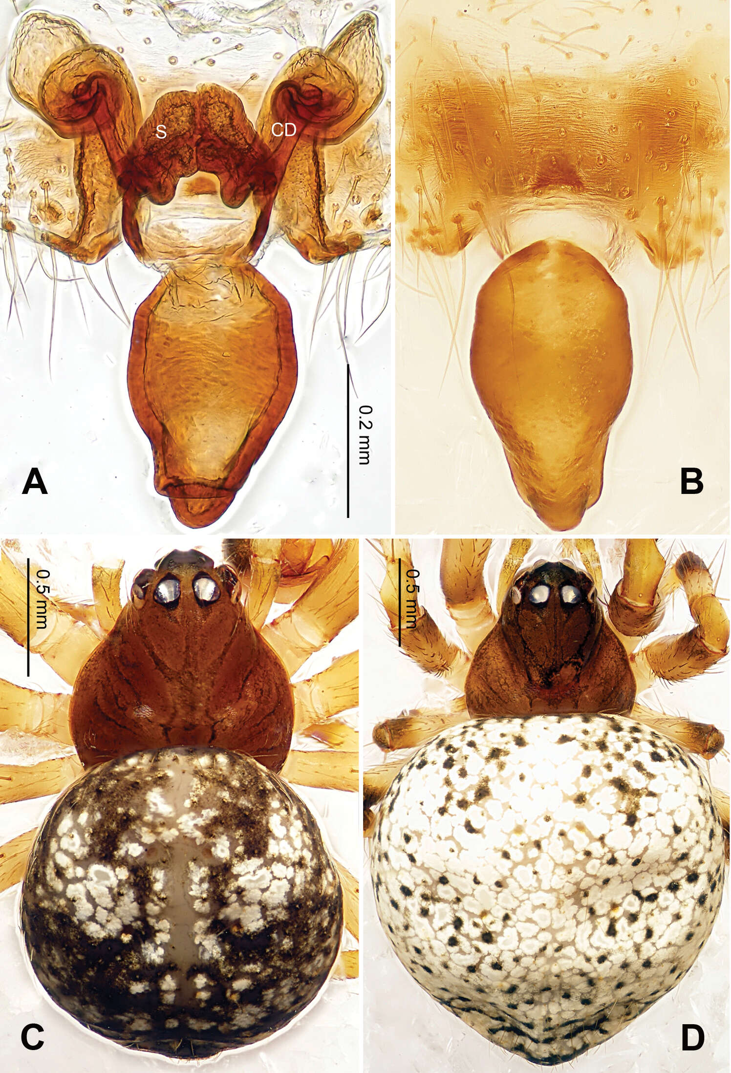
Figure 2.Alaria chengguanensis sp. n., male holotype (C) and female paratype (A–B, D). A Vulva, dorsal view B Epigyne, ventral view C Male, dorsal view D Female, dorsal view. CD copulatory duct; S spermatheca. Scale bars: B as A.
-
Centers for Disease Control/Division of Parasitic Diseases and Malaria
EOL staff
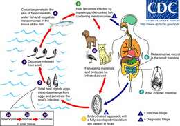
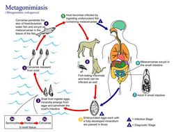
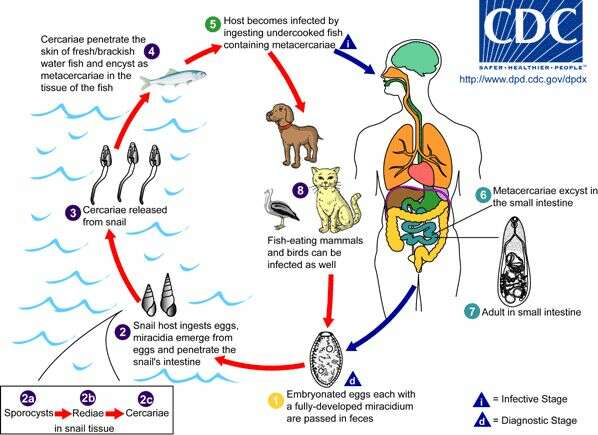
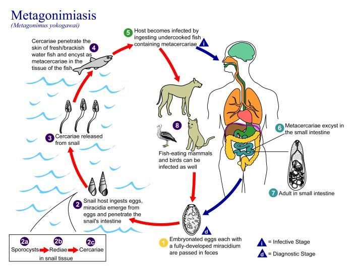
Life cycle of the trematode Metagonimus yokogawai Adult Metagonimus yokogawai release fully embryonated eggs, each with a fully-developed
miracidium, and eggs are passed in the host’s feces (1). After ingestion by a suitable snail (first intermediate host), the eggs hatch and release
miracidia, which penetrate the snail’s intestine (2). Snails of the genus Semisulcospira are the most frequent intermediate host for M. yokogawai. The
miracidia pass through several developmental stages in the snail:
sporocysts (2a),
rediae (2b), and
cercariae (2c). Many
cercariae are produced from each
redia. The
cercariae are released from the snail (3) and encyst as
metacercariae in the tissues of a suitable fresh/brackish water fish (second intermediate host) (4). The definitive host becomes infected by ingesting undercooked or salted fish containing
metacercariae (5). After ingestion, the
metacercariae excyst, attach to the mucosa of the small intestine (6), and mature into adults (which measure only 1.0 mm to 2.5 mm by 0.4 mm to 0.75 mm) (7). In addition to humans, fish-eating mammals (e.g., cats and dogs) and birds can also be infected by M. yokogawai (8).From
Centers for Disease Control Parasites and Health website
-
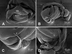
Figure 3.Alaria chengguanensis sp. n., SEM of pedipalp of a male paratype. A Prolateral view, detail showing MA B Retrolateral view C Retrolateral view, detail showingembolus D Retrolateral view, detail showing PC. Co conductor E Embolus; MA median apophysis; PC paracymbium; T tegulum.
-
Centers for Disease Control/Division of Parasitic Diseases and Malaria
EOL staff
Life cycle of Echinostoma trematode flatworms, the agents causing echinostomiasis in humans Many animals may serve as definitive hosts for various Echinostoma species, including aquatic birds, carnivores, rodents, and humans (a definitive host is the host in which the adult parasites occur). Unembryonated eggs are passed in feces (1) and develop in the water (2). On average, the ciliated
miracidium larva takes around 10 days to mature before hatching (3) and penetrating the first intermediate host, a snail (4). Several genera of snails may serve as the first intermediate host. The intramolluscan stages include a
sporocyst (4a), one or two generations of
rediae (4b), and
cercariae (4c). The
cercariae may encyst as
metacercariae within the same first intermediate host or leave the host and penetrate a new second intermediate host (5). Depending on the species, a range of animals may serve as the second intermediate host, including other snails, bivalve mollusks, fish, and tadpoles. The definitive host becomes infected after eating a second intermediate host (6).
Metacercariae excyst in the duodenum (7) and adults reside in the small intestine (8) of the definitive host.From
Centers for Disease Control Parasites and Health website.
-
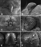
Figure 4.Alaria chengguanensis sp. n., SEM of a female paratype. A Epigyne, ventral view B Epigyne, lateral view C Spinnerets D ALS E PMS F PLS. AC aciniform gland spigot; AG aggregate gland spigot; ALS anterior lateral spinneret; CY cylindrical gland spigot; MAP major ampullate gland spigot; mAP minor ampullate gland spigot; n nubbin; PI piriform gland spigot; PLS posterior lateral spinneret; PMS posterior median spinneret; t tartipore.
-
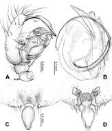
Figure 5.Alaria chengguanensis sp. n., male holotype (A–B) and female paratype (C–D). A Pedipalp, prolateral view B Embolic division, dorsal view C Epigyne, ventral view D Vulva, dorsal view. CD copulatory duct; Co conductor; MA median apophysis; S spermatheca. Scale bars: D as C.
-
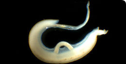
A Schistosoma mansoni male/female pair. The female is held by the male in a groove along its body.
-
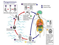
This is an illustration of the life cycle of Paragonimus westermani, one of the causal agents of Paragonimiasis.Created: 2002
-
This is an illustration of the life cycle of Heterophyes heterophyes, the causal agent of Heterophyiasis.Created: 2002
-
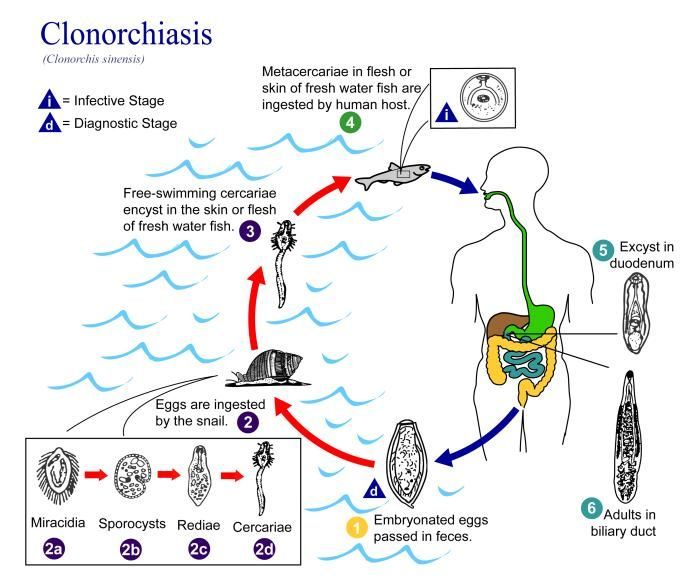
This is an illustration of the life cycle of Clonorchis sinensis, the causal agent of Clonorchiasis.Created: 2002
-
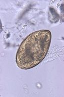
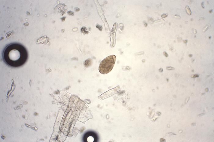
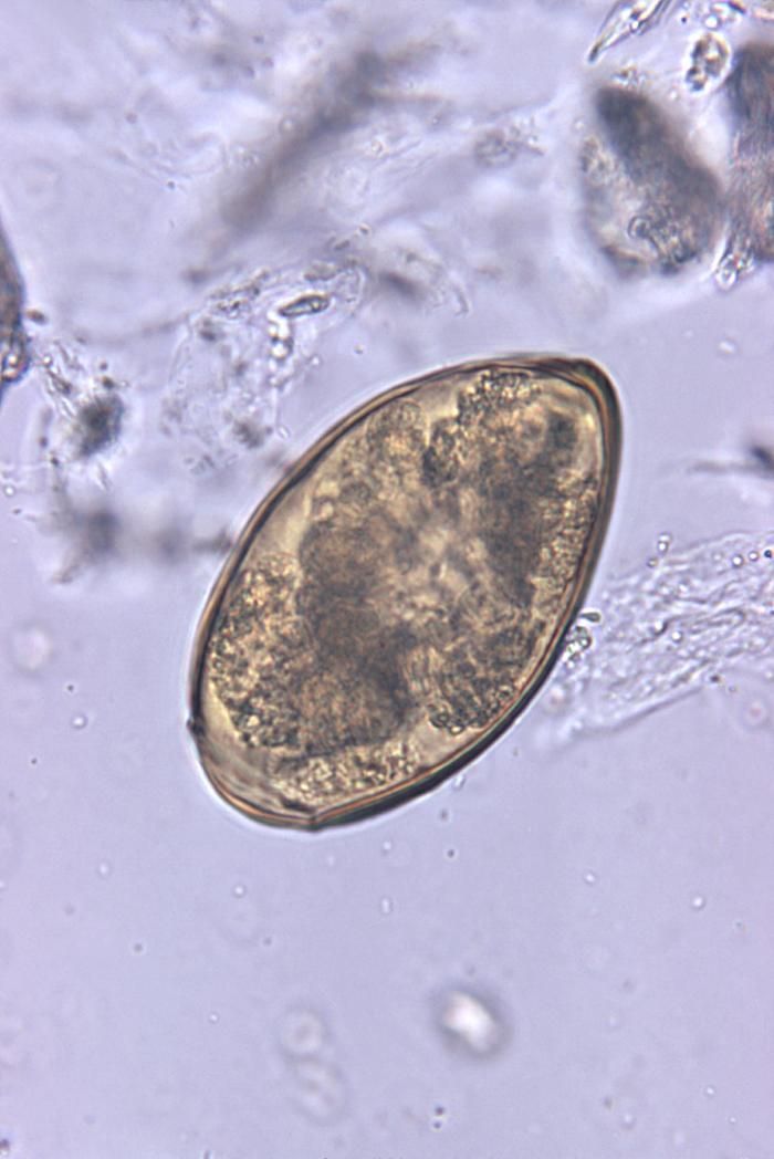
Magnified 128X, this photomicrograph revealed some of the ultrastructural morphology of a single trematode Paragonimus westermani egg. P. westermani eggs range from 80µm to 120µm long X 45µm to 70µm wide. They are yellow-brown, ovoid or elongate, with a thick shell, and often asymmetrical with one end slightly flattened. At the large end, the operculum is clearly visible. The opposite (abopercular) end is thickened. The eggs are unembryonated when passed in sputum or feces. See PHIL 1534 for an even closer view of this egg.Created: 1979
-
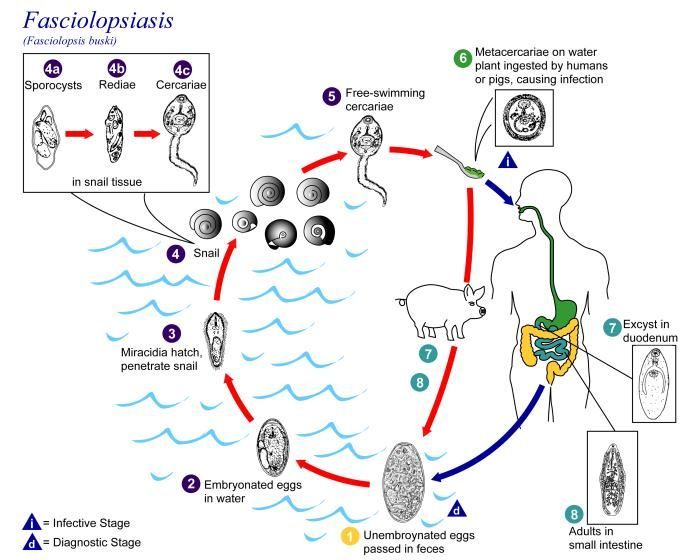
This is an illustration of the life cycle of Fasciolopsis buski, the causal agent of Fasciolopsiasis.Created: 2002
-
This is an illustration of the life cycle of Metagonimus yokogawai, the causal agent of Metagonimiasis.Created: 2002
-
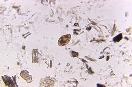
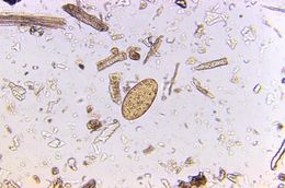
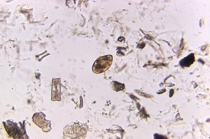
Magnified 500X, this photomicrograph of an unstained, formalin-preserved stool specimen mount, revealed the presence of a Paragonimus westermani termatode egg. See PHIL 3415 for the depiction of the following life cycle.The eggs are excreted unembryonated in the sputum, or alternately they are swallowed and passed with stool (1). In the external environment, the eggs become embryonated (2), and miracidia hatch and seek the first intermediate host, a snail, and penetrate its soft tissues (3). Miracidia go through several developmental stages inside the snail (4): sporocysts (4a), rediae (4b), with the latter giving rise to many cercariae (4c), which emerge from the snail. The cercariae invade the second intermediate host, a crustacean such as a crab or crayfish, where they encyst and become metacercariae. This is the infective stage for the mammalian host (5).Created: 1973
-
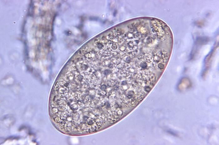
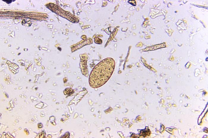
Magnified 500X, this photomicrograph revealed the presence of a Fasciolopsis buski trematode egg that was found in an unstained formalin-preserved stool sample. F. buski are the largest intestinal flukes found parasitizing human beings. These flukes inhabit Asia and the Indian subcontinent, especially in areas where humans raise pigs, and consume freshwater plants.Clinical Features:Most infections are light and asymptomatic. In heavier infections, symptoms include diarrhea, abdominal pain, fever, ascites, anasarca and intestinal obstruction.Laboratory Diagnosis:Microscopic identification of eggs, or more rarely of the adult flukes, in the stool or vomitus is the basis of specific diagnosis. The eggs are indistinguishable from those of Fasciola hepatica.Created: 1973
-
Magnified 500X, this photomicrograph of an unstained, formalin-preserved stool specimen mount, revealed the presence of a Paragonimus westermani termatode egg. See PHIL 3415 for the depiction of the following life cycle.The eggs are excreted unembryonated in the sputum, or alternately they are swallowed and passed with stool (1). In the external environment, the eggs become embryonated (2), and miracidia hatch and seek the first intermediate host, a snail, and penetrate its soft tissues (3). Miracidia go through several developmental stages inside the snail (4): sporocysts (4a), rediae (4b), with the latter giving rise to many cercariae (4c), which emerge from the snail. The cercariae invade the second intermediate host, a crustacean such as a crab or crayfish, where they encyst and become metacercariae. This is the infective stage for the mammalian host (5).Created: 1973
-
Magnified 125X, this photomicrograph revealed the presence of a Fasciolopsis buski trematode egg that was found in an unstained formalin-preserved stool sample. F. buski are the largest intestinal flukes found parasitizing human beings. These flukes inhabit Asia and the Indian subcontinent, especially in areas where humans raise pigs, and consume freshwater plants.Clinical Features:Most infections are light and asymptomatic. In heavier infections, symptoms include diarrhea, abdominal pain, fever, ascites, anasarca and intestinal obstruction.Laboratory Diagnosis:Microscopic identification of eggs, or more rarely of the adult flukes, in the stool or vomitus is the basis of specific diagnosis. The eggs are indistinguishable from those of Fasciola hepatica.Created: 1973
-
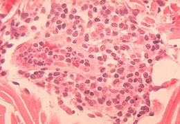
Histopathology of delayed hypersensitivity reaction to Schistosoma mansoni antigen.Created: 1972
-
Magnified 125X, this photomicrograph revealed the presence of two Fasciolopsis buski trematode eggs that were found in an unstained formalin-preserved stool sample. F. buski are the largest intestinal flukes found parasitizing human beings. These flukes inhabit Asia and the Indian subcontinent, especially in areas where humans raise pigs, and consume freshwater plants.Clinical Features:Most infections are light and asymptomatic. In heavier infections, symptoms include diarrhea, abdominal pain, fever, ascites, anasarca and intestinal obstruction.Laboratory Diagnosis:Microscopic identification of eggs, or more rarely of the adult flukes, in the stool or vomitus is the basis of specific diagnosis. The eggs are indistinguishable from those of Fasciola hepatica.Created: 1973
-
Histopathology of delayed hypersensitivity reaction to Schistosoma mansoni antigen.Created: 1972
-
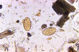
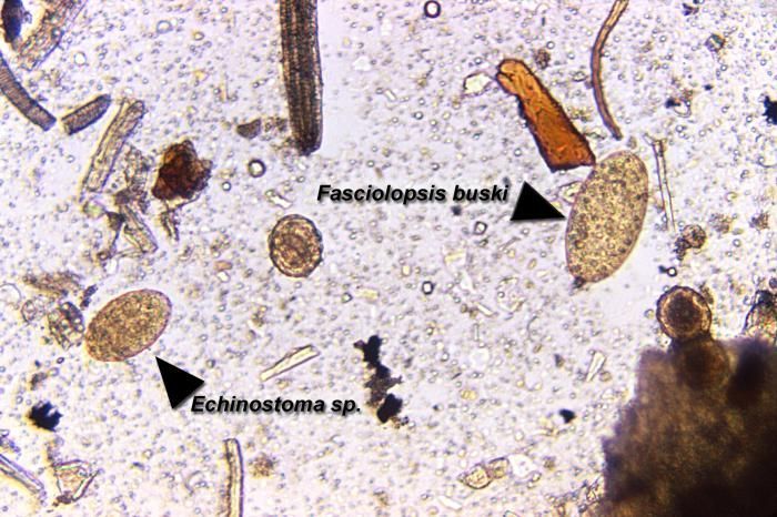
Magnified 125X, this photomicrograph revealed the presence of two trematode eggs, a Fasciolopsis buski egg on the right, and an Echinostoma sp. egg seen of the left, which were found in an unstained formalin-preserved stool sample. Note how much larger the F. buski is compared to that of the Echinostoma sp. egg. F. buski trematodes are the largest intestinal flukes found parasitizing human beings. These flukes inhabit Asia and the Indian subcontinent, especially in areas where humans raise pigs, and consume freshwater plants.Created: 1973