-
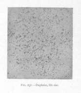
Daphniae....
-
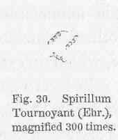
Spirillum Tournoyani (Ehr.), magnified....
-
-
-
Public Domain, U.S. Government Work 2011 Barry H. Rosen Courtesy of life.nbii.gov
NBII images
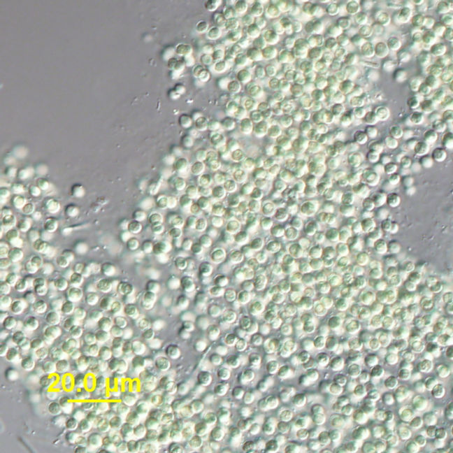
Category hierarchy: Microorganisms | AlgaeDescription: Microcystis spp. This organism cannot "fix" atmospheric nitrogen.Capture device: Olympus DP71Locality: Latitude: 2.859009900000000e+001; Longitude: -8.119031699999999e+001
-
Public Domain, U.S. Government Work 2011 Barry H. Rosen Courtesy of life.nbii.gov
NBII images
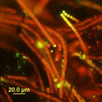
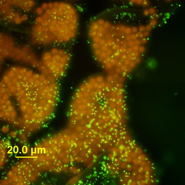
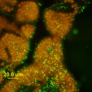
Category hierarchy: Microorganisms | AlgaeDescription: Phormidium in SYTOX® Green nucleic acid stain. Yellow/green indicates that the cells are dead. Red is chlorophyll a fluorescence in live cells.Locality: Latitude: 2.859009900000000e+001; Longitude: -8.119031699999999e+001
-
Public Domain, U.S. Government Work 2011 Barry H. Rosen Courtesy of life.nbii.gov
NBII images
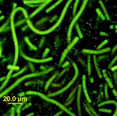
Category hierarchy: Microorganisms | AlgaeDescription: Phormidium in SYTOX® Green nucleic acid stain. The green color indicates that these cells are dead.Locality: Latitude: 2.859009900000000e+001; Longitude: -8.119031699999999e+001
-
Public Domain, U.S. Government Work 2011 Barry H. Rosen Courtesy of life.nbii.gov
NBII images
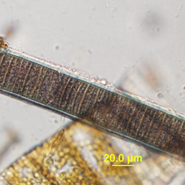
Category hierarchy: Interactions Among Species | Defense: Poisons & MoreDescription: Lyngbya wollei shealth micrograph.. Sample was collected from Silver Springs, Florida.Capture device: Olympus DP71Locality: Latitude: 2.859009900000000e+001; Longitude: -8.119031699999999e+001
-
Public Domain, U.S. Government Work 2011 Barry H. Rosen Courtesy of life.nbii.gov
NBII images
Category hierarchy: Microorganisms | AlgaeDescription: Microcystis wesenbergii light micrograph. Sample was collected from Pinto Lake, California.Capture device: Olympus DP71Locality: Latitude: 2.859009900000000e+001; Longitude: -8.119031699999999e+001
-
Public Domain, U.S. Government Work 2011 Barry H. Rosen Courtesy of life.nbii.gov
NBII images
Category hierarchy: Microorganisms | AlgaeDescription: Microcystis wesenbergii epifluoresecent microscopy. SYTOX® green staining (no evidence of cell death as cells are not green) Epifluorescence using FITC Excitation/Emission cube. Sample was collected from Pinto Lake, California.Capture device: Olympus DP71Locality: Latitude: 2.859009900000000e+001; Longitude: -8.119031699999999e+001
-
Public Domain, U.S. Government Work 2011 Barry H. Rosen Courtesy of life.nbii.gov
NBII images
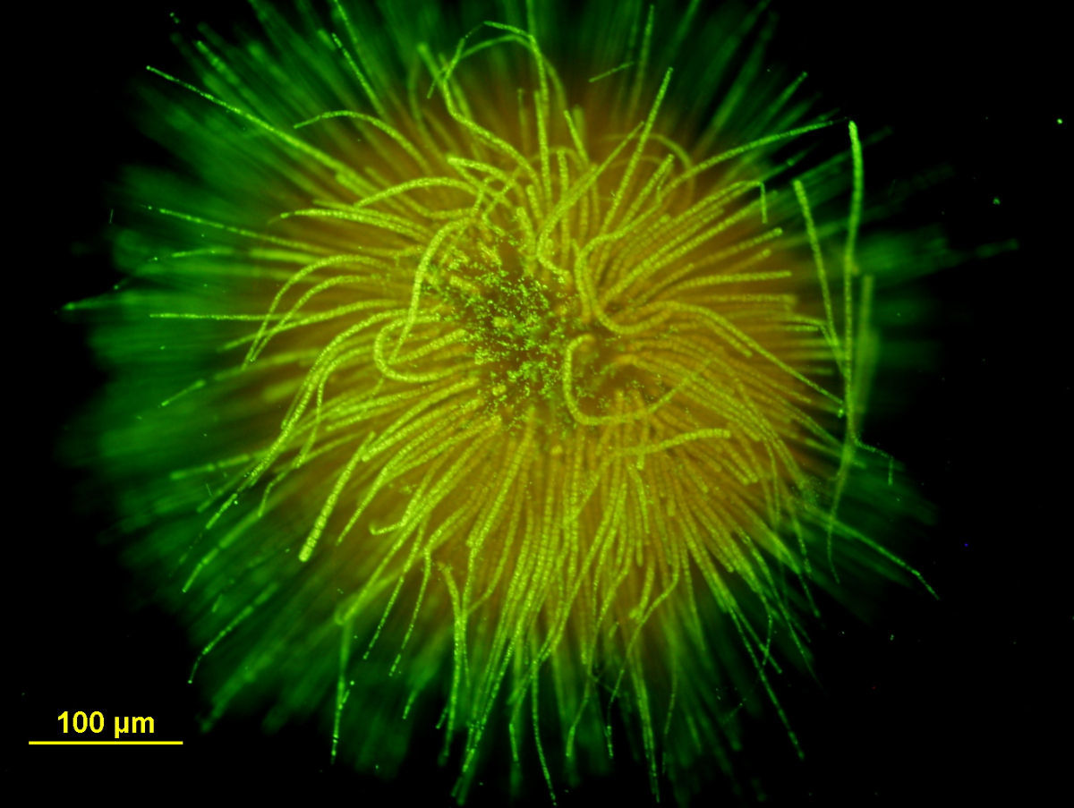
Category hierarchy: Microorganisms | AlgaeDescription: Gloeotrichia in SYTOX® Green nucleic acid stain. This cyanobacteria has radiating filaments and basal heterocysts.Capture device: Olympus DP71Locality: Latitude: 2.859009900000000e+001; Longitude: -8.119031699999999e+001
-
Public Domain, U.S. Government Work 2011 Barry H. Rosen Courtesy of life.nbii.gov
NBII images
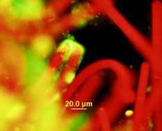
Category hierarchy: Interactions Among Species | Defense: Poisons & MoreDescription: A toxin-producing cyanobacteria with natural chlorophyll fluorescence (red) and SYTOX® Green nucleic acid stain (green). Note the filament tips and extracellular material is staining positive (green) with the nucleic acid stain.Locality: Latitude: 2.859009900000000e+001; Longitude: -8.119031699999999e+001
-
Public Domain, U.S. Government Work 2011 Barry H. Rosen Courtesy of life.nbii.gov
NBII images
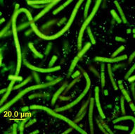
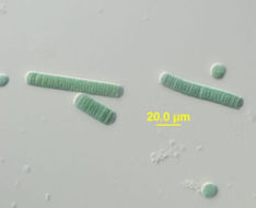
Category hierarchy: Microorganisms | AlgaeDescription: Lyngbya with normal illumination.Locality: Latitude: 2.859009900000000e+001; Longitude: -8.119031699999999e+001
-
Public Domain, U.S. Government Work 2011 Barry H. Rosen Courtesy of life.nbii.gov
NBII images
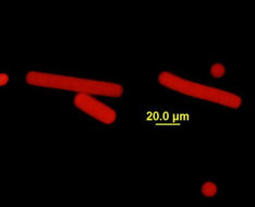
Category hierarchy: Microorganisms | AlgaeDescription: Lyngbya with epifluorescence illumination.Locality: Latitude: 2.859009900000000e+001; Longitude: -8.119031699999999e+001
-
Public Domain, U.S. Government Work 2011 Barry H. Rosen Courtesy of life.nbii.gov
NBII images
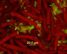
Category hierarchy: Microorganisms | AlgaeDescription: Lyngbya with epifluorescence illumination and SYTOX® Green nucleic acid stain. Note the filament tips and extracellular material is staining positive (green) with the nucleic acid stain.Locality: Latitude: 2.859009900000000e+001; Longitude: -8.119031699999999e+001
-
Public Domain, U.S. Government Work Barry H. Rosen Courtesy of life.nbii.gov
NBII images
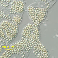
Category hierarchy: Microorganisms | BacteriaDescription: Gomphosphaeria light micrograph. Sample was collected from Cassidy Lake, Washington.Capture device: Olympus DP71Locality: Latitude: 2.859009900000000e+001; Longitude: -8.119031699999999e+001
-
Public Domain, U.S. Government Work Barry H. Rosen Courtesy of life.nbii.gov
NBII images
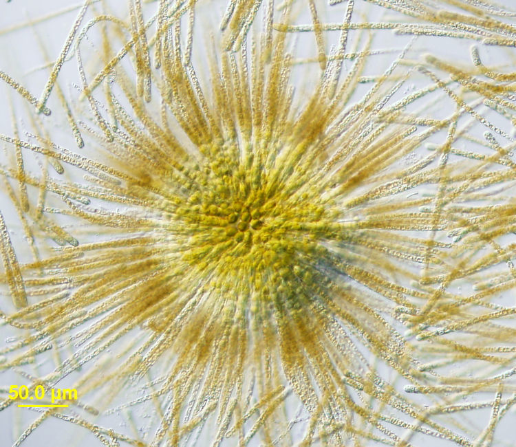
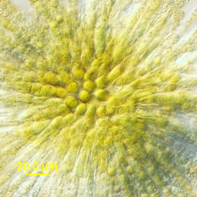
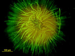
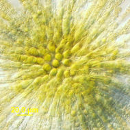
Category hierarchy: Microorganisms | AlgaeDescription: Gloeotrichia whole colony micrograph. Sample was collected from Klamath Lake, Oregon.Capture device: Olympus DP71Locality: Latitude: 2.859009900000000e+001; Longitude: -8.119031699999999e+001
-
Public Domain, U.S. Government Work 2011 Barry H. Rosen Courtesy of life.nbii.gov
NBII images
Category hierarchy: Microorganisms | AlgaeDescription: Gloeotrichia heterocystis micrograph. Sample was collected from Klamath Lake, Oregon.Capture device: Olympus DP71Locality: Latitude: 2.859009900000000e+001; Longitude: -8.119031699999999e+001
-
Public Domain, U.S. Government Work 2011 Barry H. Rosen Courtesy of life.nbii.gov
NBII images
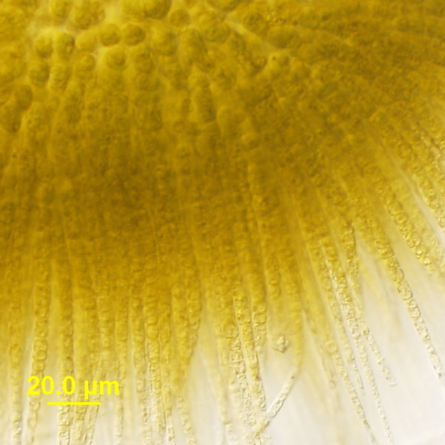
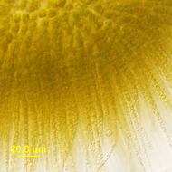
Category hierarchy: Microorganisms | AlgaeDescription: Gloeotrichia filaments, light microscopy. Sample was collected from Klamath Lake, Oregon.Capture device: Olympus DP71Locality: Latitude: 2.859009900000000e+001; Longitude: -8.119031699999999e+001
-
Public Domain, U.S. Government Work 2011 Barry H. Rosen Courtesy of life.nbii.gov
NBII images
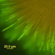
Category hierarchy: Microorganisms | AlgaeDescription: Gloeotrichia filaments epifluoresecent microscopy. SYTOX® green staining (no evidence of cell death as cells are not green) Sample was collected from Klamath Lake, Oregon.Capture device: Olympus DP71Locality: Latitude: 2.859009900000000e+001; Longitude: -8.119031699999999e+001
-
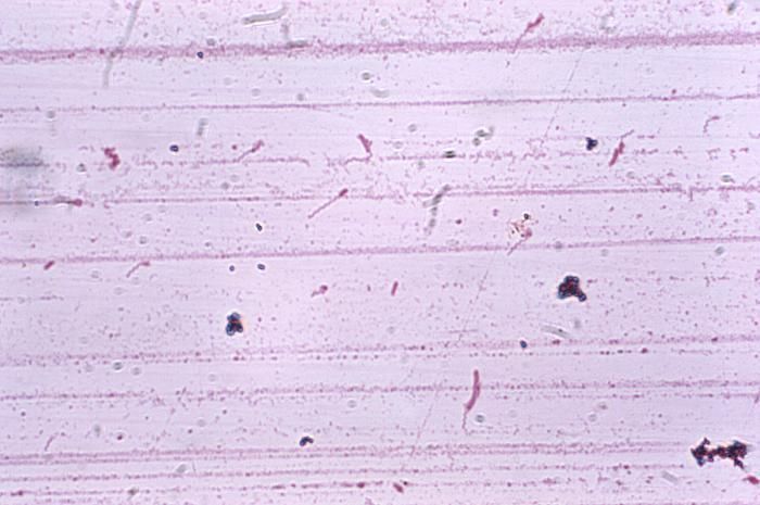
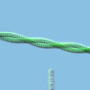
Magnified 1000X, this Liefsons flagella stained photomicrograph revealed the presence of a number of flagellated Brevundimonas diminuta, formerly known as Pseudomonas diminuta.Created: 1975
-
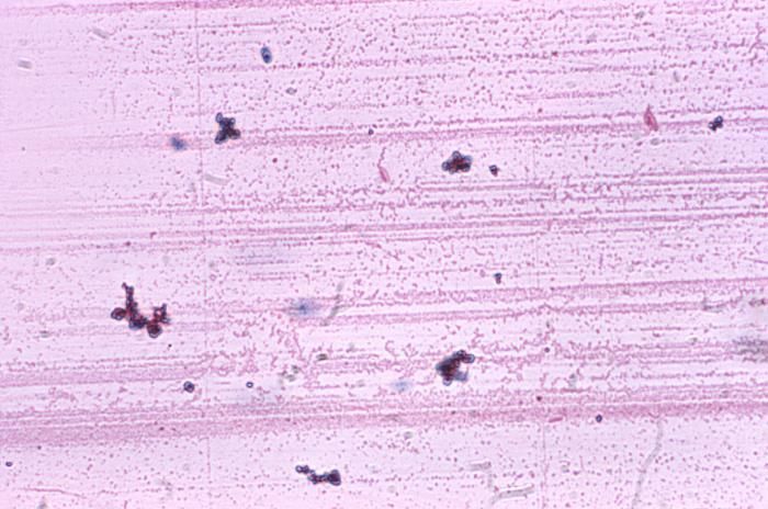
Magnified 1000X, this Liefsons flagella stained photomicrograph revealed the presence of a number of flagellated Brevundimonas diminuta, formerly known as Pseudomonas diminuta.Created: 1975
-
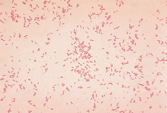
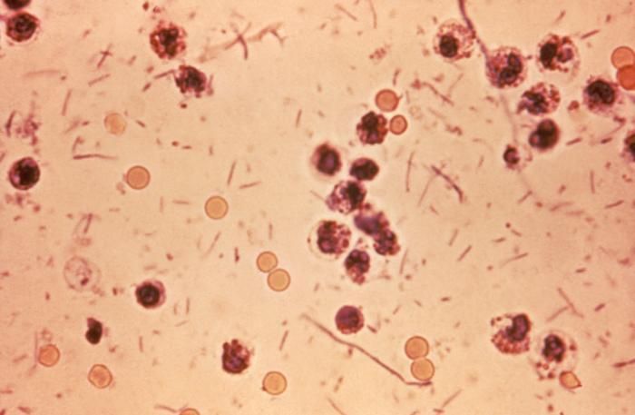
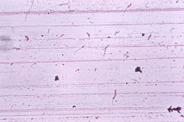
This photomicrograph revealed stool exudates in a patient with shigellosis, which is also known as Shigella dysentery, or Bacterial dysentery.Created: 1980
-
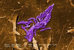
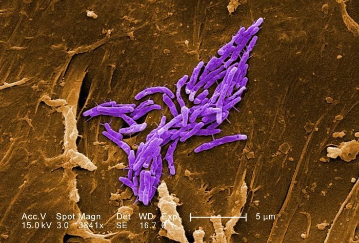
Under a magnification of 3841X, this scanning electron micrograph SEM) revealed some of the ultrastructural morphologic details exhibited by a number of Gram-positive bacilli, or rod-shaped, Mycobacterium fortuitum bacteria. See PHIL 11032 for a black and white version of this image.M. fortuitum is classified as a rapidly-growing Mycobacterium, due to the fact that it can be grown on laboratory culture medium in less than 7 days. As a human pathogen, this organism has been determined to be the cause of skin infections, including furunculosis, i.e., boils, on the legs of people receiving pedicures in nail salons.Created: 2009


