-
-
-
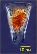
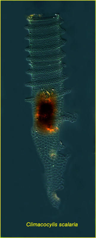
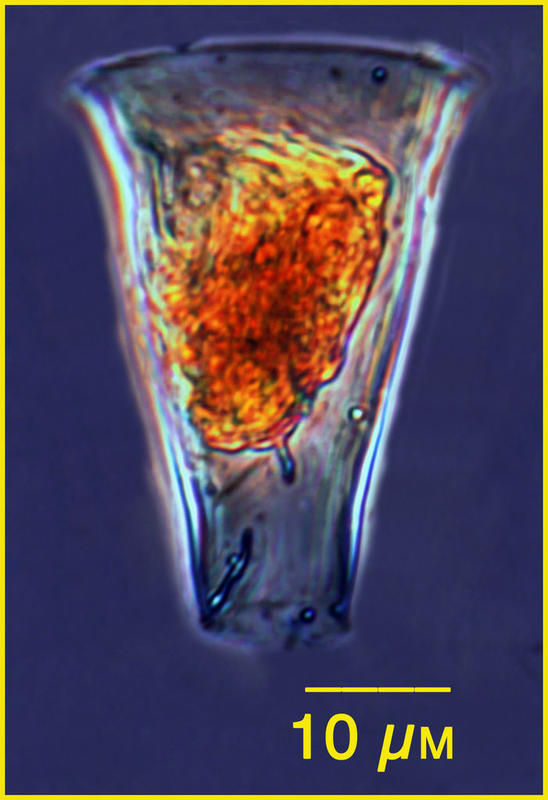
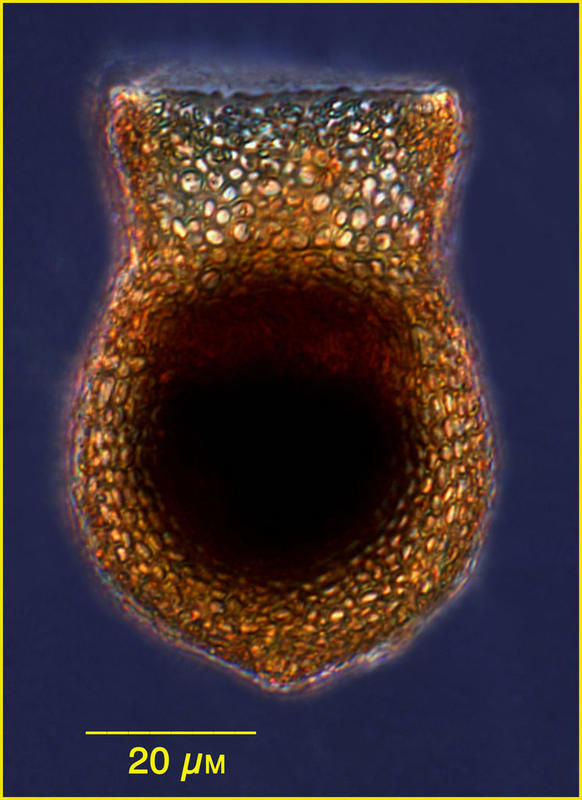
Specimen from the Bay of Vilefranche in Oct 2010, Lugol's-fixed. Images taken with a 20x objective & complied with Helico Focus
-
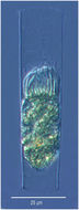
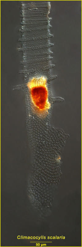
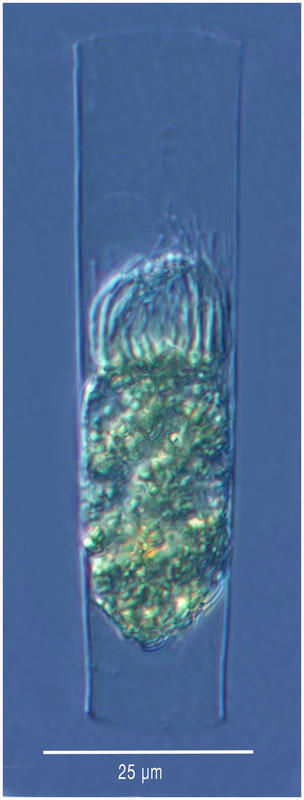
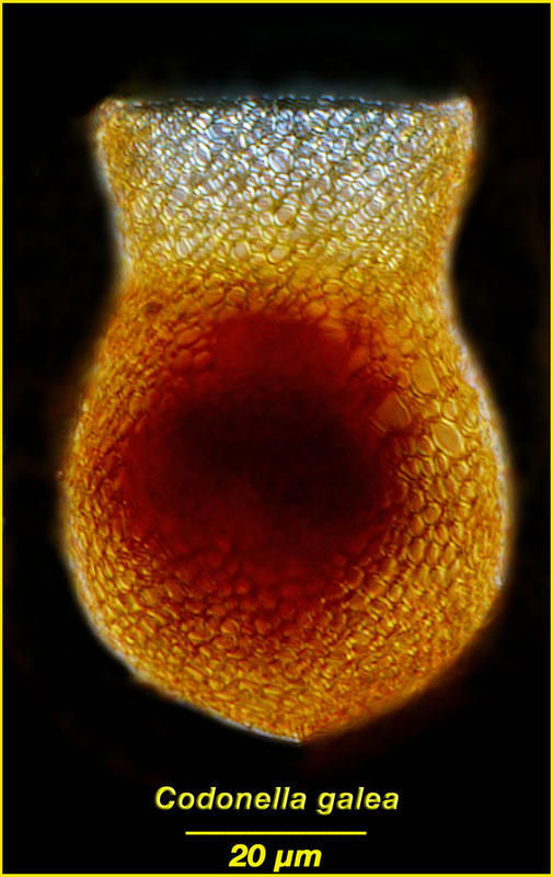
Specimen from the Bay of Vilefranche in Jan 2011, Lugol's-fixed
-
-
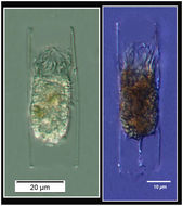
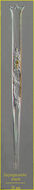
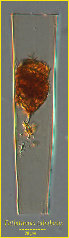
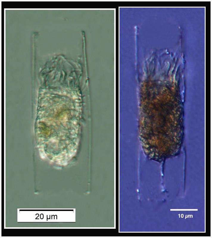
This might be a morph of Eutintinnus tubulosus. Imaged using a 40x objective, sample from the Etang de Thau (Sète, France) in May 2012, lugol's-fixed.
-
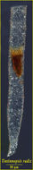
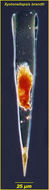
Eutintinnus sp. These very small Eutintinnus specimens are from the Ganges River estuary.
-
-
-
-
-
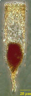
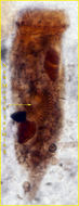
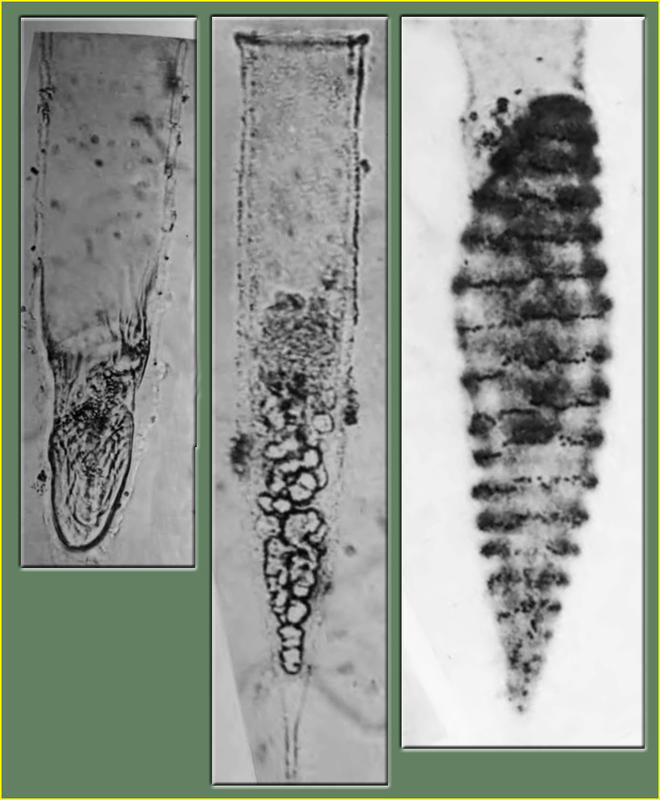
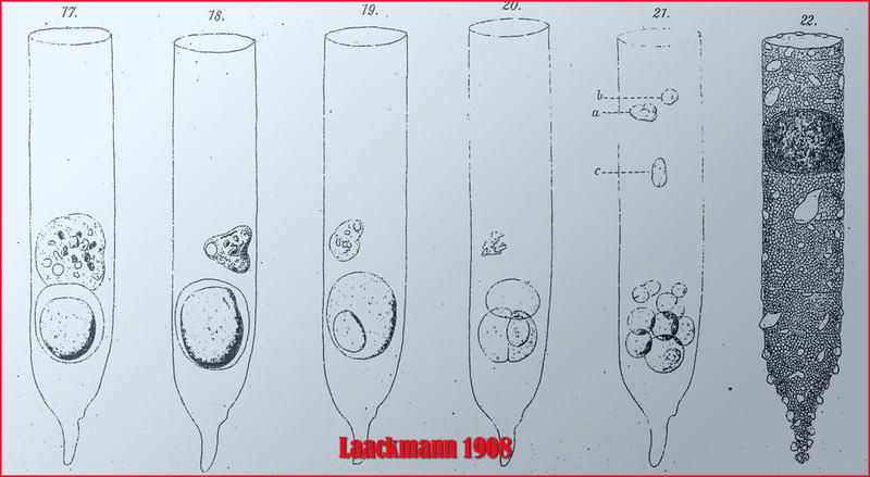
These tintinnid ciliates made unfortunate dietary choices having ingested a parasite. Likely among the first photos published of a parasitic dinoflagellate. Images are from Jean Cachon's 1964 monograph.
-
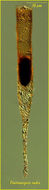
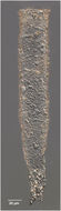
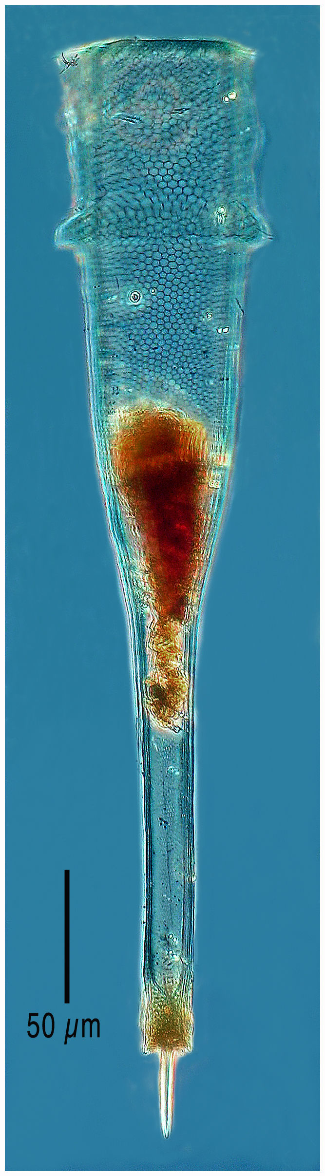
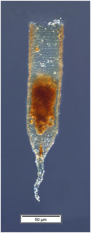
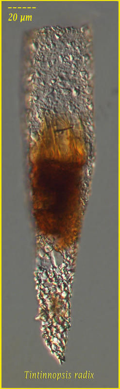
Tintinnopsis radix from Pt B in the Bay of Villefranche. Specimen collected July 8 2013 and fixed with Lugol's; Z-stack of images made using a 20x objective and DIC optics.
-
-
Lugol's-fixed specimen from the Bay of Vilefranche in Sept 2010. Images taken using a 20x objective and compiled with Helico Focus
-
Lugol's-fixed specimen from the bay of Villefranche in Jan 2011.
-
Lugol's-fixed specimen from the Bay of Villefranche in Jan 2011.
-
Lugol's-fixed specimen from Pt B in Villefranche in December 2010.
-
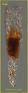
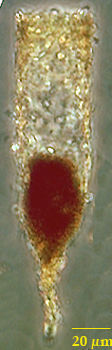
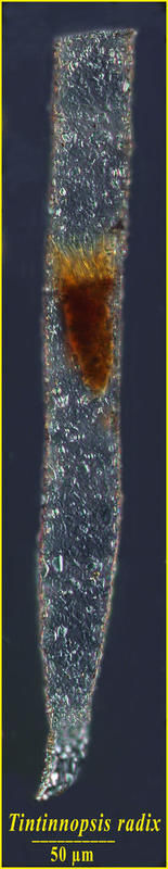
Tintinnopsis radix FRom the Ganges River, estuarine portion (st ganga) in Jan 2013
-
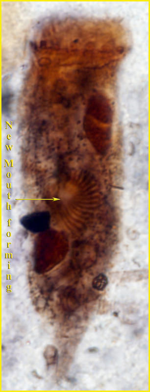
This is an early stage of cell division in which the daughter cell is being formed with the development of a new mouth. The cell is an Protargol preparation of a sample from the Chesapeake Bay in 1987.
-
Parasites were mistaken for cysts, spores, embryos and gametes in early studies. The Laackmann 1908 paper is available on the Aquaparadox site on the page 'Classic Monographs'.
-
Specimen from the East Med
-
Specimen from the Ionian Sea
-
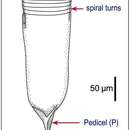
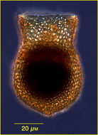
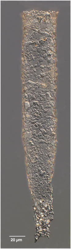
The lorica wall has a mesh appearance.