Description
provided by Zookeys
Body firm, muscular, elongated, gaining regularly in width in caudal direction, dorso-ventral depressed, sides nearly parallel from mid length to point just anterior to caudal sucker, BL 30.5 mm, BW 2.5 mm (Fig. 2). Caudal sucker ventral, oval, its diameter smaller than BW (Figs 2B, 3D). In life, dorsal surface brown, ventral surface grayish white. Color faded in preservative, without any dark lines (Fig. 2)
Somite I completely merged with prostomium (Fig. 3A). Somites II–IV uniannulate (Fig. 3A). Somite V biannulate, (a1+a2) = a3 (Fig. 3A), V a3 forming posterior margin of oral sucker (Fig. 3B). Somites VI and VII triannulate (Fig. 3A–B). Somites VIII–XXIV quadrannulate, a1 = a2 = b5 = b6 (Fig. 3A–B, E). Somites XXV and XXVI triannulate (Fig. 3C–D), XXVI a3 being last complete annulus on venter (Fig. 3D). Somite XXVII uniannulate with one slight furrow on dorsal; anus behind it with no post-anal annulus (Fig. 3C).
Anterior ganglionic mass in VI a2 and a3. Ganglion VII in a2. Ganglion VIII in a2 and b5. Ganglion IX in a2. Ganglia X–XII in a2 and b5 of each somite (Fig. 4A). Ganglion XIII in b5 (Fig. 4A). Ganglia XIV and XV in a2 and b5 of each somite (Fig. 4A). Ganglia XVI–XXI in a2 of each somite (Fig. 4A). Ganglia XXII–XXIV in a1 and a2 of each somite. Ganglia XXV and XXVI in a1 of each somite. Posterior ganglionic mass in XXVII a2 and a3.
Eyes two pairs, first pair dorsally on posterior margin of II, second pair dorsolaterally on middle of V (a1 + a2) (Fig. 3A). Nephridiopores in 17 pairs, ventrally at posterior margin of a1 of each somite of VIII–XXIV (Fig. 3A, E). Papillae numerous, minute, hardly visible, one row on every annulus.
Pharynx agnathous, euthylaematous, reaching to XIII/XIV (Fig. 3F). Crop tubular, acaecate, in XIII/XIV to XIX b5/b6. Gastropore conspicuous, ventral, located slightly posterior to middle of XIII a2 (Fig. 3E, G). Gastroporal duct, winding and bulbous at junction with gastropore, tubular but bulbous at junction with crop, joining with crop in XIV a1 (Fig. 3F). Intestine tubular, acaecate, in XIX b5/b6 to XXIII a2. Rectum, tubular, thin-walled.
Male gonopore at middle of XI b6 (Fig. 3E). Female gonopore located slightly posterior to middle of XIII a2, inconspicuous, located behind gastropore (Fig. 3G). Gonopores separated by 1/2 + 4 + 1/2 annuli (Fig. 3E). Testisacs multiple, one or two testisacs on each side in each annulus, in XIX a2/b5 to XXIV a1 (Fig. 4A). Paired epididymides in XVI a2/b5 to XIX a2/b5 (Fig. 4A). Ejaculatory bulbs absent. Ejaculatory ducts in XI a2/b5 to XVI a2/b5, loosely coiled, each winding from each junction with epididymis, narrowing at junction with atrial cornu, then turning sharply inward toward atrial cornu without pre-atrial loop (Fig. 4A–D). Pair of atrial cornua in XI b5 and b6, muscular, ovate (Fig. 4B). Atrium short, muscular, globular in XI b6 (Fig. 4B–D). Penis sheath and penis absent. Ovisacs one pair, thin-walled, globular, in XIII a2 and b5 (Fig. 4A, E). Oviducts thin-walled, left oviduct crossing ventrally beneath nerve cord, both oviducts converging into common oviduct in XIII a2 (Fig. 4A, E). Common oviduct thin-walled, short, directly ascending to female gonopore (Fig. 4E).
- bibliographic citation
- Nakano T (2012) A new species of Orobdella (Hirudinida, Arhynchobdellida, Gastrostomobdellidae) and redescription of Orobdella kawakatsuorum from Hokkaido, Japan with the phylogenetic position of the new species ZooKeys 169: 9–30
- author
- Takafumi Nakano
Distribution
provided by Zookeys
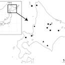
Known in mountainous regions of the central part of Hokkaido, Japan (Fig. 1).
- bibliographic citation
- Nakano T (2012) A new species of Orobdella (Hirudinida, Arhynchobdellida, Gastrostomobdellidae) and redescription of Orobdella kawakatsuorum from Hokkaido, Japan with the phylogenetic position of the new species ZooKeys 169: 9–30
- author
- Takafumi Nakano