-
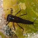
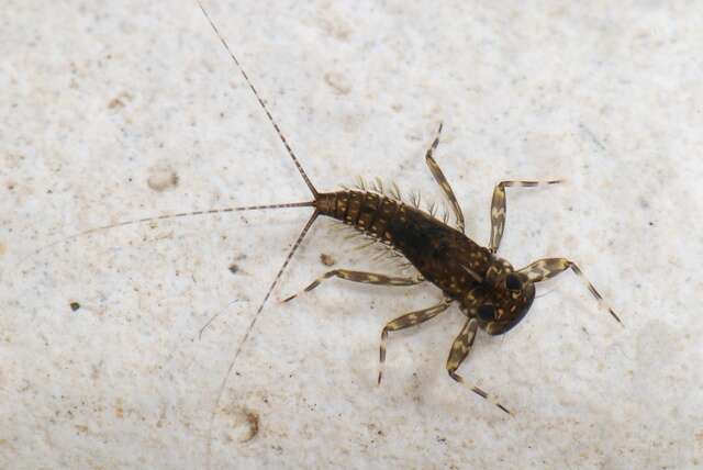
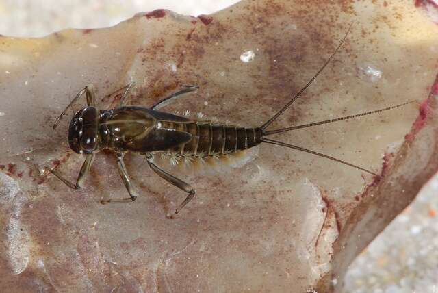
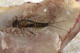
Mattrup Å ved Stidsmølle, Jylland, Danmark
-
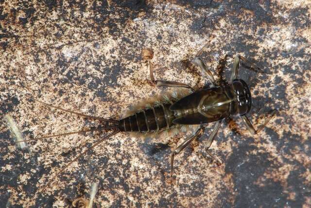
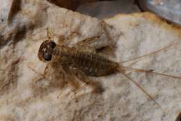
Møgelbæk, tilløb til Højen Bæk, Vejle, Danmark
-
Boonsatien Boonsoong, Dietrich Braasch
Zookeys
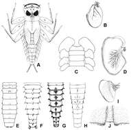
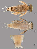
Figure 1.A Habitus of Epeorus aculeatus Braasch, 1990 B lamellae of gills 7 of Epeorus khayengensis Boonsoong & Braasch, 2010 C–E ventral view of abdomen (C), abdominal gills 1 (D) and abdominal terga of (E) Epeorus thailandensis sp. n. F abdominal terga of Epeorus unicornutus Braasch, 2006 G abdominal terga of Epeorus khayengensis Boonsoong & Braasch, 2010 H–J abdominal terga (H), lamellae of gills 1 (I) and tergum VII (J) of Epeorus inthanonensis Braasch & Boonsoong, 2010.
-
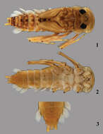
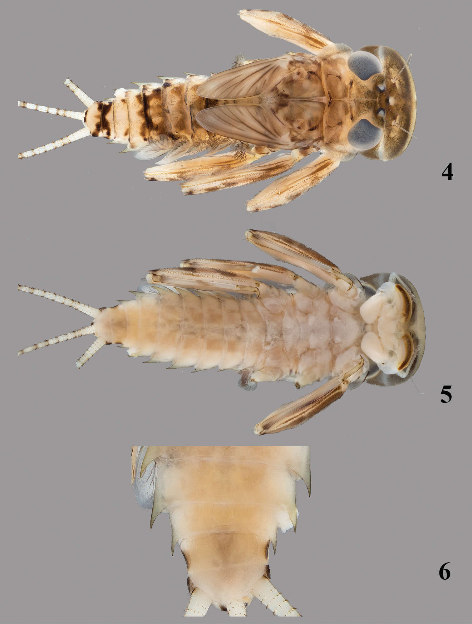
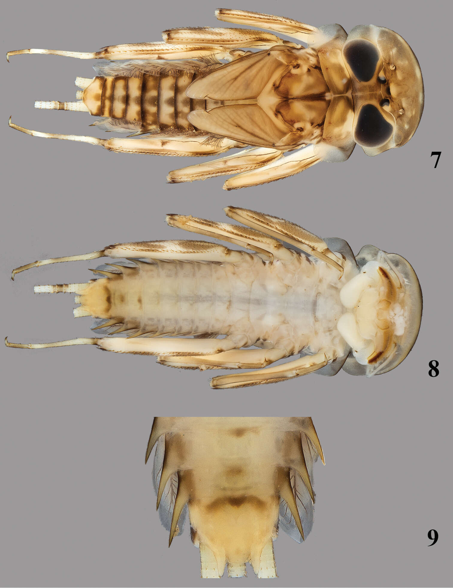
Figures 1–3.Thalerosphyrus determinatus (Walker, 1853). 1 Habitus in dorsal view 2 Habitus in ventral view 3 Detail of abdominal segments VI–IX in ventral view.
-
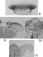
Figures 1–4.Rhithrogeniella ornata Ulmer, 1939. 1 Genitalia of the male imago (holotype) in ventral view 2 Foreleg of a male subimago (paratype) 3 Hindleg of a male subimago (paratype) 4 Penis lobes of a male subimago (paratype): plain line, cuticular structures of the subimago; dotted line, outline of the imago penis lobes.
-
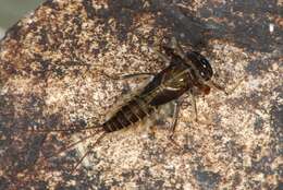
Catacol Isle of Arran. NR915495
-
Mattrup Å ved Stidsmølle, Jylland, Danmark
-
Møgelbæk, tilløb til Højen Bæk, Vejle, Danmark
-
Boonsatien Boonsoong, Dietrich Braasch
Zookeys
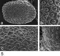
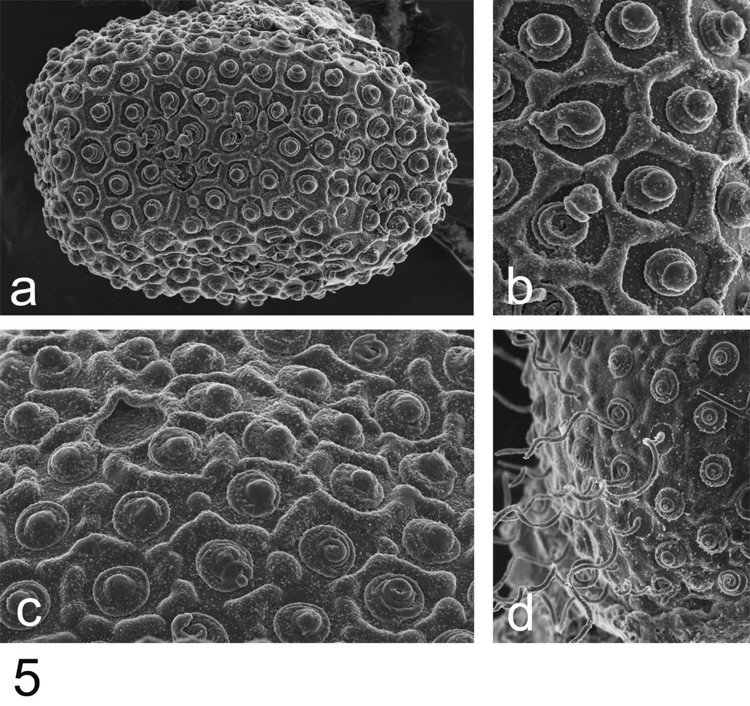
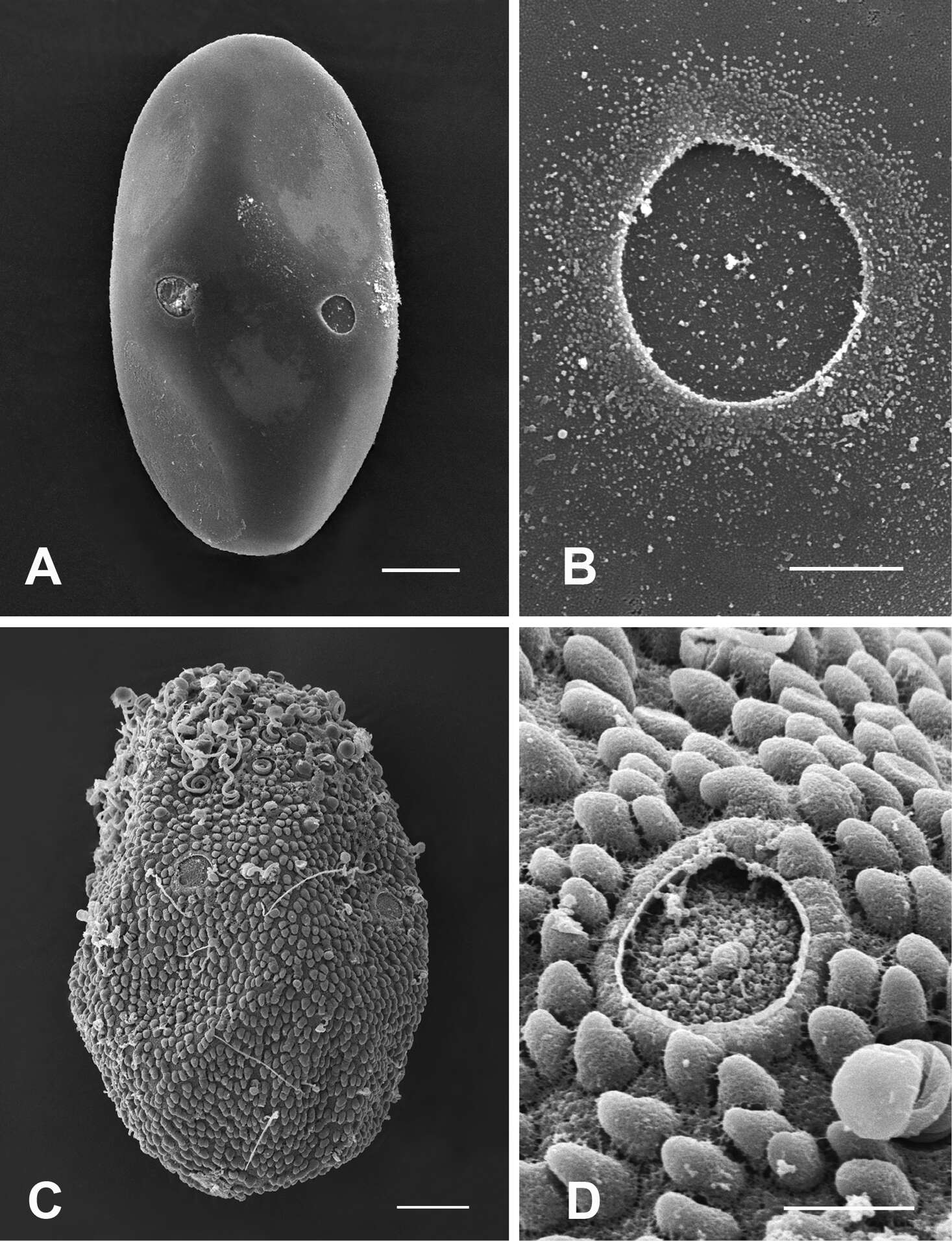
Figure 5.A–B General outline (A) and micropyle (B) of the egg of Epeorus khayengensis Boonsoong & Braasch, 2010 C-D General outline (C) and micropyle (D) of the egg of Rhithrogena siamensis Braasch & Boonsoong, 2009. Scale bars 20 µm for A and C; 5 µm for B and D.
-
Figures 4–6.Thalerosphyrus sinuosus (Navás, 1933). 4 Habitus in dorsal view 5 Habitus in ventral view 6 Detail of abdominal segments VI–IX in ventral view.
-
Figure 5.Rhithrogeniella ornata Ulmer, 1939, SEM pictures of egg structures. 5a Egg extracted from a female subimago paratype from Padang, Sumatra 5b Details of the chorionic structure of a female nymph from Ombilin River, Sumatra 5c Details of the chorionic structure and micropyle of a female subimago paratype from Buitenzorg [Bogor], Java 5d chorionic surface of the female allotype from Buitenzorg [Bogor], Java.
-
Gudenå opstrøms Klostermølle, Mattrup Å ved Stidsmølle
-
Midtjylland
-
Boonsatien Boonsoong, Dietrich Braasch
Zookeys
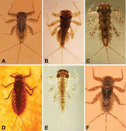
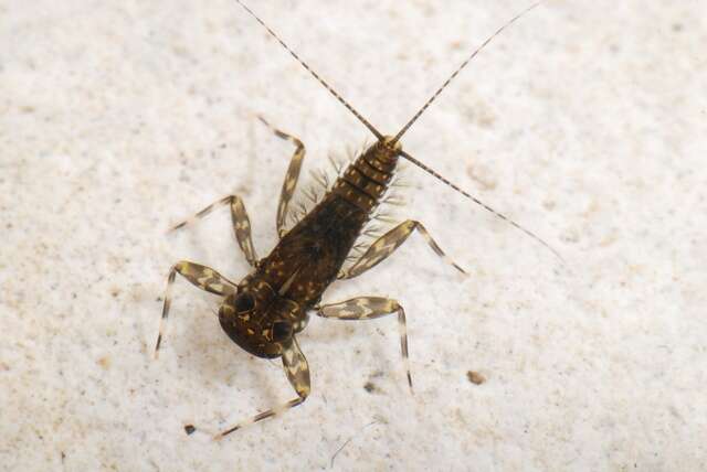
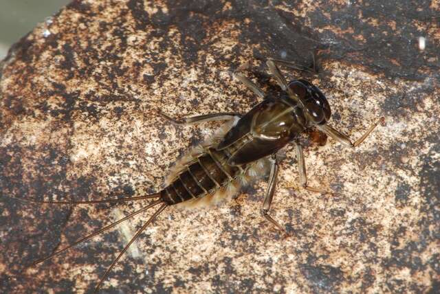
Figure 9.A Habitus of Asionurus namnaoensis Braasch & Boonsoong, 2010 B habitus of Asionurus primus Braasch & Soldán, 1986 Chabitus of Compsoneuria thienemanniUlmer, 1939 Dhabitus of Epeorus khayengensis Boonsoong & Braasch, 2010 E habitus of Rhithrogena tonkinensis Soldán & Braasch, 1986 F habitus of Thalerosphyrus sinuosus Navás, 1933.
-
Figures 7–9.Thalerosphyrus lamuriensis Sartori, 2014. 7 Habitus in dorsal view 8 Habitus in ventral view 9 Detail of abdominal segments VI–IX in ventral view.
-
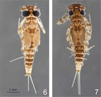
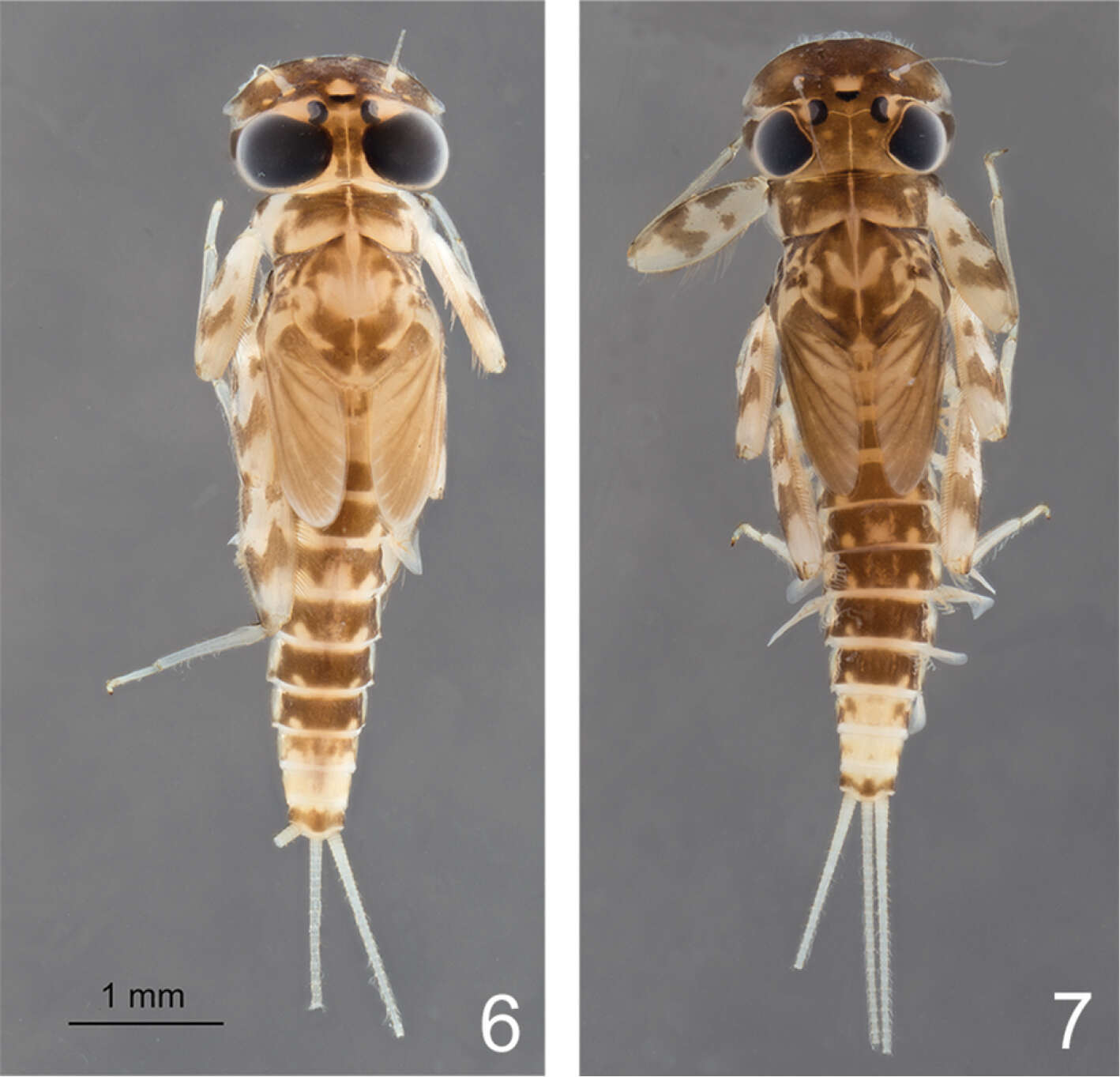
Figures 6–7.Rhithrogeniella ornata Ulmer, 1939. 6 Male nymph 7 Female nymph with slight color variations.
-
Gudenå opstrøms Klostermølle, Mattrup Å ved Stidsmølle
-
Midtjylland
-
Boonsatien Boonsoong, Dietrich Braasch
Zookeys
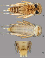
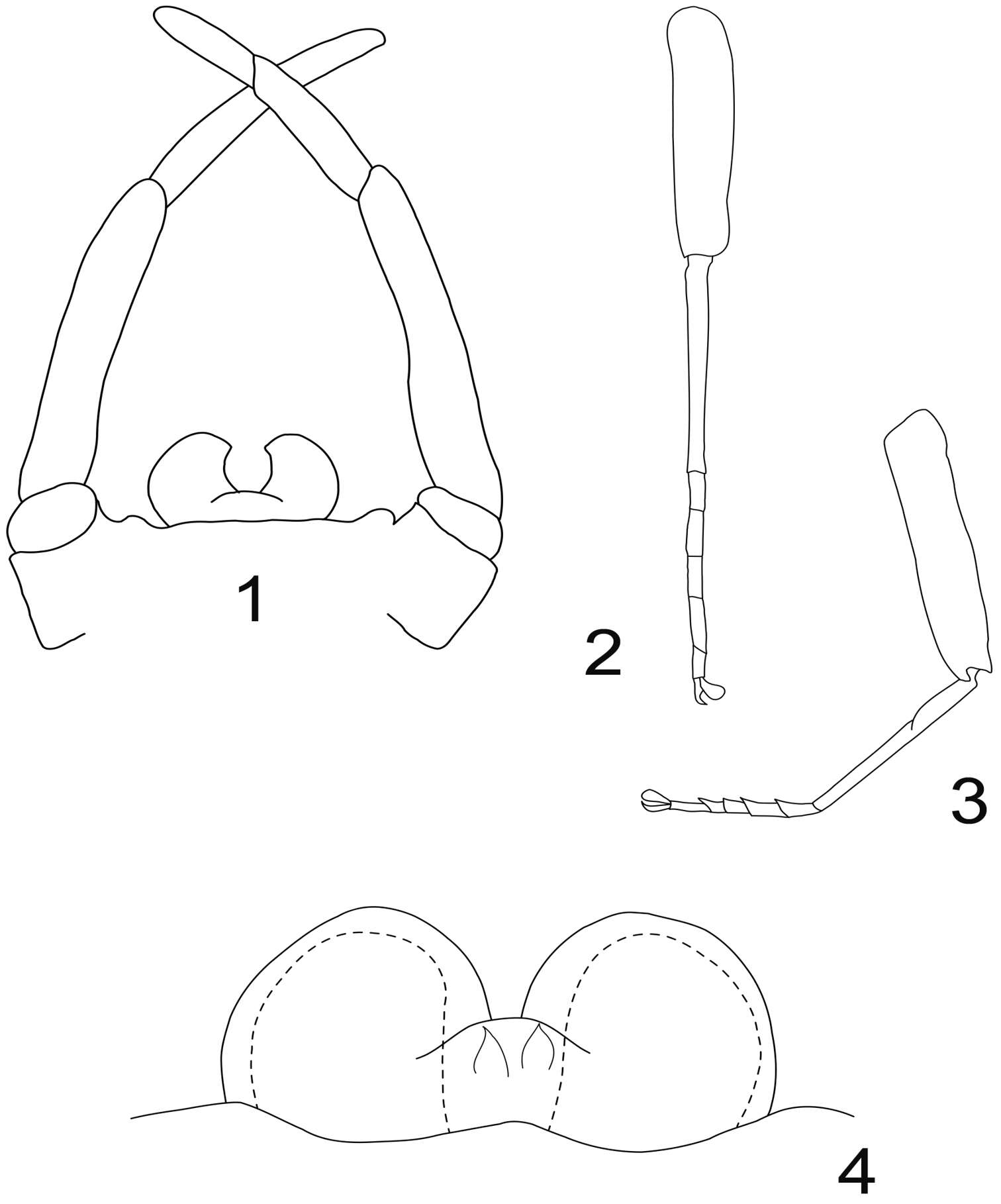
Figure 2.A Ventral view of abdomen of Rhithrogena siamensis Braasch & Boonsoong, 2009 B–E lamellae of gills 1 (B), 3 (C), 5 (D) and 7 (E) of Trichogenia maxillaris Braasch & Soldán, 1988 F ventral view of left maxilla of Trichogenia maxillaris Braasch & Soldán, 1988 G bristles on dorsal face of abdominal terga of Trichogenia maxillaris Braasch & Soldán, 1988 H ventral view of left maxilla of Compsoneuria langensis Braasch & Boonsoong, 2010 I-J abdominal terga (I) and tergum VII (J) of Notacanthurus baei Braasch & Boonsoong, 2009.
-
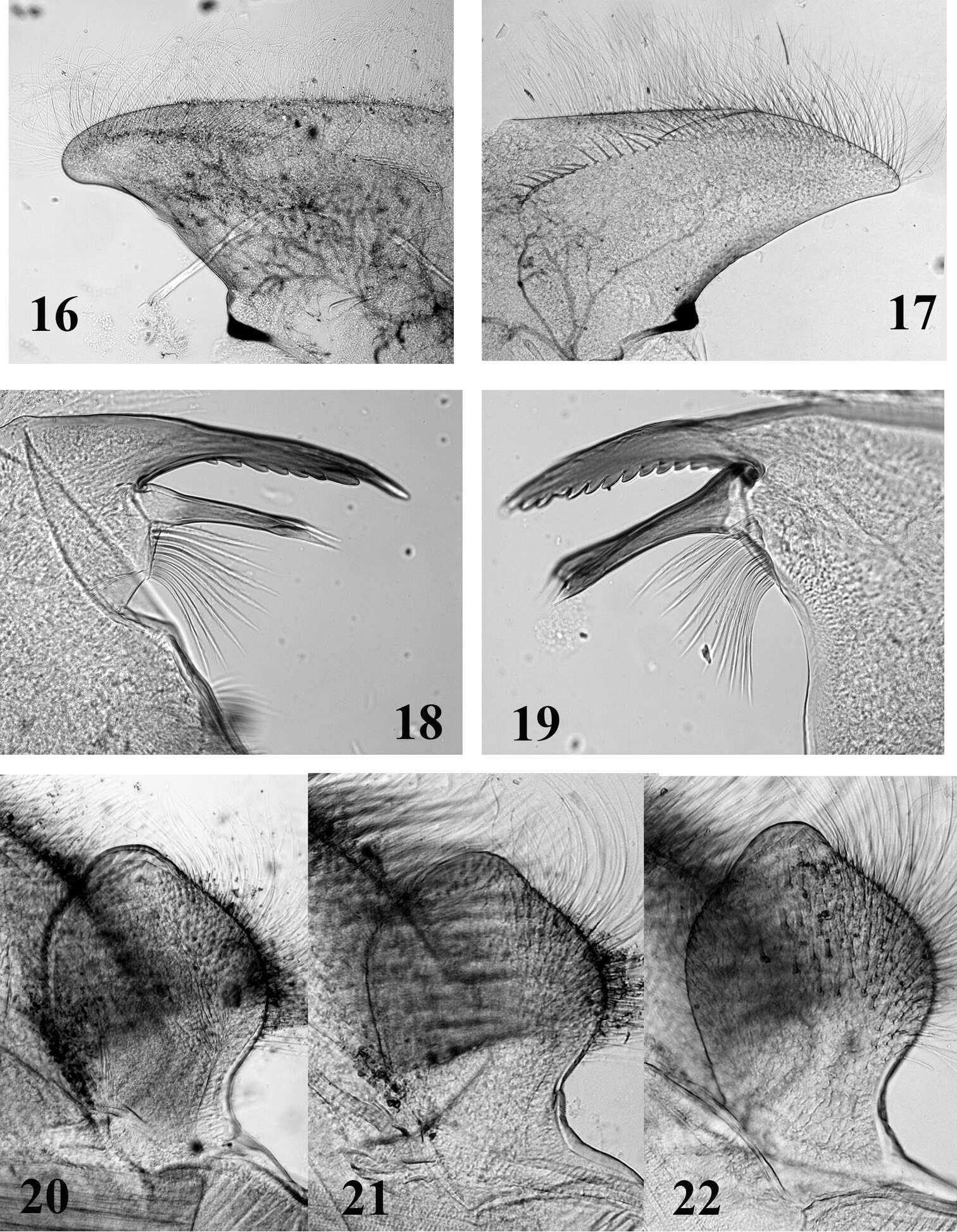
Figures 16–22.Mouthparts structure of Thalerosphyrus determinatus (20), Thalerosphyrus sinuosus (16, 21) and Thalerosphyrus lamuriensis (17, 18, 19, 22). 16–17 Hemi-labrum 18 Left mandible 19 Right mandible 20–22 Labial glossa.
-
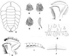
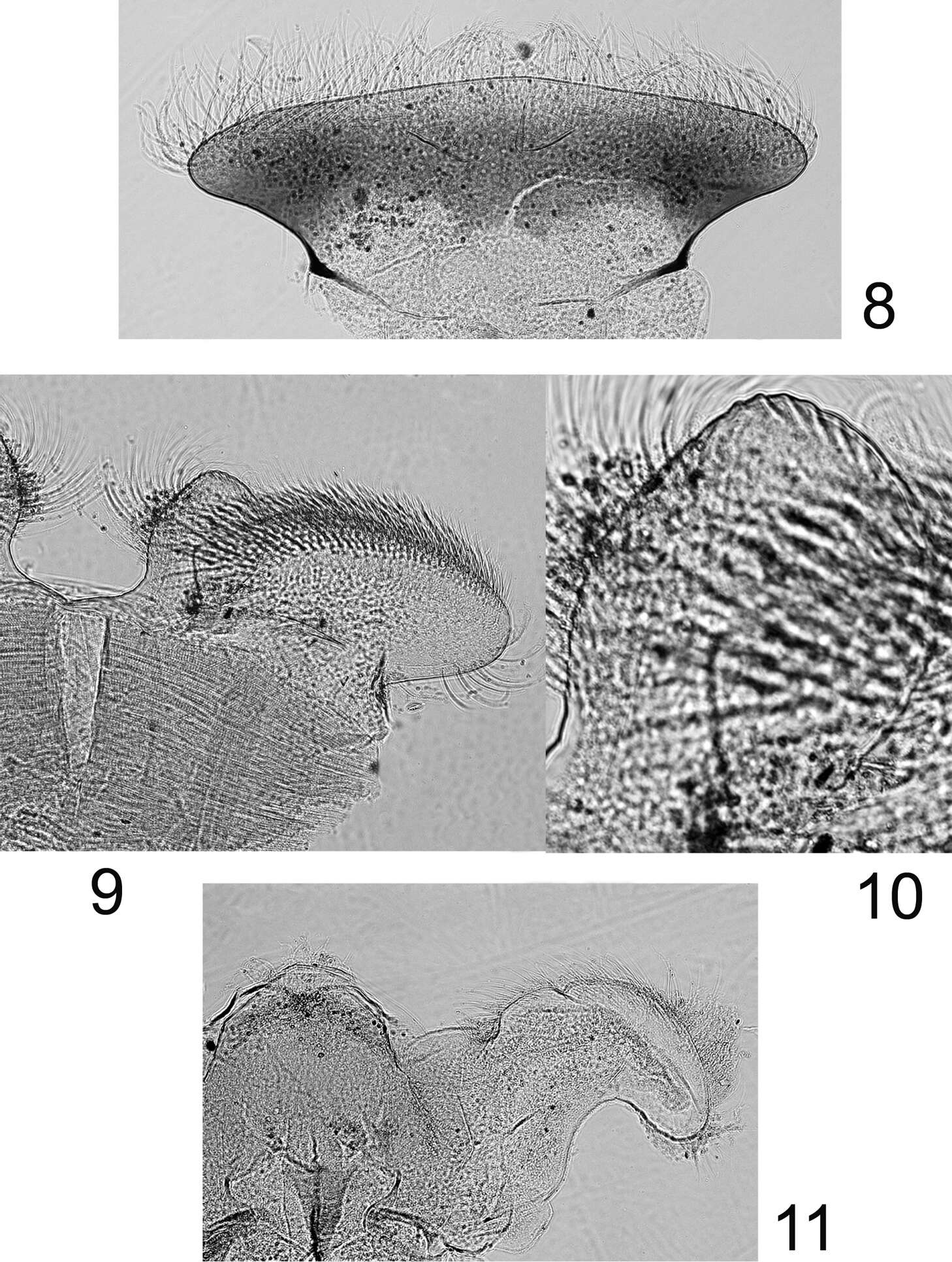
Figures 8–11.Rhithrogeniella ornata Ulmer, 1939, nymphal mouthparts. 8 Labrum in dorsal view 9 Left glossae and paraglossae of the labium 10 Detail of the glossae from 9 11 Hypopharynx, ventral view lingua and left superlingua.
-
Gudenå opstrøms Klostermølle, Mattrup Å ved Stidsmølle
-
Midtjylland
-
Boonsatien Boonsoong, Dietrich Braasch
Zookeys
Figure 5.A–B General outline (A) and micropyle (B) of the egg of Epeorus khayengensis Boonsoong & Braasch, 2010 C-D General outline (C) and micropyle (D) of the egg of Rhithrogena siamensis Braasch & Boonsoong, 2009. Scale bars 20 µm for A and C; 5 µm for B and D.