Malassezia furfur resembles most other unicellular yeasts until it begins to produce conidia; phialides are formed with distinct collarettes that gives them their bowling pin appearance. M. furfur is a lipophilic yeast that requires oil and fatty acids for growth in culture. This characteristic is a good diagnostic tool as many other types of yeast do not have lipid dependence.
Malassezia furfur is found worldwide but is more prevalent in tropical or subtropical climate regions. The disease pityriasis versicolor may be acquired by visiting one of these areas.
Malassezia furfur is a lipophilic yeast, surviving on fats that are exuded from human skin. Oddly enough it is closely related to the smuts, such as corn smut.
a.k.a. Pityrosporum ovale, P. orbiculare, Malassezia ovalis

Picture from Tom Volk’s Medical Mycology (UW – La Crosse) Spring 2009 lecture notes.
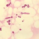
Microscopic Characteristics – Reproductive cell size is about 5 micrometers, with cells shaped like medicine capsules or sometimes bowling pins. Each produces a single phialoconidium followed by successive budding at a single location. Hyphae, very rarely produced in culture, are approximately 2 – 3 micrometers wide. In direct examination of a smear of infected tissue, hyphae are septate and hyaline usually not branched. Conidia resemble budding yeast cells and are approximately 3 micrometers in diameter. Together the hyphae and conidia are said to look like ‘spaghetti and meatballs.’
Macroscopic Characteristics – Slow growing colonies, appearing at temperatures of 35-37ºC. Colonies begin shiny and white to cream later becoming dull and beige, resembling bacteria-like colonies. Growth takes around one to two weeks on modified SDA which must be supplemented with fatty acids (usually done by covering medium with a thin layer of olive oil).
Malassezia furfur is part of the normal flora of human skin. It has been documented to be found on the skin of up to 90% of adults. It is mostly isolated from the upper chest, back, arms and neck. It can also be found on the skin of domestic animals and birds.
Malassezia furfur must be differentiated from Candida species and also other varied species of Malassezia. Helpful diagnostics are the requirement for oil and fatty acids for growth, the “spaghetti and meatballs” hyphal appearance of direct tissue smears, and the phialides and collarettes that appear upon budding.
Malassezia furfur is the etiological agent of pityriasis versicolor a.k.a. tinea versicolor. This condition is a superficial infection of the skin characterized by hypo- or hyperpigmentation of the patient’s skin. It is most commonly seen on the upper chest, back, arms, and neck. Conditions that reduce the shedding of the epidermal cells of the skin can contribute to this infection as well as poor nutrition, pregnancy, and excessive sweating. M. furfur has also been isolated as the cause of folliculitis, catheter-acquired sepsis, and even dandruff. However, M. globosa is the most commonly associated Malassezia species with dandruff as Procter and Gamble (makers of Head & Shoulders dandruff shampoo) have even sequenced the genome to better understand its high lipase activity and its role in dandruff causation. M. globosa and M. sympodalis have also been shown to cause pityriasis versicolor in humans.
However, it should be noted that as M. furfur is a common organism in the normal flora of human skin, simply isolating this species does not necessarily mean that it caused the disease the patient has incurred. Appropriate actions must be taken to insure that M. furfur was indeed the causative agent.

Malassezia furfur (formerly known as Pityrosporum ovale) is a species of yeast (a type of fungus) that is naturally found on the skin surfaces of humans and some other mammals. It is associated with a variety of dermatological conditions caused by fungal infections, notably seborrhoeic dermatitis and tinea versicolor. As an opportunistic pathogen, it has further been associated with dandruff, malassezia folliculitis, pityriasis versicolor (alba), and malassezia intertrigo,[1] as well as catheter-related fungemia and pneumonia in patients receiving hematopoietic transplants. The fungus can also affect other animals, including dogs.
Malassezia furfur is a fungus that lives on the superficial layers of the dermis. It generally exists as a commensal organism forming a natural part of the human skin microbiota, but it can gain pathogenic capabilities when morphing from a yeast to a hyphal form during its life cycle, through unknown molecular changes.[2] This can lead to its uncontrolled proliferation and a subsequent imbalance of the residential skin flora. Some virulence factors or properties which may increase the fungus' ability to acquire an infectious nature include the formation of biofilms, increased adherence to surfaces, and hydrophobicity and also can form hyphae (long, cylindrical filaments)[3]
Infections with pathogenic M. furfur occur on the trunk or the limbs and present clinically as pigmented macules that can merge in the form of scaling plaques. Many of these lesions resolve spontaneously in most patients.[2] The pathogen most frequently affects children compared to people of other age groups.[4] It has been associated with numerous dermatological conditions, including seborrhoeic dermatitis, dandruff, pityriasis versicolor, and tinea circinata, all of which affect the skin.[5] Some other diseases can also arise due to an infection with the fungus, such as catheter-related fungemia and pneumonia in patients receiving hematopoietic cell transplants.[6]
Malassezia furfur is a unicellular organism which varies in size between 1.5–4.5 × 2.0–6.5 micrometers. The cells have a bottle-like shape due to a small protrusion visible at the end of each cell. Cells are difficult to grow in a lab since they require specific conditions.[7]
Topical application of antifungal medications such as ketoconazole, ciclopirox olamine, piroctone-olamine, zinc pyrithione, or sulfur compounds are commonly prescribed to treat diseases caused by Malassezia furfur.[5]
{{cite journal}}: CS1 maint: url-status (link) Malassezia furfur (formerly known as Pityrosporum ovale) is a species of yeast (a type of fungus) that is naturally found on the skin surfaces of humans and some other mammals. It is associated with a variety of dermatological conditions caused by fungal infections, notably seborrhoeic dermatitis and tinea versicolor. As an opportunistic pathogen, it has further been associated with dandruff, malassezia folliculitis, pityriasis versicolor (alba), and malassezia intertrigo, as well as catheter-related fungemia and pneumonia in patients receiving hematopoietic transplants. The fungus can also affect other animals, including dogs.