Description
provided by Zookeys
Body length (from apex of vertex to tip of forewings): ♂ 6.8 mm (N=1).
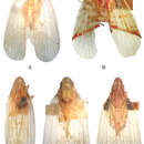
Colour. General colour tawny yellow, forewings (Figs 1D, 4D) with two fuscous elongate marks near bases of sutural margins, nodal line suffused with pale brown marks, many fuscous spots marked from nodal line to apex, tips of spines on hind tibiae and tarsi black.
Head and thorax. Head (Figs 1D, 4A) projecting before eyes approximately 3/5 median length of eye, not strongly dorsoventrally depressed. Vertex (Figs 1D, 4A, 4B) distinctly shorter in middle than the widest breadth (0.6: 1), distinctly longer than pronotum at midline (1.7: 1), anterior margin convex, broadly callused, uniting with base of frons to form smooth surface, lateral margins ridged and converged anteriorly, median carina long and percurrent, thickened and broad, posterior margin straight. Frons (Fig. 4C) slightly longer medially than greatest width (1.3: 1), disc flat and smooth, covered with sparsely microsetae (Fig. 4B), lateral margins diverging to below level of eyes, distinctly callused; median carina with basal part strongly broad and thickened, not reaching to frontoclypeal suture, obsolete on level of antennae. Clypeus (Fig. 4C) triangular, with distinctly broad median carina. Pronotum (Figs 1D, 4A) distinctly shorter than mesonotum in midline (0.2: 1), carinae broadly ridged, lateral carinae diverging posteriorly, median carina distinctly thickened and broad, reaching posterior margin. Pronotum and mesonotum together medially 3.0 times as long as median length of vertex. Hind tibiae each with 2 distinct lateral spines; spinal formula of hind leg 5–5–2. Forewings (Figs 1D, 4D) relatively elongate and narrow, 3.0 times as long as maximum breadth, with corium smooth, not granulate, Sc+R forking at apical 2/5, Cu1 forking after level of junction of claval veins, with 11 apical cells and 6 subapical cells, claval veins uniting at about middle of clavus.
Male genitalia. Pygofer (Figs 4E–G) irregular subquadrate in lateral view, anterior margin concave on dorsal 1/3, posterior margin produced caudad in lateral view. Anal tube (Figs 4E, 4F) relatively elongate, ventral margin slightly bent ventrad in lateral view; lateral margins convex medially then narrowing distad, apical margin slightly concave in dorsal view; anal styles relatively long and narrow, surpassing apex of anal tube in dorsal view. Gonostylus (Figs 4E, 4G) elongate, but not surpassing to apex of gonostylus, apical half narrow and basal half broad in lateral view; median conical process very small, sclerotized in ventral view. Periandrium (Fig. 4E) distinctly elongate and slender, tube-like, distinctly sclerotized, with a short process directed caudad at dorsal apex, surrounding aedeagus subapically. Aedeagus (Figs 4E, 4F) with shaft thin and tubular, arched and its apex directed ventrad in lateral view, endosoma membranous, moderately expanded, with two, anteroventrally directed, spinous processes on right side in lateral view.
- license
- cc-by-3.0
- copyright
- Rong-rong Wang, Ai-Ping Liang
- bibliographic citation
- Wang R, Liang A (2011) Taxonomic review of the genus Tambinia Stål (Hemiptera, Fulgoromorpha, Tropiduchidae) with descriptions of four new species from the Pacific region ZooKeys 132: 13–31
- author
- Rong-rong Wang
- author
- Ai-Ping Liang