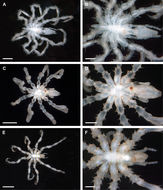
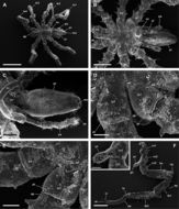
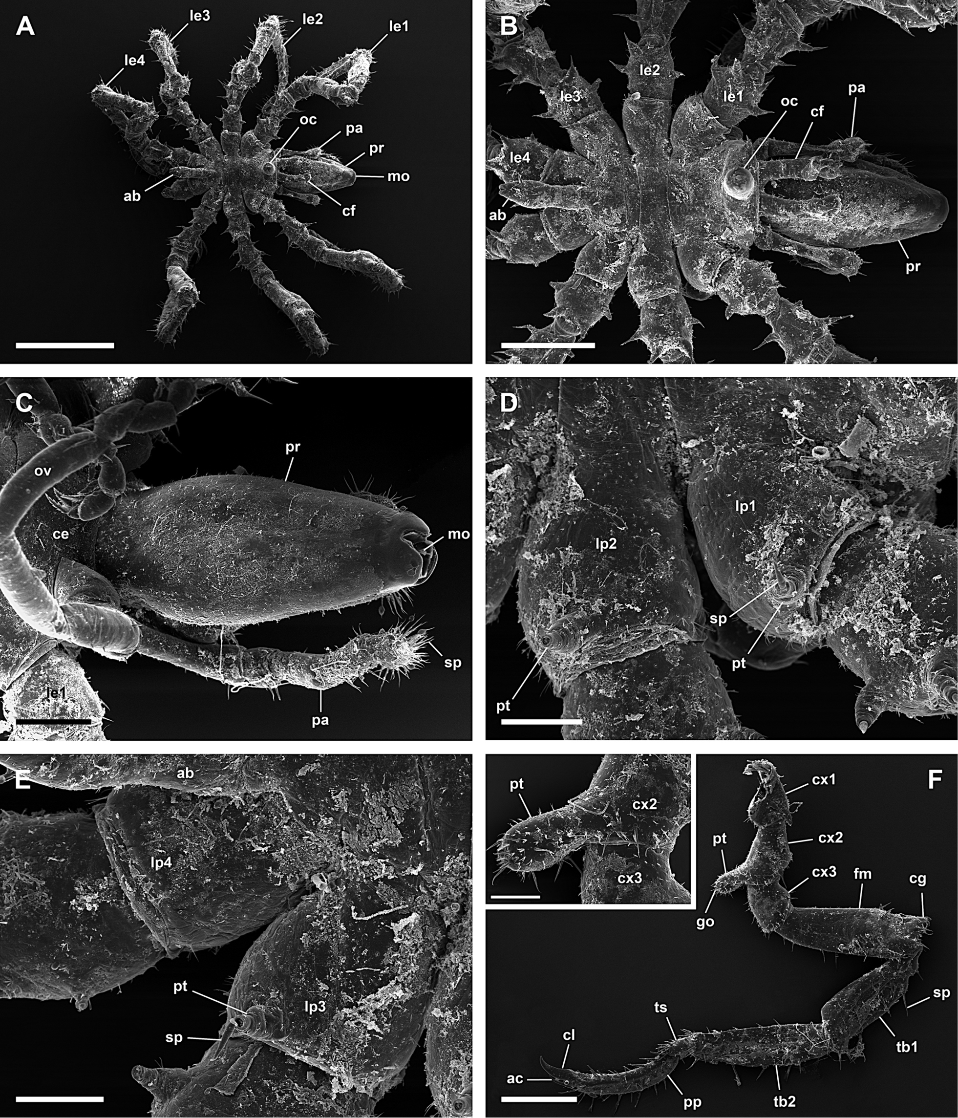
Figure 9.Achelia langi, male; A: Dorsal view; scale 1 mm; B: Dorsal view of trunk; scale 400 µm; C: Ventral view of proboscis; scale 200 µm; D: Dorsal view of right lateral processes 1 and 2, 2 protuberances with spine on lateral process 1 and 1 on lateral process 2; scale 100 µm; E: Dorsal view of right lateral processes 3 and 4, 1 protuberance with spine on lateral process 3 and lateral process 4 without protuberance or spine; scale 100 µm; F: Left 3rd leg; scale 400 µm; insert: Genital protuberance; scale 100 µm.
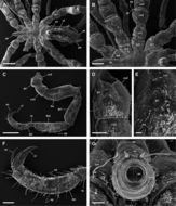
Figure 10.Achelia langi, female; A: Dorsal view of trunk; scale 400 µm; B: Dorsal view of left lateral processes, 1 protuberance with spine on lateral process 1-3, lateral process 4 without protuberance or spine; scale 200 µm; C: Right 3rd leg; scale 400 µm; D: Ventral view of coxa 2 with genital opening, distal is down (right 3rd leg); scale 100 µm; E: Genital opening; scale 20 µm; F: Tarsus, propodus, and claw, auxiliary claws about 2/3 of length of claw; scale 100 µm; G: Abdomen with anus; scale 40 µm.