-
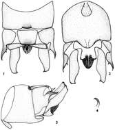
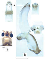
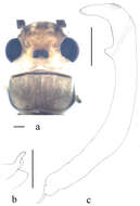
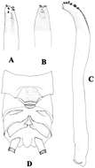
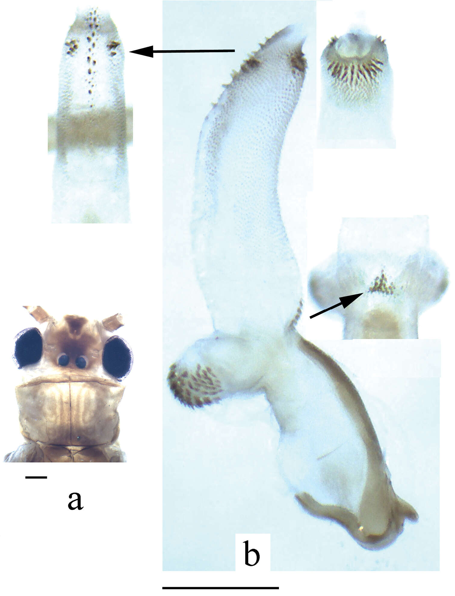
Figures 1–4.Rhopalopsole exiguspina male structures 1 Male terminal, dorsal aspect 2 Male terminal, ventral aspect 3 Male terminal, lateral aspect 4 Epiproct, lateral aspect.
-
Li Wei-Hai, Wang Guo-Quan, Qin Xue-Feng
Zookeys
Figure 1.Neoperla furcostyla Li and Qin,sp. n. (male). A Head and pronotum, dorsal view B Terminalia, dorsal view C Aedeagus, lateral view.
-
Hong–Liang Wang, Guo–Quan Wang, Wei–Hai Li
Zookeys
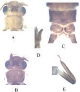
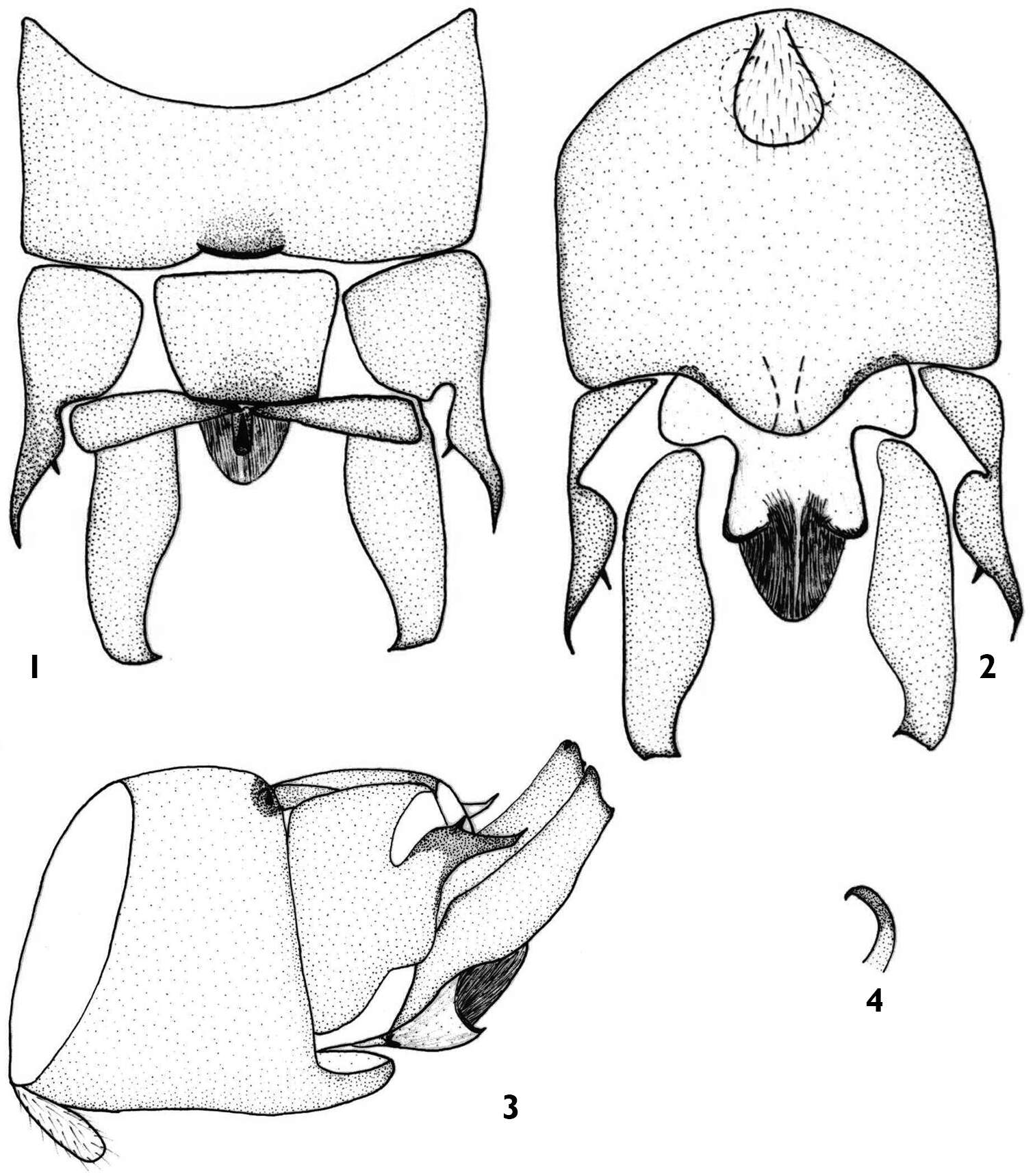
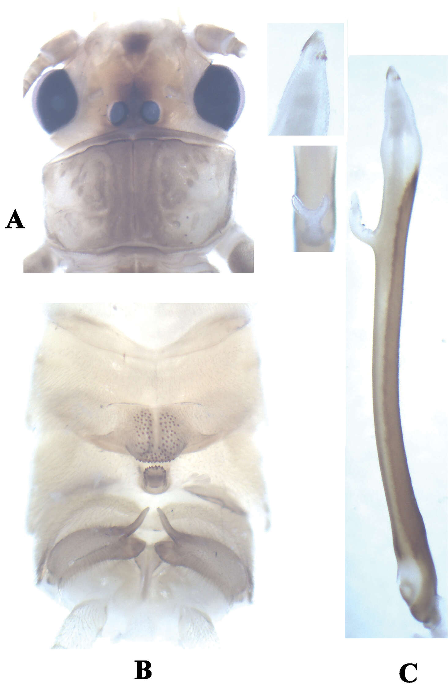
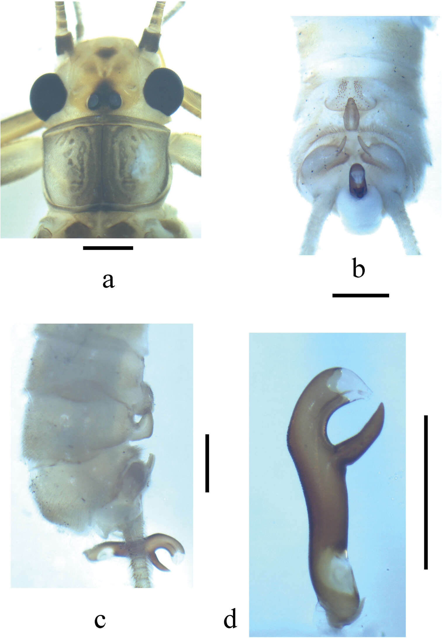
Figure 1.Kamimuria guangxia Li & Wang, sp. n. (male). A Head and pronotum, dorsal view (teneral specimen) B Head and pronotum, dorsal view (older specimen) C Terminalia, dorsal view D Hemitergal process, lateral view E Foreleg, lateral view.
-
Xue-Feng Qin, Dávid Murányi, Guo-Quan Wang, Wei-Hai Li
Zookeys
Figure 8.Neoperla xuansongae. Male a Head and pronotum, dorsal view b Aedeagus, lateral view with details in dorsal and ventral views. Scale bars: 0.5 mm.
-
Scott A. Grubbs, Boris C. Kondratieff, Bill P. Stark, R. Edward DeWalt
Zookeys
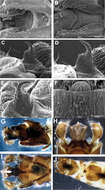
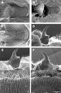
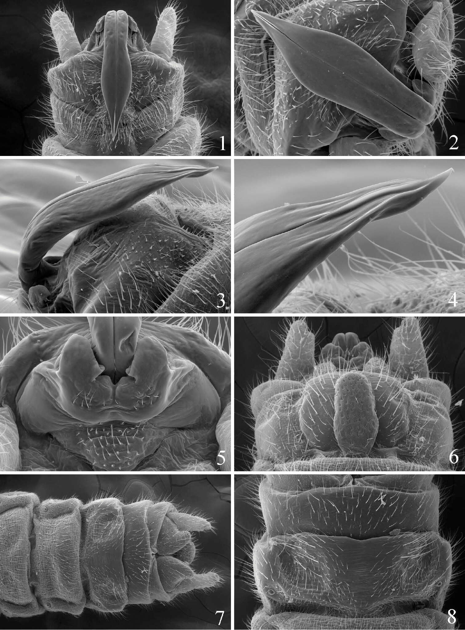
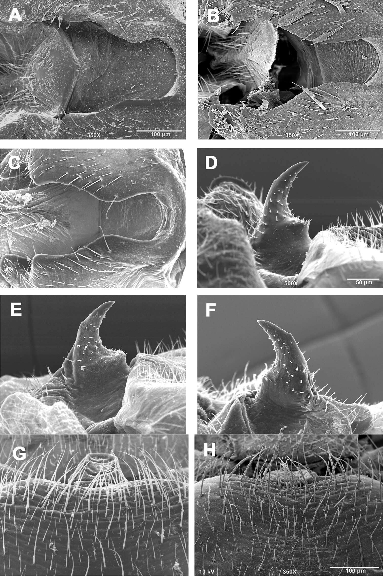
Figures 8.Zealeuctra ukayodi, sp. n., scanning electron micrographs, USA, Alabama, Jackson Co., Poplar Spring, 16 March 2008 (A–D, F–J), USA, Tennessee, Grundy Co., tributary to Elk River, 12 February 2007 (E). A–B male, cleft, dorsal view, 200× C–E male, epiproct, lateral view, 350× or 500× F female, posteromedial portion of seventh abdominal sternite, 350× G male terminalia, lateral H male terminalia, dorsal I male terminalia, ventral J female terminalia, ventral.
-
Scott A. Grubbs, Richard W. Baumann, R. Edward DeWalt, Tari Tweddale
Zookeys
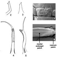
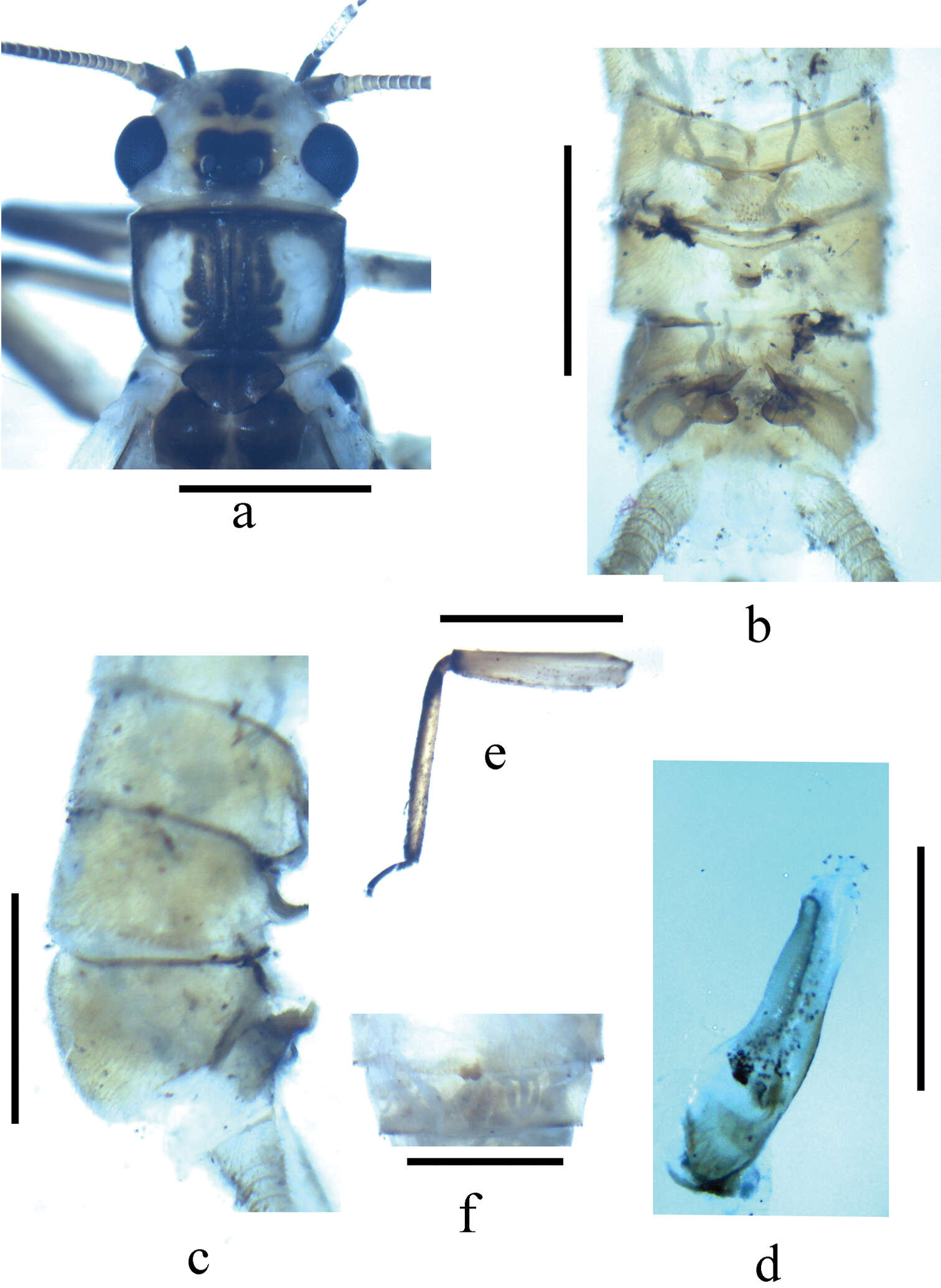
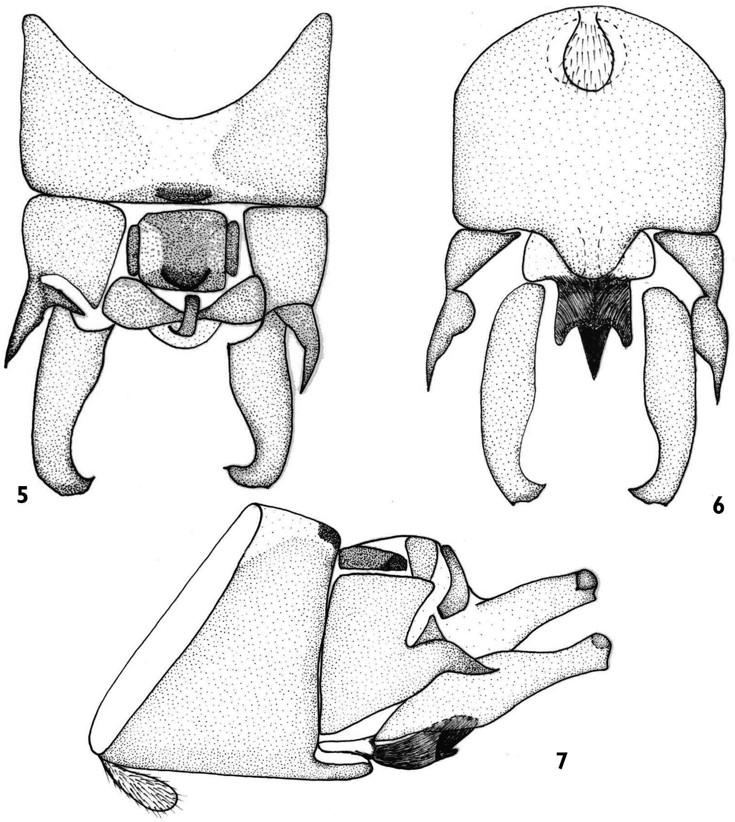
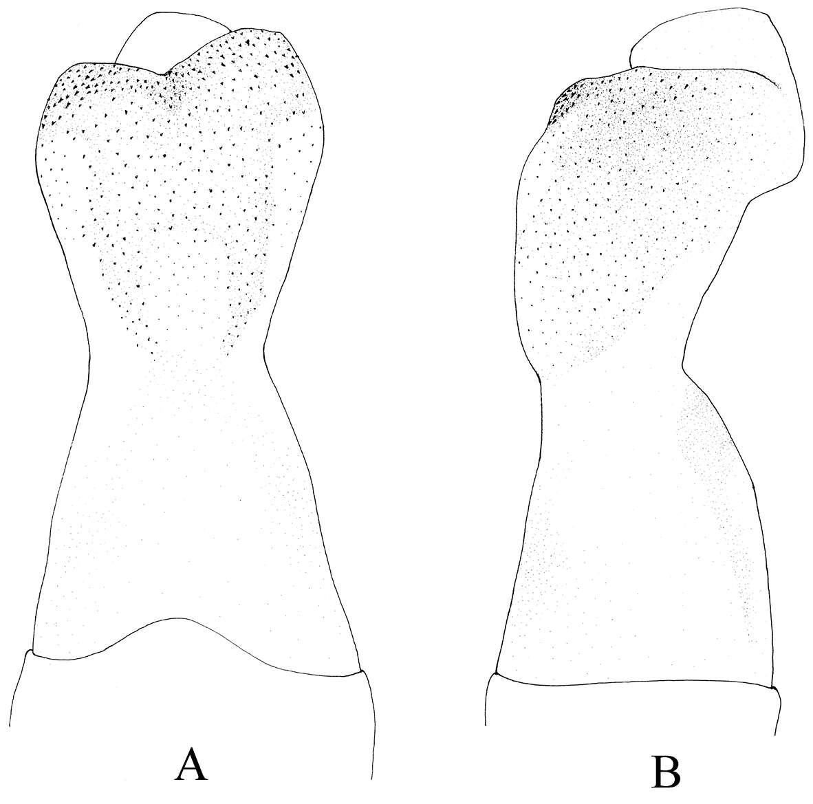
Figures 1–8.Prostoia besametsa, scanning electron micrographs, 1 USA, Utah, Monroe Creek, male, epiproct, dorsal view 2 USA, Montana, Gallatin River, male, epiproct, dorsal view 3 USA, South Dakota, Iron Creek, male, epiproct, lateral view 4 USA, South Dakota, Iron Creek, male, epiproct, lateral view 5 USA, Utah, Monroe Creek, male, abdominal terminalia, caudal view 6 USA, South Dakota, Iron Creek, male, abdominal terminalia, ventral view 7 USA, South Dakota, Iron Creek, female, abdominal terminalia, ventral view 8 USA, South Dakota, Iron Creek, female, abdominal terminalia, ventral view.
-
Wei-Hai Li, Sheng-Quan Zhang
Zookeys
Figure 1.Neoperla nigromarginata Li & Zhang, sp. n. Male (a–e) a Head and pronotum, dorsal view b Terminalia, dorsal view c Terminalia, lateral view d Aedeagus before eversion, lateral view e Hindleg f Female subgenital fig, ventral view.
-
Figures 5–7.Rhopalopsole ampulla male structures 5 Male terminal, dorsal aspect 6 Male terminal, ventral aspect 7 Male terminal, lateral aspect.
-
Li Wei-Hai, Wang Guo-Quan, Qin Xue-Feng
Zookeys
Figure 2.A–C Neoperla furcostyla Li and Qin,sp. n. (male). A Terminalia, dorsal view B Aedeagus, lateral view C Aedeagus of Neoperla forcipata Yang and Yang, lateral view.
-
Hong–Liang Wang, Guo–Quan Wang, Wei–Hai Li
Zookeys
Figure 2.Kamimuria guangxia Li & Wang, sp. n. (male). A Terminalia, dorsal view B Hemitergal process, lateral view C Aedeagus before eversion, ventral view.
-
Xue-Feng Qin, Dávid Murányi, Guo-Quan Wang, Wei-Hai Li
Zookeys
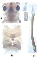
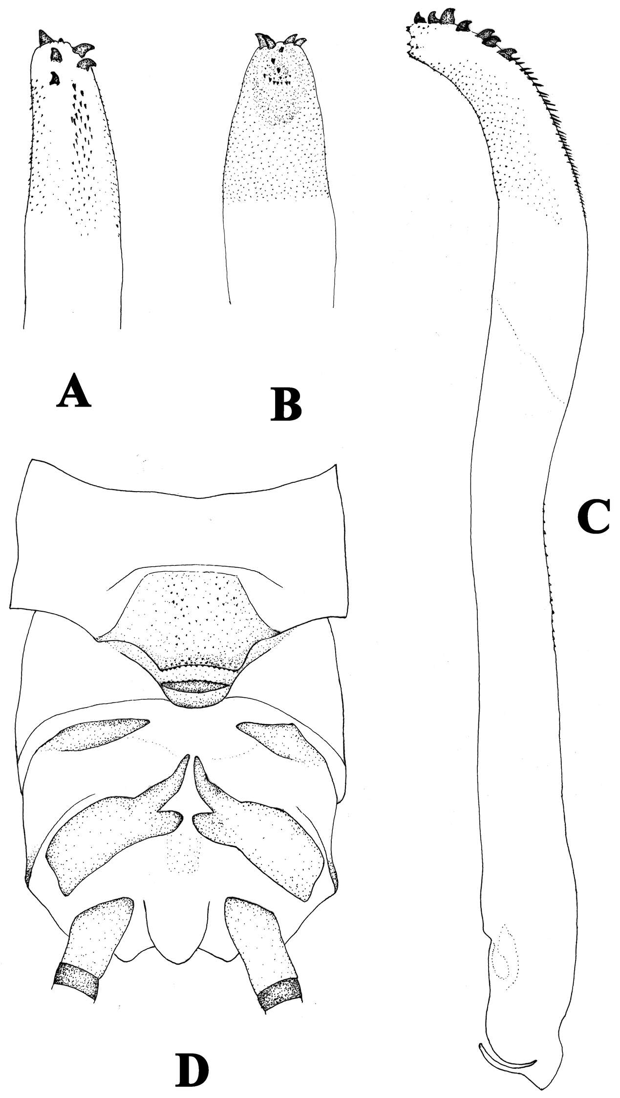
Figure 1.Neoperla brevistyla Li & Murányi, sp. n. Male a Head and pronotum, dorsal view b Hemitergal process, dorsal view c Aedeagus, lateral view. Scale bars: 0.5 mm.
-
Scott A. Grubbs, Boris C. Kondratieff, Bill P. Stark, R. Edward DeWalt
Zookeys
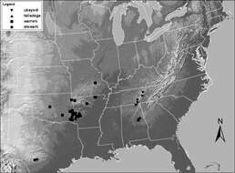
Figure 13.Distribution map for Zealeuctra stewarti, Zealeuctra talladega, Zealeuctra ukayodi sp. n., and Zealeuctra warreni.
-
Scott A. Grubbs, Richard W. Baumann, R. Edward DeWalt, Tari Tweddale
Zookeys
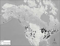
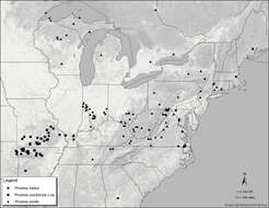
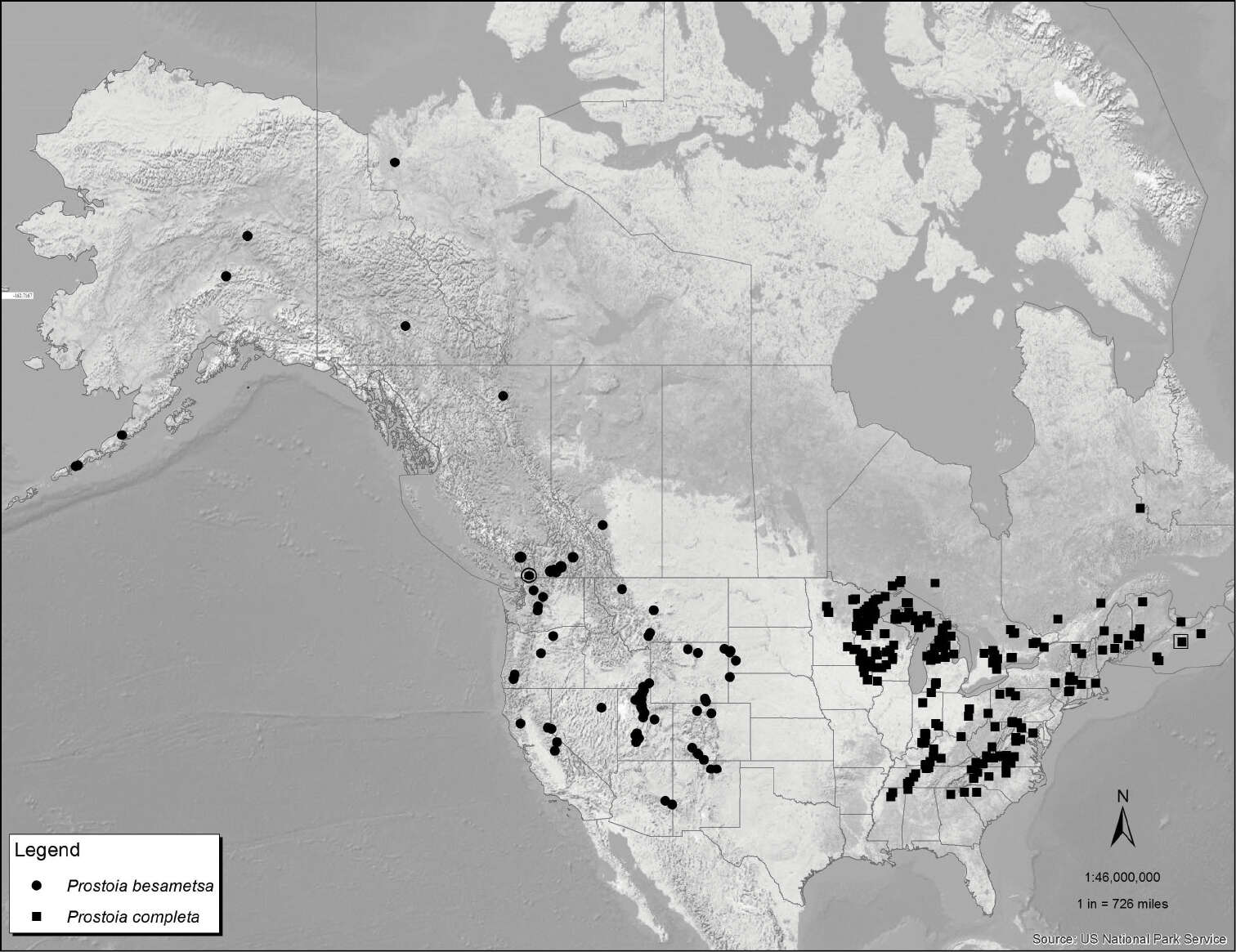
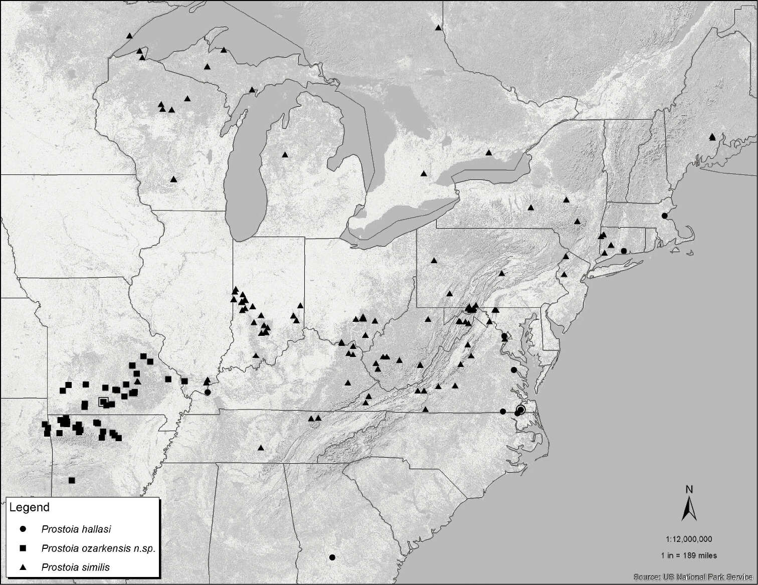
Figure 41.Distribution map for Prostoia besametsa (circles) and Prostoia completa (squares). The open symbols enclosing the solid symbols refer to the type localities for the two species.
-
Wei-Hai Li, Sheng-Quan Zhang
Zookeys
Figure 2.Neoperla nigromarginata Li & Zhang, sp. n. Male. a Dorsal aspect of aedeagal sac, top view b Aedeagus, lateral view. Note that the spines in b appear lightly pigmented and unclear, actually they are located on the lower surface of the sac, and are seen from beneath through the cuticle.
-
Scott A. Grubbs, R. Edward DeWalt
Zookeys
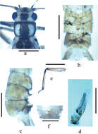
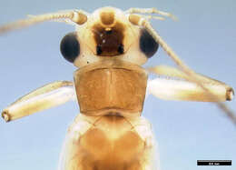
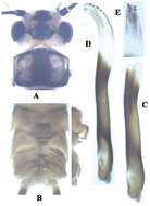
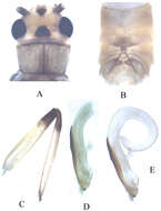
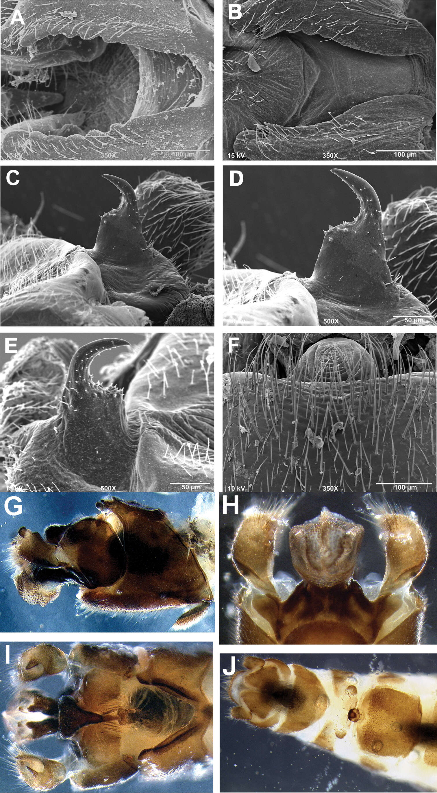
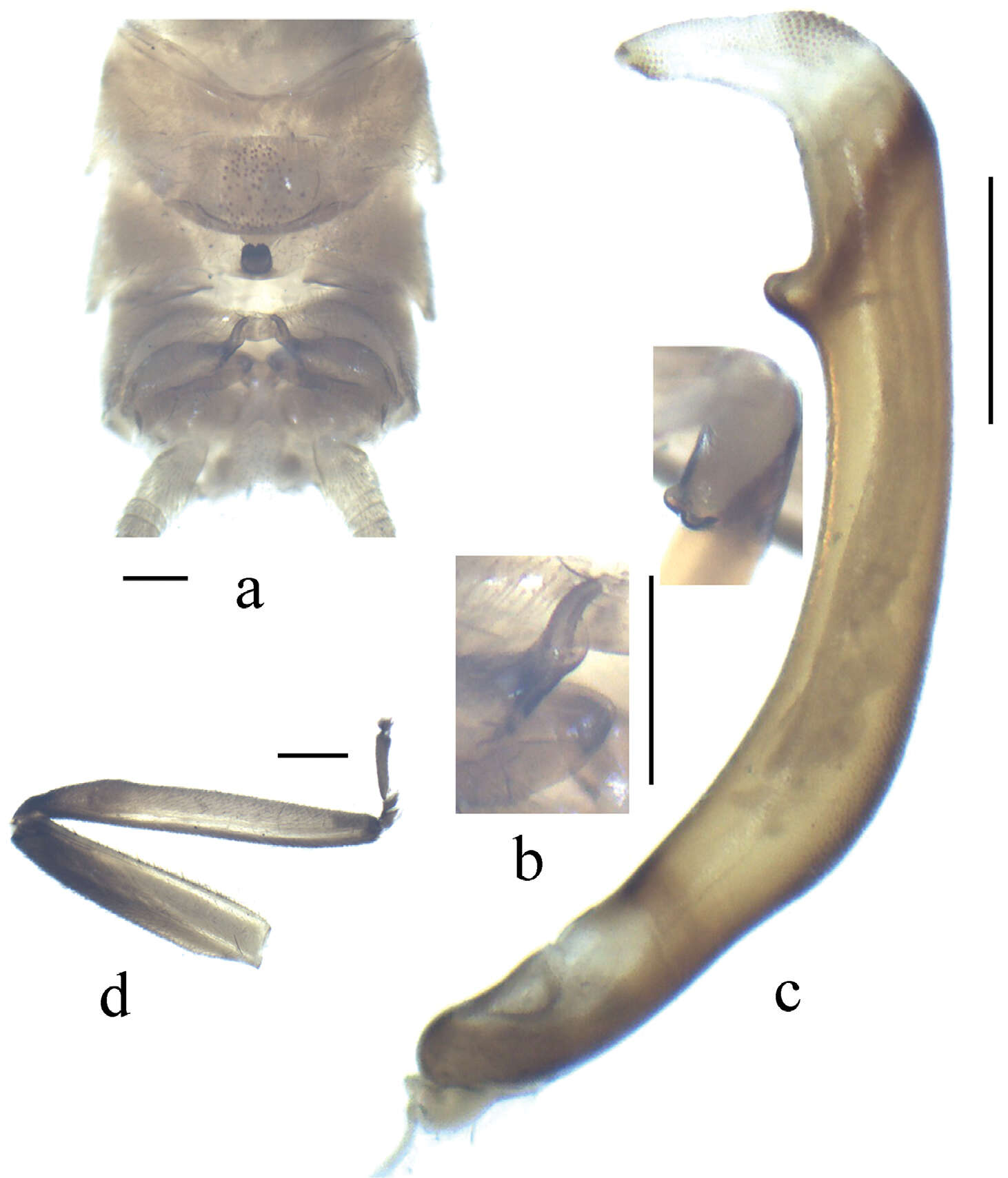
Figure 1.Perlesta ephelida. 1. Adult head and pronotum, dorsal.
-
Li Wei-Hai, Wang Guo-Quan, Qin Xue-Feng
Zookeys
Figure 3.Neoperla similidella Li and Wang, sp. n. (male). A Head and pronotum, dorsal view B Terminalia, dorsal view C Aedeagus before eversion, lateral view D Aedeagus, lateral view E Aedeagal sac, dorsal view.
-
Hong–Liang Wang, Guo–Quan Wang, Wei–Hai Li
Zookeys
Figure 3.Kamimuria guangxia Li & Wang, sp. n. (male). A Aedeagus, dorsal view B Aedeagus, lateral view.
-
Xue-Feng Qin, Dávid Murányi, Guo-Quan Wang, Wei-Hai Li
Zookeys
Figure 2.Neoperla brevistyla Li & Murányi,sp. n. Male a Terminalia, dorsal view b Hemitergal process, dorsal view c Aedeagus, lateral view d Foreleg, lateral view. Scale bars: 0.5 mm.
-
Scott A. Grubbs, Boris C. Kondratieff, Bill P. Stark, R. Edward DeWalt
Zookeys
Figures 7.Zealeuctra talladega, scanning electron micrographs, USA, Alabama, Clay Co., tributary to Swept Creek, 24 January 2006 (A–B, D–E, G), USA, Alabama, Clay Co., tributary to West Fork Hatchet Creek, 25 January 2006 (C, F, H). A–C male, cleft, dorsal view, 350× D–F male, epiproct, lateral view, 500× G–H female, posteromedial portion of seventh abdominal sternite, 350×.
-
Scott A. Grubbs, Richard W. Baumann, R. Edward DeWalt, Tari Tweddale
Zookeys
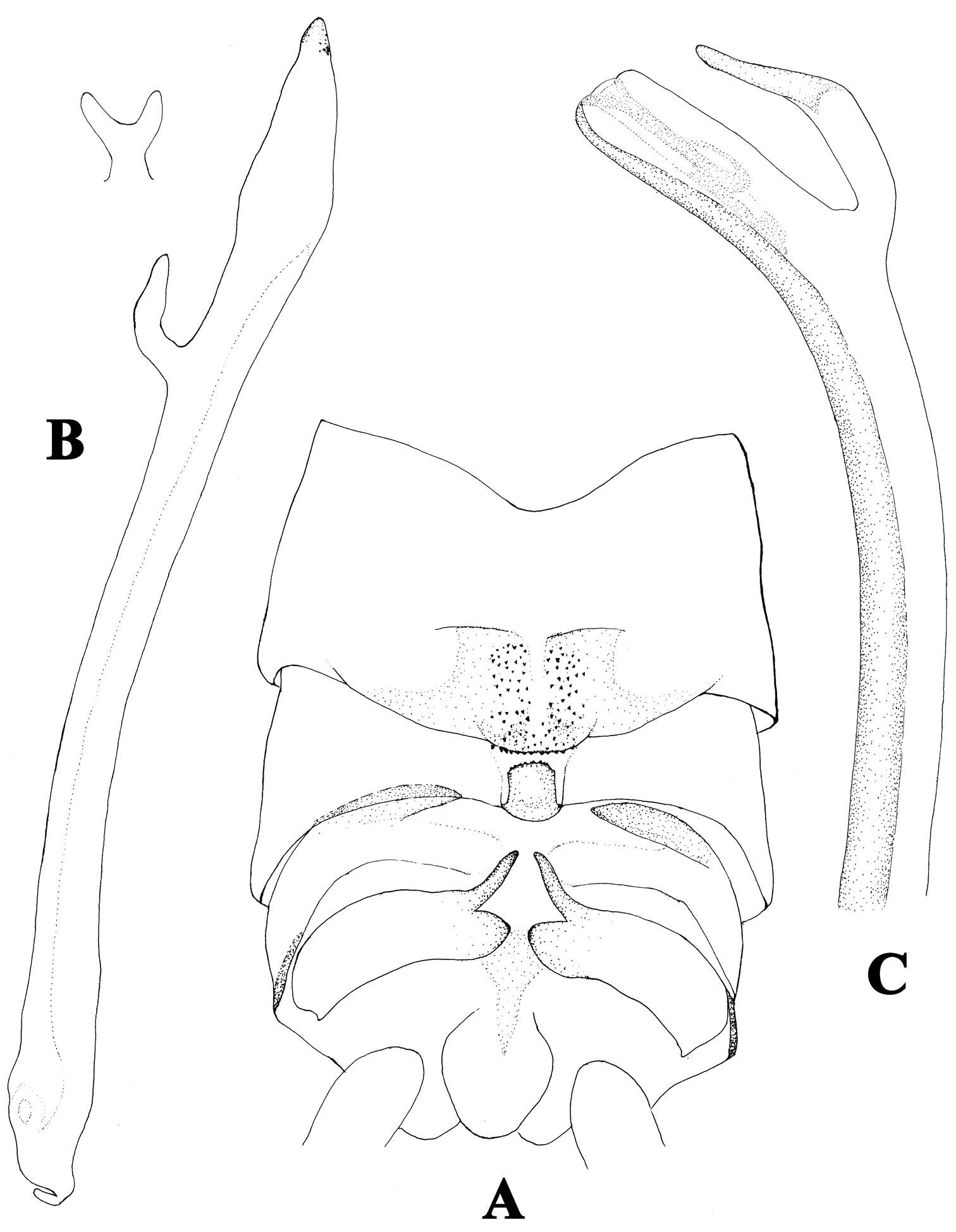
Figure 42.Distribution map for Prostoia hallasi (circles), Prostoia ozarkensis sp. n. (squares), and Prostoia similis (triangles). The open symbols enclosing the solid symbols refer to the type localities for the three species.
-
Wei-Hai Li, Sheng-Quan Zhang
Zookeys
Figure 3.Neoperla similiflavescens Li & Zhang, sp. n. Male. a Head and pronotum, dorsal view b Terminalia, dorsal view c Terminalia, lateral view d Aedeagus, lateral view.
-
Scott A. Grubbs, R. Edward DeWalt
Zookeys
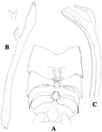
Figures 2–7.Perlesta ephelida. 2 Left paraproct, lateral 3 Left paraproct, caudal 4 Aedeagus, dorsal 5 Aedeagus, lateral 6 SEM micrograph, aedeagus, dorsal, 500X 7 SEM micrograph, female terminalia, ventral, 150X.
-
Li Wei-Hai, Wang Guo-Quan, Qin Xue-Feng
Zookeys
Figure 4.Neoperla similidella Li and Wang, sp. n. (male). A Aedeagal sac, dorsal view B Aedeagal sac, ventral view C Aedeagus, lateral view D Terminalia, dorsal view.
-
Hong–Liang Wang, Guo–Quan Wang, Wei–Hai Li
Zookeys
Figure 4.Neoperla mesostyla Li & Wang, sp. n. (male). A Head and pronotum, dorsal view B Terminalia, dorsal view C Hindleg (part of tarsi in this leg missing), lateral view D Aedeagus before eversion, lateral view E Aedeagus, lateral view.