About
Education
Discuss
TraitBank
Sign In
Sign Up
Language
Deutsch
English
Español
français
italiano
Nederlands
Piemontèis
Português do Brasil
suomi
Türkçe
Čeština
Ελληνικά
македонски
Українська
العربية
简体中文
繁體中文
names in breadcrumbs
vernacular
scientific
About
Education
Discuss
TraitBank
Sign In
Sign Up
en
Deutsch
English
Español
français
italiano
Nederlands
Piemontèis
Português do Brasil
suomi
Türkçe
Čeština
Ελληνικά
македонски
Українська
العربية
简体中文
繁體中文
names in breadcrumbs
vernacular
scientific
Biota
»
…
»
Animals
»
…
»
Arthropods
»
Chelicerates
»
Sea Spiders
»
…
»
Ammotheidae
»
…
Biota
»
Cellular
»
Eukaryotes
»
Opisthokonts
»
Animals
»
Bilaterians
»
Protostomes
»
Ecdysozoans
»
Arthropods
»
Chelicerates
»
Sea Spiders
»
Sea Spiders
»
Ascorhynchoidea
»
Ammotheidae
»
Ammothella
«
Ammothella biunguiculata (Dohrn 1881)
collect
overview
data
media
articles
maps
names
license
any license
CC-BY
CC-BY-NC
provider
any provider
iNaturalist
Zoosystematics and Evolution
cc-by-3.0
trusted
cc-by-3.0
trusted
cc-by-3.0
trusted
cc-by-nc-4.0
trusted
cc-by-nc-4.0
trusted
cc-by-nc-4.0
trusted
""
cc-by-3.0
Tobias Lehmann, Martin Heß, Roland R. Melzer
Zoosystematics and Evolution
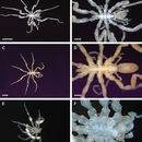
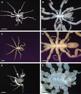
Figure 3.Ammotheidae 2; A, B: Ammothella appendiculata, female, dorsal view; scales 1 mm and 250 µm, respectively; C, D: Ammothella biunguiculata, male, dorsal view; scales 1 mm and 250 µm, respectively; E, F: Ammothella longipes, female, dorsal view; scales 1 mm and 250 µm, respectively.
""
cc-by-3.0
Tobias Lehmann, Martin Heß, Roland R. Melzer
Zoosystematics and Evolution
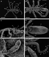
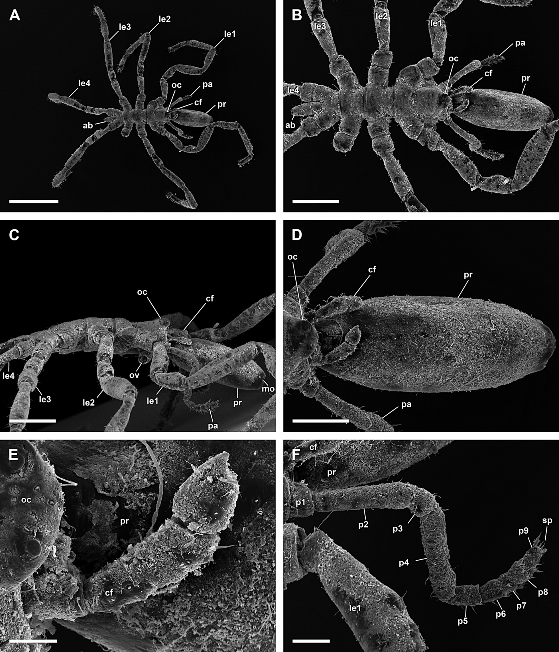
Figure 15.Ammothella biunguiculata, male; A: Dorsal view; scale 1 mm; B: Dorsal view of trunk; scale 400 µm; C: Lateral view of trunk, oviger dissected; scale 400 µm; D: Dorsal view of proboscis; scale 100 µm; E: Right chelifore with reduced chela; scale 40 µm; F: Right 9-articled palp; scale 100 µm.
""
cc-by-3.0
Tobias Lehmann, Martin Heß, Roland R. Melzer
Zoosystematics and Evolution
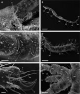
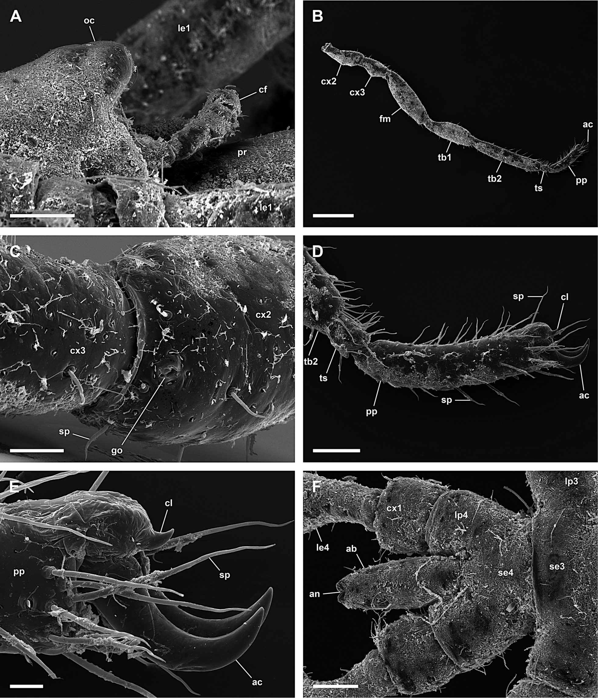
Figure 16.Ammothella biunguiculata, male; A: Lateral view of ocular tubercle; scale 100 µm; B: Right 3rd leg; scale 400 µm; C: Ventral view of coxa 2 with genital opening, distal is left (right 3rd leg); scale 40 µm; D: Tarsus and propodus with reduced claw and dominating auxiliary claws; scale 100 µm; E: Reduced claw and dominating auxiliary claws; scale 20 µm; F: Dorsal view of trunk segment 4 and abdomen; scale 100 µm.
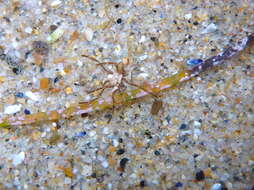
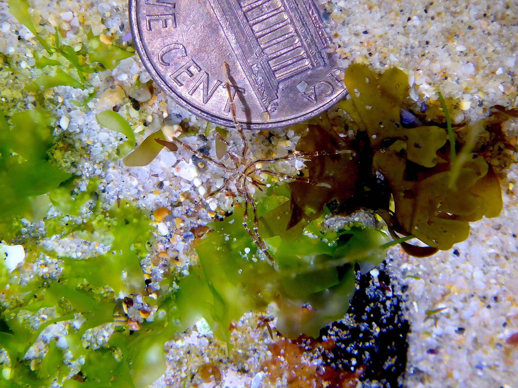
"
cc-by-nc-4.0
ponkyjoe
iNaturalist
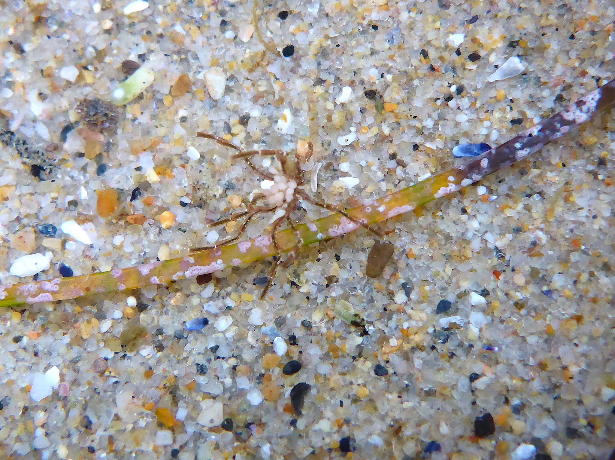
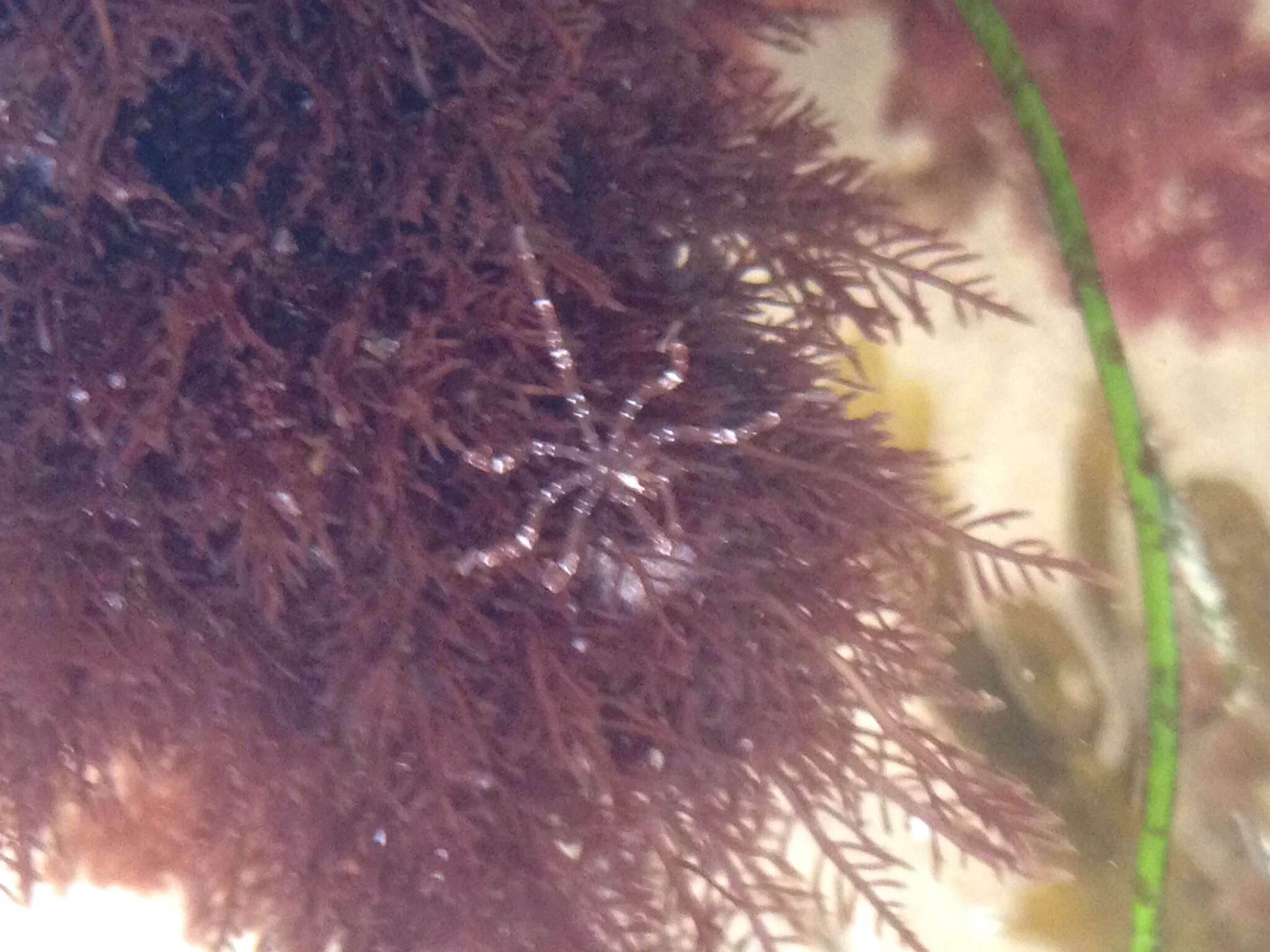
"
cc-by-nc-4.0
ponkyjoe
iNaturalist
"
cc-by-nc-4.0
ponkyjoe
iNaturalist