nomes no trilho de navegação
Caulobacter crescentus is a Gram-negative, oligotrophic bacterium widely distributed in fresh water lakes and streams. The taxon is more properly known as Caulobacter vibrioides (Henrici and Johnson 1935).[1]
C. crescentus is an important model organism for studying the regulation of the cell cycle, asymmetric cell division, and cellular differentiation. Caulobacter daughter cells have two very different forms. One daughter is a mobile "swarmer" cell that has a single flagellum at one cell pole that provides swimming motility for chemotaxis. The other daughter, called the "stalked" cell, has a tubular stalk structure protruding from one pole that has an adhesive holdfast material on its end, with which the stalked cell can adhere to surfaces. Swarmer cells differentiate into stalked cells after a short period of motility. Chromosome replication and cell division only occurs in the stalked cell stage.
C. crescentus derives its name from its crescent shape, which is caused by the protein crescentin. It is an interesting organism to study because it inhabits nutrient-poor aquatic environments. Their ability to thrive in low levels of nutrients is facilitated by its dimorphic developmental cycle. The swarmer cell has a flagellum that protrudes from a single pole and is unable to initiate DNA replication unless differentiated into a stalked cell. The differentiation process includes a morphological transition characterized by ejection of its flagellum and growth of a stalk at the same pole. Stalked cells can elongate and replicate their DNA while growing a flagellum at the opposite pole, giving rise to a pre-divisional cell. Although the precise function of stalks is still being investigated, it is likely that the stalks are involved in the uptake of nutrients in nutrient-limited conditions.[2] Its use as a model originated with developmental biologist Lucy Shapiro.[3][4]
In the laboratory, researchers distinguish between C. crescentus strain CB15 (the strain originally isolated from a freshwater lake) and NA1000 (the primary experimental strain). In strain NA1000, which was derived from CB15 in the 1970s,[5] the stalked and predivisional cells can be physically separated in the laboratory from new swarmer cells, while cell types from strain CB15 cannot be physically separated. The isolated swarmer cells can then be grown as a synchronized cell culture. Detailed study of the molecular development of these cells as they progress through the cell cycle has enabled researchers to understand Caulobacter cell cycle regulation in great detail. Due to this capacity to be physically synchronized, strain NA1000 has become the predominant experimental Caulobacter strain throughout the world. Additional phenotypic differences between the two strains have subsequently accumulated due to selective pressures on the NA1000 strain in the laboratory environment. The genetic basis of the phenotypic differences between the two strains results from coding, regulatory, and insertion/deletion polymorphisms at five chromosomal loci.[6] C. crescentus is synonymous with Caulobacter vibrioides.[1]
The Caulobacter CB15 genome has 4,016,942 base pairs in a single circular chromosome encoding 3,767 genes.[7] The genome contains multiple clusters of genes encoding proteins essential for survival in a nutrient-poor habitat. Included are those involved in chemotaxis, outer membrane channel function, degradation of aromatic ring compounds, and the breakdown of plant-derived carbon sources, in addition to many extracytoplasmic function sigma factors, providing the organism with the ability to respond to a wide range of environmental fluctuations. In 2010, the Caulobacter NA1000 strain was sequenced and all differences with the CB15 "wild type" strain were identified.[6]
The Caulobacter stalked cell stage provides a fitness advantage by anchoring the cell to surfaces to form biofilms and or to exploit nutrient sources. Generally, the bacterial species that divides fastest will be most effective at exploiting resources and effectively occupying ecological niches. Yet, Caulobacter has the swarmer cell stage that results in slower population growth. The swarmer cell is thought to provide cell dispersal, so that the organism constantly seeks out new environments. This may be particularly useful in severely nutrient-limited environments when the scant resources available can be depleted very quickly. Many, perhaps most, of the swarmer daughter cells will not find a productive environment, but the obligate dispersal stage must increase the reproductive fitness of the species as a whole.
The Caulobacter cell cycle regulatory system controls many modular subsystems that organize the progression of cell growth and reproduction. A control system constructed using biochemical and genetic logic circuitry organizes the timing of initiation of each of these subsystems. The central feature of the cell cycle regulation is a cyclical genetic circuit—a cell cycle engine—that is centered around the successive interactions of five master regulatory proteins: DnaA, GcrA, CtrA, SciP, and CcrM whose roles were worked out by the laboratories of Lucy Shapiro and Harley McAdams.[8][9][10] These five proteins directly control the timing of expression of over 200 genes. The five master regulatory proteins are synthesized and then eliminated from the cell one after the other over the course of the cell cycle. Several additional cell signaling pathways are also essential to the proper functioning of this cell cycle engine. The principal role of these signaling pathways is to ensure reliable production and elimination of the CtrA protein from the cell at just the right times in the cell cycle.
An essential feature of the Caulobacter cell cycle is that the chromosome is replicated once and only once per cell cycle. This is in contrast to the E. coli cell cycle where there can be overlapping rounds of chromosome replication simultaneously underway. The opposing roles of the Caulobacter DnaA and CtrA proteins are essential to the tight control of Caulobacter chromosome replication.[11] The DnaA protein acts at the origin of replication to initiate the replication of the chromosome. The CtrA protein, in contrast, acts to block initiation of replication, so it must be removed from the cell before chromosome replication can begin. Multiple additional regulatory pathways integral to cell cycle regulation and involving both phospho signaling pathways and regulated control of protein proteolysis[12] act to assure that DnaA and CtrA are present in the cell just exactly when needed.
Each process activated by the proteins of the cell cycle engine involve a cascade of many reactions. The longest subsystem cascade is DNA replication. In Caulobacter cells, replication of the chromosome involves about 2 million DNA synthesis reactions for each arm of the chromosome over 40 to 80 min depending on conditions. While the average time for each individual synthesis reaction can be estimated from the observed average total time to replicate the chromosome, the actual reaction time for each reaction varies widely around the average rate. This leads to a significant and inevitable cell-to-cell variation time to complete replication of the chromosome. There is similar random variation in the rates of progression of all the other subsystem reaction cascades. The net effect is that the time to complete the cell cycle varies widely over the cells in a population even when they all are growing in identical environmental conditions. Cell cycle regulation includes feedback signals that pace progression of the cell cycle engine to match progress of events at the regulatory subsystem level in each particular cell. This control system organization, with a controller (the cell cycle engine) driving a complex system, with modulation by feedback signals from the controlled system creates a closed loop control system.
The rate of progression of the cell cycle is further adjusted by additional signals arising from cellular sensors that monitor environmental conditions (for example, nutrient levels and the oxygen level) or the internal cell status (for example, presence of DNA damage).[13]
The control circuitry that directs and paces Caulobacter cell cycle progression involves the entire cell operating as an integrated system. The control circuitry monitors the environment and the internal state of the cell, including the cell topology, as it orchestrates activation of cell cycle subsystems and Caulobacter crescentus asymmetric cell division. The proteins of the Caulobacter cell cycle control system and its internal organization are co-conserved across many alphaproteobacteria species, but there are great differences in the regulatory apparatus' functionality and peripheral connectivity to other cellular subsystems from species to species.[14][15] The Caulobacter cell cycle control system has been exquisitely optimized by evolutionary selection as a total system for robust operation in the face of internal stochastic noise and environmental uncertainty.
The bacterial cell's control system has a hierarchical organization.[16] The signaling and the control subsystem interfaces with the environment by means of sensory modules largely located on the cell surface. The genetic network logic responds to signals received from the environment and from internal cell status sensors to adapt the cell to current conditions. A major function of the top level control is to ensure that the operations involved in the cell cycle occur in the proper temporal order. In Caulobacter, this is accomplished by the genetic regulatory circuit composed of five master regulators and an associated phospho-signaling network. The phosphosignaling network monitors the state of progression of the cell cycle and plays an essential role in accomplishing asymmetric cell division. The cell cycle control system manages the time and place of the initiation of chromosome replication and cytokinesis as well as the development of polar organelles. Underlying all these operations are the mechanisms for production of protein and structural components and energy production. The “housekeeping” metabolic and catabolic subsystems provide the energy and the molecular raw materials for protein synthesis, cell wall construction and other operations of the cell. The housekeeping functions are coupled bidirectionally to the cell cycle control system. However, they can adapt, somewhat independently of the cell cycle control logic, to changing composition and levels of the available nutrient sources.
The proteins of the Caulobacter cell cycle control system are widely co-conserved across the alphaproteobacteria, but the ultimate function of this regulatory system varies widely in different species. These evolutionary changes reflect enormous differences between the individual species in fitness strategies and ecological niches. For example, Agrobacterium tumefaciens is a plant pathogen, Brucella abortus is an animal pathogen, and Sinorhizobium meliloti is a soil bacterium that invades, and becomes a symbiont in, plant root nodules that fix nitrogen yet most of the proteins of the Caulobacter cell cycle control are also found in these species. The specific coupling between the protein components of the cell cycle control network and the downstream readout of the circuit differ from species to species. The pattern is that the internal functionality of the network circuitry is conserved, but the coupling at the “edges” of the regulatory apparatus to the proteins controlling specific cellular functions differs widely among the different species.
Caulobacter crescentus is a member of a group of bacteria that possess the stalk structure, a tubular extension from the cell body. However, the positioning of the stalk is not necessarily conserved at the pole of the cell body in different closely related species. Specifically, research has shown that not only the position of the stalk can change, but the number can vary as well in the closely related genus Asticcacaulis.[17][18] SpmX, a polarly localized protein in Caulobacter crescentus, has been shown to manipulate stalk positioning in these Asticcacaulis species.[17] Presumably, It does so by a gain of function after protein expansion from around 400 amino acids in Caulobacter crescentus to more than 800 amino acids in Asticcacaulis species.
Caulobacter was the first asymmetric bacterium shown to age. Reproductive senescence was measured as the decline in the number of progeny produced over time.[19][20] On the basis of experimental evolution studies in C. crescentus, Ackermann et al.[19] suggested that aging is probably a fundamental property of all cellular organisms. A similar phenomenon has since been described in the bacterium Escherichia coli, which gives rise to morphologically similar daughter cells.[21]
In C. crescentus, cell polarity is readily apparent by the assembly of polar organelles and by the polarization of the division plane, which results in the generation of stalked progeny that are longer than swarmer progeny. The formation of new cell poles at division implies that cell polarity must be re-established in the stalked progeny and reversed in the swarmer progeny.[22]
The C. crescentus life cycle is governed by regulators such as TipN, a cell cycle protein. Yale University's data strongly suggest a model in which TipN regulates the orientation of the polarity axis by providing a positional cue from the preceding cell cycle. In this model TipN specifies the site of the most recent division by identifying the new pole. The cell uses this positional information as a source of intracellular asymmetry to establish and maintain the orientation of the polarity axis, which is crucial for polar morphogenesis and division. Recruitment of TipN to the nascent poles at the end of the division cycle redefines the identity of the poles and resets the correct polarity in both future daughter cells (with a polarity reversal in the swarmer cell).[22] The cell cycle–regulated synthesis and removal of these polarly localized structures have provided a rich playground for the identification of landmark proteins important for their proper localization.[23] TipN has two transmembrane regions in the N-terminal region and a large C-terminal coiled-coil domain. TipN homologues are present in other alpha-proteobacteria. TipN localizes to the new pole in both daughter cells after division and relocalizes to the cell division site in the late predivisional cell. Therefore, both daughter cells have TipN at the new pole after division.[23]
The landmark protein TipN is essential for the proper placement of the flagellum. [24] Mutants lacking TipN make serious mistakes in development. Instead of making a single flagellum at the correct cell pole , the cell makes multiple flagella at various locations, even on the stalk.[22]
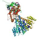
Cell development involves many such proteins working together. Fig#1 shows how TipN interact with two other polar proteins : the flagellar marker PodJ , and the stalk marker DivJ. [25]
Caulobacter crescentus is a Gram-negative, oligotrophic bacterium widely distributed in fresh water lakes and streams. The taxon is more properly known as Caulobacter vibrioides (Henrici and Johnson 1935).
C. crescentus is an important model organism for studying the regulation of the cell cycle, asymmetric cell division, and cellular differentiation. Caulobacter daughter cells have two very different forms. One daughter is a mobile "swarmer" cell that has a single flagellum at one cell pole that provides swimming motility for chemotaxis. The other daughter, called the "stalked" cell, has a tubular stalk structure protruding from one pole that has an adhesive holdfast material on its end, with which the stalked cell can adhere to surfaces. Swarmer cells differentiate into stalked cells after a short period of motility. Chromosome replication and cell division only occurs in the stalked cell stage.
C. crescentus derives its name from its crescent shape, which is caused by the protein crescentin. It is an interesting organism to study because it inhabits nutrient-poor aquatic environments. Their ability to thrive in low levels of nutrients is facilitated by its dimorphic developmental cycle. The swarmer cell has a flagellum that protrudes from a single pole and is unable to initiate DNA replication unless differentiated into a stalked cell. The differentiation process includes a morphological transition characterized by ejection of its flagellum and growth of a stalk at the same pole. Stalked cells can elongate and replicate their DNA while growing a flagellum at the opposite pole, giving rise to a pre-divisional cell. Although the precise function of stalks is still being investigated, it is likely that the stalks are involved in the uptake of nutrients in nutrient-limited conditions. Its use as a model originated with developmental biologist Lucy Shapiro.
Caulobacter crescentus
Caulobacter vibrioides (anc. Caulobacter crescentus)[5] est une espèce de bactéries gram-négatives vivant dans les milieux aquatiques comme les lacs et les rivières. C’est une bactérie oligotrophe capable de croître dans les habitats pollués, grâce à des gènes impliqués dans la résistance aux métaux de transition et aux stress oxydants[6],[7].
Cette bactérie se présente sous deux formes très différentes : une forme non réplicative mobile grâce à un flagelle et une forme réplicative fixée munie d'un pédoncule. Alors que la forme mobile permet la dissémination et la recherche de nutriments, la forme fixée est le siège de la réplication. Lorsque les conditions sont favorables, la forme mobile se différencie vers la forme fixée pour se diviser de manière asymétrique et polarisée : une cellule mère fixée se diviser alors en une cellule fille fixée et une cellule fille mobile[7].
C. vibrioides est utilisée comme modèle dans l’étude de la régulation du cycle cellulaire procaryote, de la division asymétrique et de la différenciation cellulaire. Le pédoncule confère à la cellule fixée une propriété particulière, l'adhésion, qui met en jeu des macromolécules appelées adhésines à structure polysaccharidique[8].
Il existe deux souches de Caulobacter crescentus : la souche originelle CB15 et la souche utilisée en laboratoire NA1000. NA1000 est une souche dérivée de CB15 associée à une pression de sélection induit par l’environnement du laboratoire. Au niveau de la taille du génome, la souche CB15 fait 4,02 mégabases et la souche NA1000 fait 4,04 mégabases. Les deux souches sont génétiquement différentes par la présence de 8 polymorphismes codants, 2 non codants et 1 site d’insertions/suppressions (26 kb)[9].
Ces différences génétiques ont induit des différences phénotypiques entre les deux souches notamment au niveau de l’adhérence, la vitesse de croissance et la synchronisation. La synchronisation est la capacité de séparer physiquement les deux formes cellulaires, flagellées et pédonculées par centrifugation. Alors que la souche CB15 est adhérente (forme fixée), se multiplie lentement et est incapable de se synchroniser, la souche NA1000 n’est pas adhérente, se multiplie plus rapidement et possède la capacité de synchronisation. Cette dernière caractéristique fait de la souche NA1000, la souche prédominante en laboratoire[9].
La forme flagellée mobile, permettant la dispersion, est limitée à la phase G1 de l'interphase. En conditions favorables, la bactérie se différencie en forme pédonculée fixée et permet la réplication du chromosome en phase S. Après la réplication du chromosome, la cellule s'allonge et s'apprête à se diviser en phase G2. La division cellulaire est asymétrique et permet de générer une cellule fixée qui va de nouveau répliquer le chromosome, et une cellule mobile qui va permettre la dissémination dans l'environnement. L'initiation de la réplication, la ségrégation des chromosomes et la cytokinèse constituent les trois grandes étapes du cycle cellulaire[7].
La première étape, l'initiation de la réplication ne se fait que lorsque la cellule est en phase S quand l'origine de réplication, ori, est accessible. En plus d'être un régulateur transcriptionnel, la protéine CtrA régule le cycle cellulaire : lorsque celle-ci est phosphorylée, elle se lie au niveau de la région ori et inhibe la réplication du chromosome. La protéine CtrA phosphorylée est fortement présente dans les cellules flagellées, puis est déphosphorylée et dégradée après la différenciation en cellule pédonculée, permettant l'initiation de la réplication par la protéine DnaA[10].
Après la réplication, le chromosome originel et sa copie doivent être distribués aux deux cellules filles. Cette étape est appelée ségrégation des chromosomes et est régulée par le système de partition ParABS. parS est une séquence d'ADN située près de l'ori correspondant à un centromère où se fixe la protéine de fixation ParB. La protéine ParA se fixe au complexe et permet l'hydrolyse de l'ATP entraînant la migration des chromosomes d'une part et d'autre de la cellule bactérienne[11].
La cytokinèse, étape de division en deux cellules filles, débute par l'assemblage de l'anneau Z au centre de la cellule. L'anneau Z est constitué de la protéine FtsZ qui se polymérise et permet de générer les forces constriction essentielles à la division de la cellule. Afin que l'assemblage de l'anneau Z soit bien localisé, il est nécessaire que la polymérisation de FtsZ soit inhibée dans le reste de la cellule. Cette inhibition est exercée par la protéine MipZ recrutée par la protéine ParB créant un gradient de concentration : MipZ est plus important au niveau des pôles et moins important au centre de la cellule[12]. La séparation des deux cellules filles est assurées par plusieurs protéines : FtsEX, un transporteur de type ABC permet l'ancrage de la protéine FtsZ dans la membrane, FtsK, une translocase à ADN permet de transloquer l’ADN qui serait au niveau du septum et le complexe FtsQL permet la stabilité du divisome. Enfin, les protéines FtsN, FtsW et FtsI permettent la biosynthèse et le remodelage du peptidoglycane induisant ainsi la septation et la séparation des deux cellules filles[13].
Le cycle cellulaire chez Caulobacter crescentus permet de générer deux formes cellulaires différentes. Au-delà de la différence structurale, l'assemblage excentré de l'anneau Z permet d'obtenir une cellule fille pédonculée légèrement plus grande que la cellule fille flagellée[7]. Cette asymétrie du cycle est intrinsèquement liée à l'accumulation de différentes protéines à chaque pôle de la cellule : on parle de polarité cellulaire[14].
Au niveau protéique, une des différences entre la cellule mobile et la cellule fixée est la concentration en CtrA phosphorylée, protéine régulant la réplication cellulaire. La phosphorylation de la protéine CtrA est contrôlée par un complexe de phosphorelais, dont une unité diverge entre la future cellule flagellée et la future cellule pédonculée. Basiquement, la phosphorylation de CtrA est initiée par la protéine régulatrice Ccka et passe par l'intermédiaire d'une protéine ChpT. L'activation de Ccka dépend de son interaction avec la protéine DivL, complexe qui peut être inhibée par la protéine DivK. Enfin, la phosphorylation de DivK est contrôlée par la phosphatase PleC au pôle de la future cellule mobile et par la kinase DivJ au pôle de la future cellule fixée. La protéine PleC désactive DivK, DivK n'inhibe plus Ccka-DivL, Ccka en tant que kinase est activée et permet la phosphorylation de CtrA, ce qui empêche l'initiation de la réplication dans la future cellule mobile. La protéine DivJ active DivK, DivK peut inhiber Ccka-DivL, Ccka permet la déphosphorylation de CtrA, ce qui permet l'initiation de la réplication dans la future cellule fixée[15].
La combinaison des protéines ZitP, PopZ et CpaM permet la distinction majeure entre les deux cellules : la présence d'un flagelle ou d'un pédoncule. La protéine doigt de zinc ZitP possède deux fonctions, dépendant du pôle de la cellule[14]. Au pôle de la future cellule mobile, ZitP recrute la protéine effectrice CpaM et permet l'assemblage du flagelle alors qu'au pôle de la future cellule fixée, ZitP s'associe à la protéine PopZ afin de contrôler sa position lors du cycle cellulaire. PopZ est une protéine présente dans les deux pôles de la cellule en division qui interagit directement avec le système ParABS[16]. L'association des protéines ZitP et CpaM au pôle de la future cellule mobile et non au pôle fixée est influencée par le système régulant la réplication cellulaire, DivJ-PleC-DivK[17].
Caulobacter crescentus
Caulobacter vibrioides (anc. Caulobacter crescentus) est une espèce de bactéries gram-négatives vivant dans les milieux aquatiques comme les lacs et les rivières. C’est une bactérie oligotrophe capable de croître dans les habitats pollués, grâce à des gènes impliqués dans la résistance aux métaux de transition et aux stress oxydants,.
Cette bactérie se présente sous deux formes très différentes : une forme non réplicative mobile grâce à un flagelle et une forme réplicative fixée munie d'un pédoncule. Alors que la forme mobile permet la dissémination et la recherche de nutriments, la forme fixée est le siège de la réplication. Lorsque les conditions sont favorables, la forme mobile se différencie vers la forme fixée pour se diviser de manière asymétrique et polarisée : une cellule mère fixée se diviser alors en une cellule fille fixée et une cellule fille mobile.
C. vibrioides est utilisée comme modèle dans l’étude de la régulation du cycle cellulaire procaryote, de la division asymétrique et de la différenciation cellulaire. Le pédoncule confère à la cellule fixée une propriété particulière, l'adhésion, qui met en jeu des macromolécules appelées adhésines à structure polysaccharidique.
Caulobacter crescentus è un batterio Gram-Negativo, oligotrofico diffuso ampiamente nelle acque di laghi e fiumi. Il taxon è meglio conosciuto come Caulobacter vibrioides (Henrici e Jonson 1935).
Caulobacter è un importante organismo modello per lo studio della regolazione del ciclo cellulare, della divisione cellulare asimmetrica e della differenziazione cellulare. Le cellule figlie di Caulobacter presentano due forme molto diverse. Una di esse è una cellula mobile detta "sciamante" con un singolo flagello situato su un polo cellulare che fornisce motilità per chemiotassi. L'altra cellula, chiamata cellula "peduncolata", possiede una struttura a gambo di forma tubolare sporgente da un polo. Su di esso è presente una sostanza adesiva resistente, con cui la cellula può aderire alle superfici. Le cellule sciamanti si differenziano in cellule peduncolate dopo un breve periodo di motilità. La duplicazione del cromosoma e la divisione cellulare avvengono solo nello stadio peduncolato. Il nome crescentus deriva dalla forma a mezzaluna dovuta dalla presenza della proteina crescentina.[1]
Il suo uso come organismo modello ha avuto origine dalla biologa evolutiva Lucy Shapiro.[2][3]
Il genoma di Caulobacter CB15 possiede 4.016.942 paia di basi racchiuse in un unico cromosoma circolare contenente 3.767 geni.[4] Il genoma contiene molteplici gruppi di geni che codificano per proteine essenziali alla sopravvivenza in un ambiente a basso contenuto nutritivo. Tra di essi ritroviamo quelli coinvolti nella chemiotassi, nella funzione delle proteine canale esterne alla membrana, nella degradazione dei composti contenenti anelli aromatici e nella scomposizione dei composti del carbonio derivati dalle piante oltre a molti fattori sigma che agiscono fuori dalla membrana citoplasmatica, permettendo così all'organismo di rispondere ad una vasta gamma di fluttuazioni ambientali. Nel 2010 è stato sequenziato il ceppo di Caulobacter NA1000 dando la possibilità di differenziarlo dal ceppo CB15 "wild type".[5]
Caulobacter crescentus è un batterio Gram-Negativo, oligotrofico diffuso ampiamente nelle acque di laghi e fiumi. Il taxon è meglio conosciuto come Caulobacter vibrioides (Henrici e Jonson 1935).


