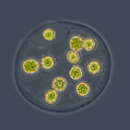
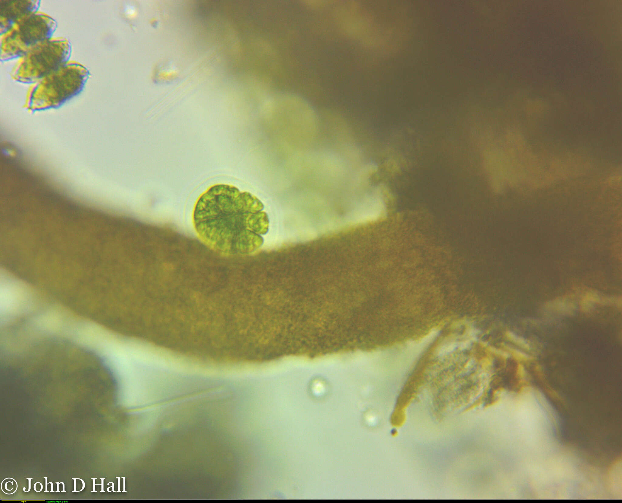
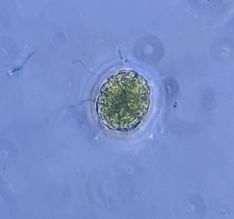
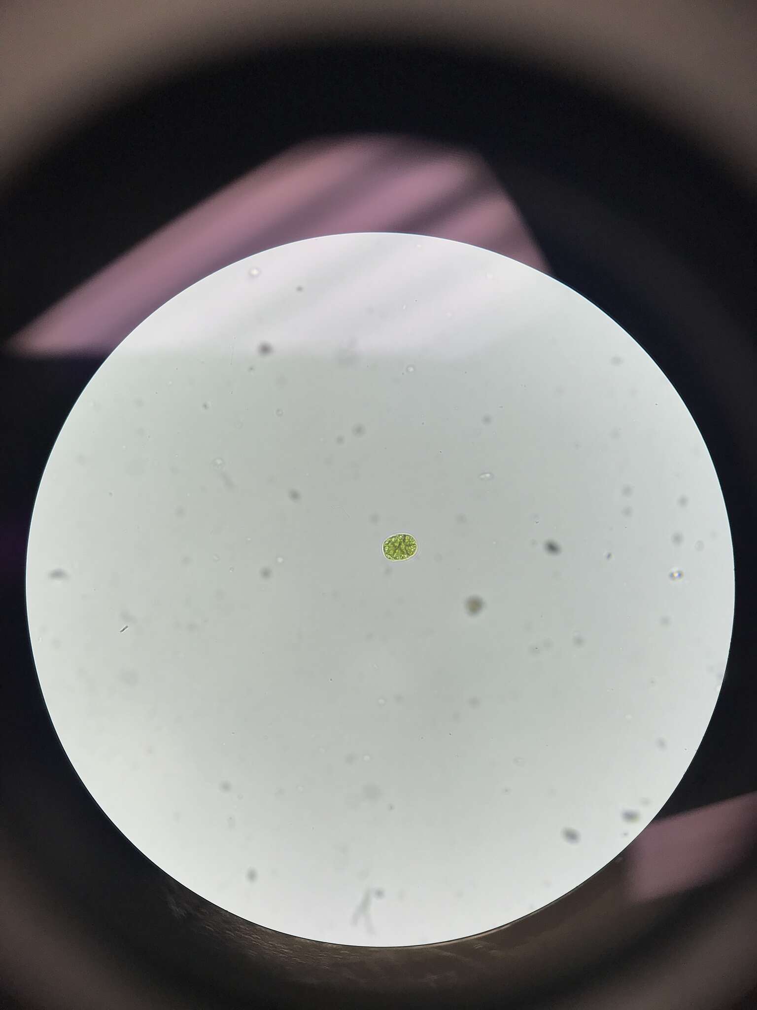
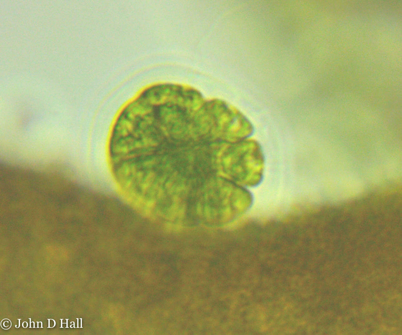
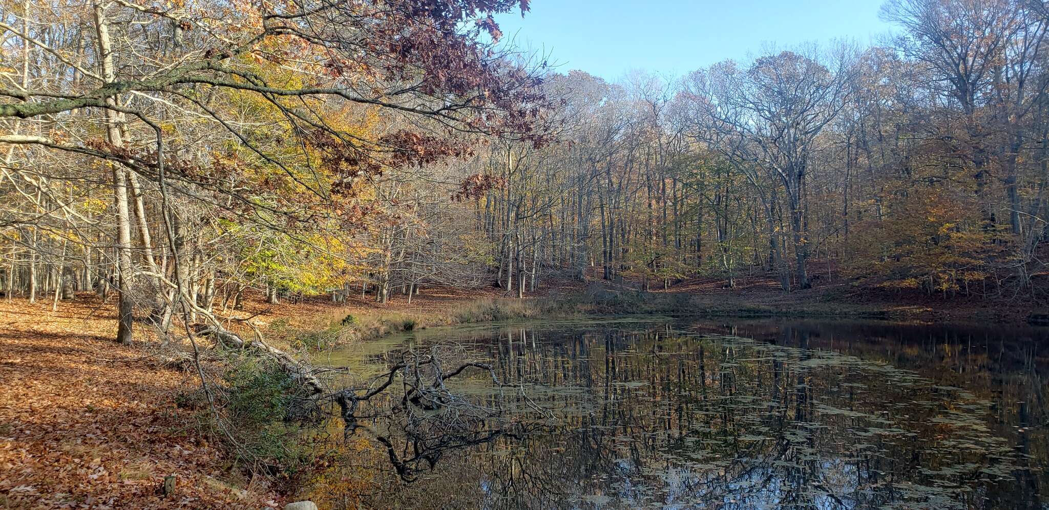
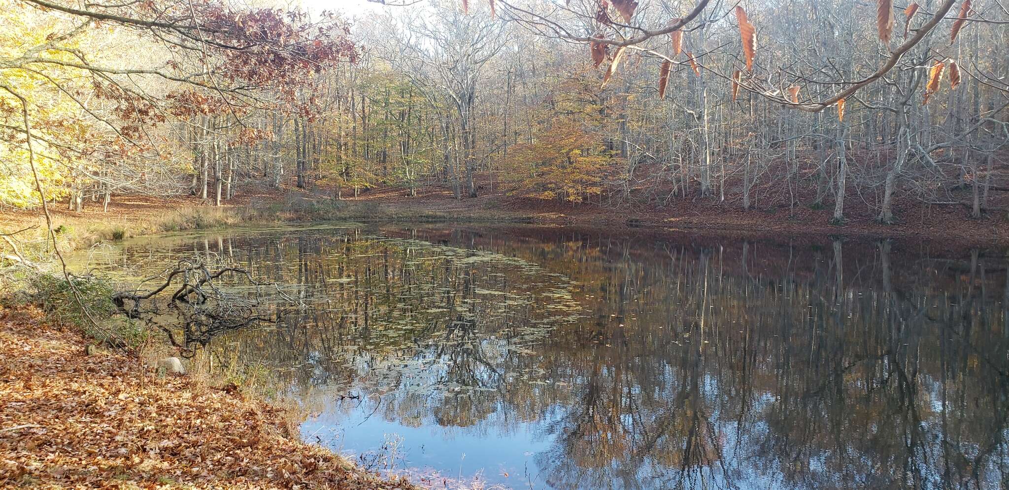
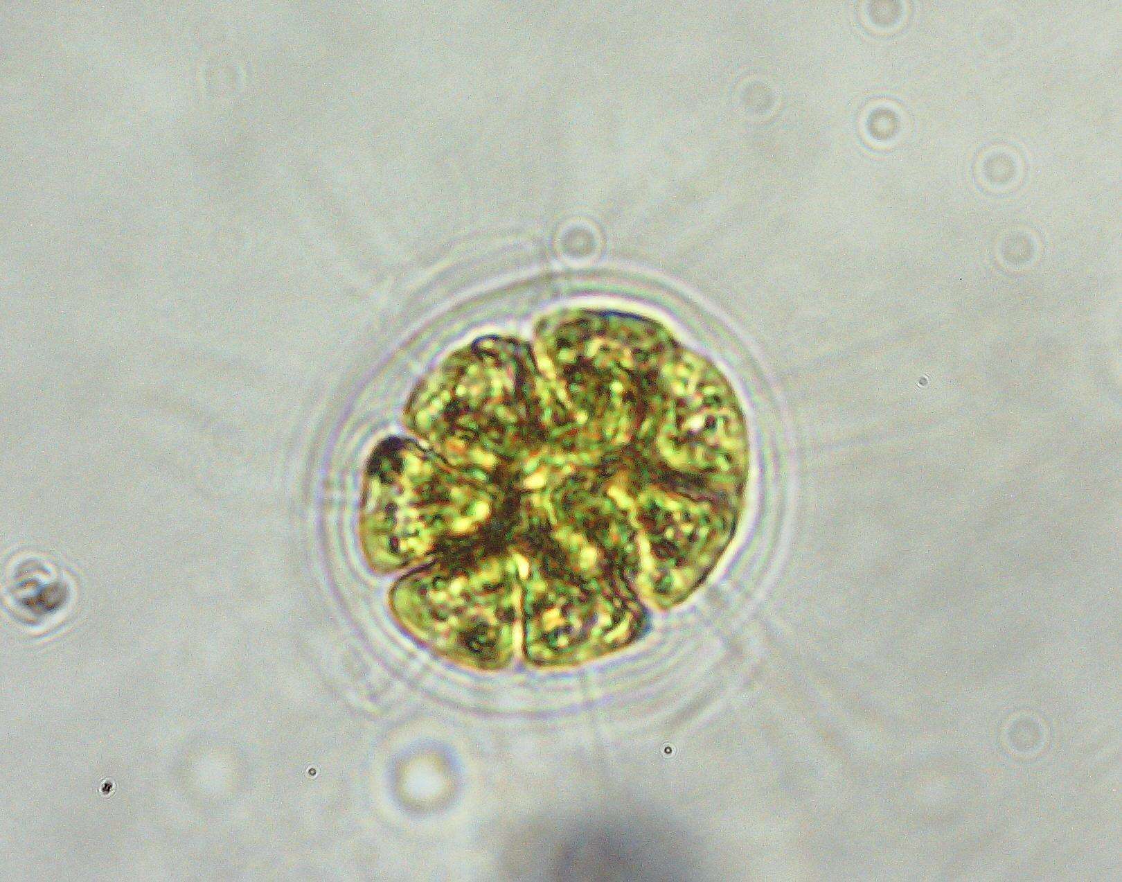
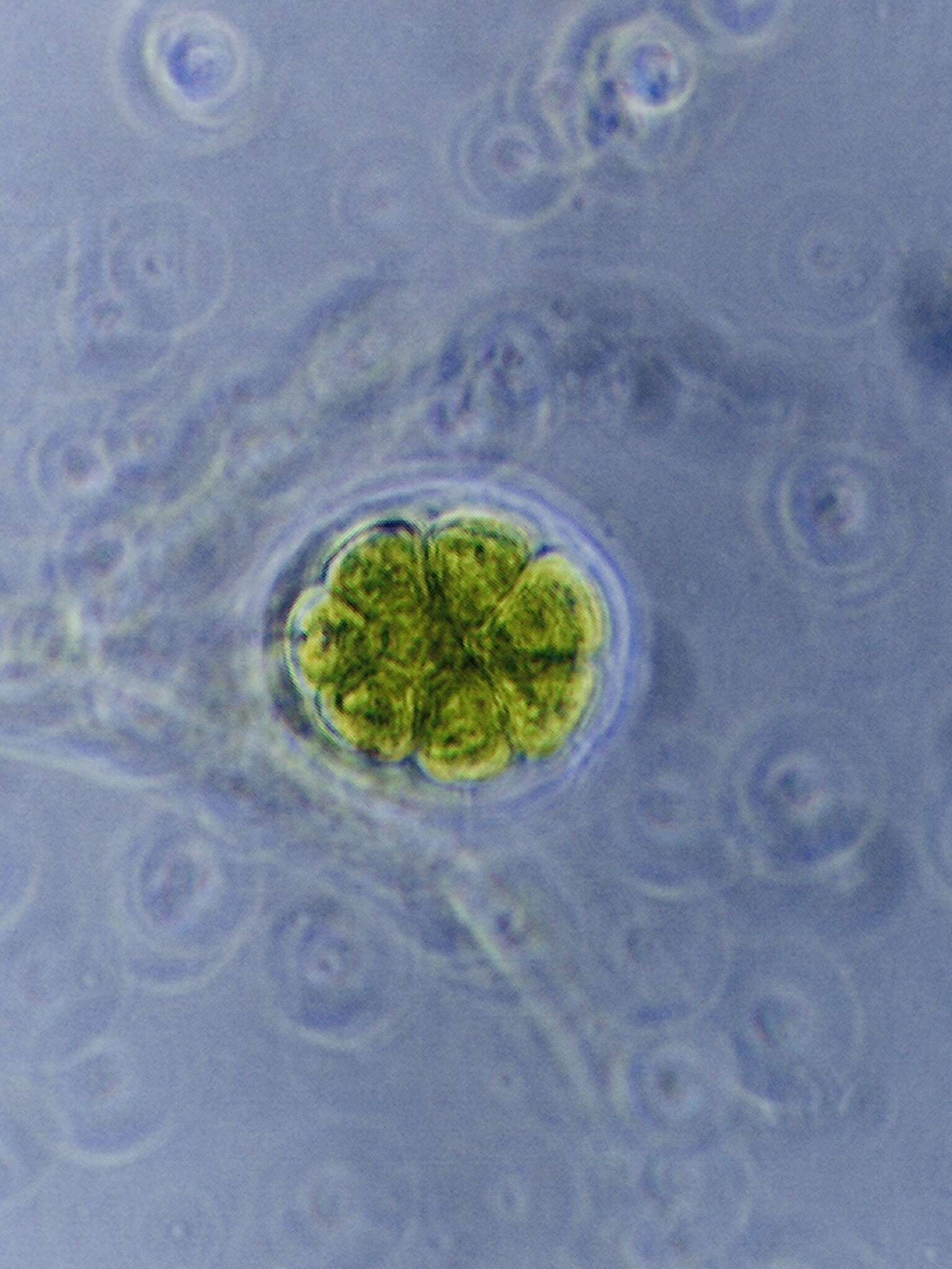
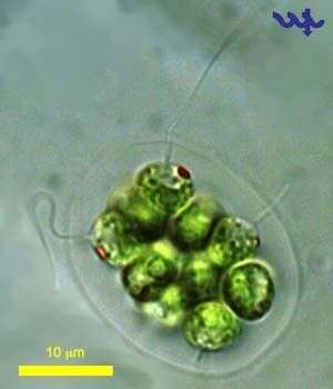
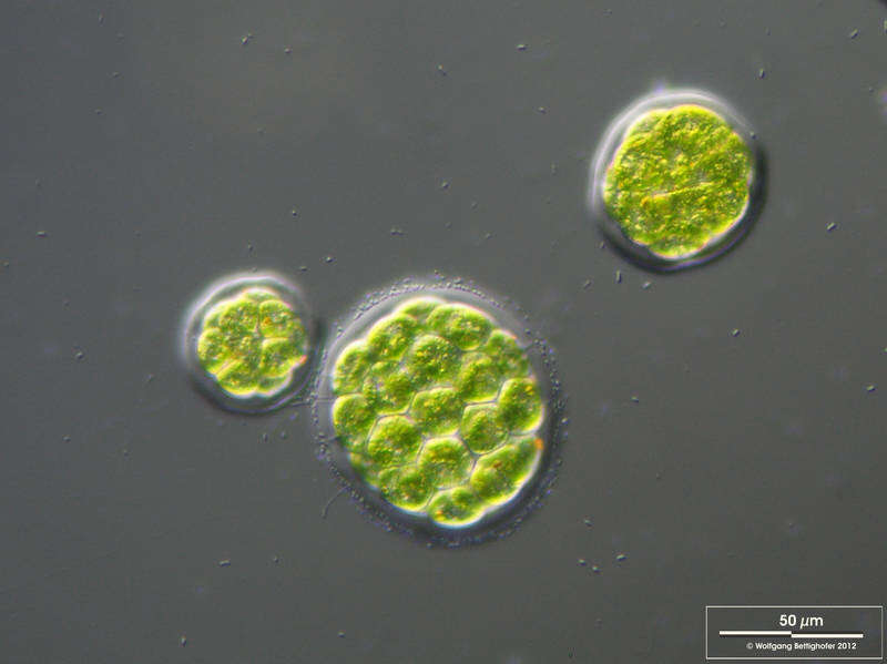
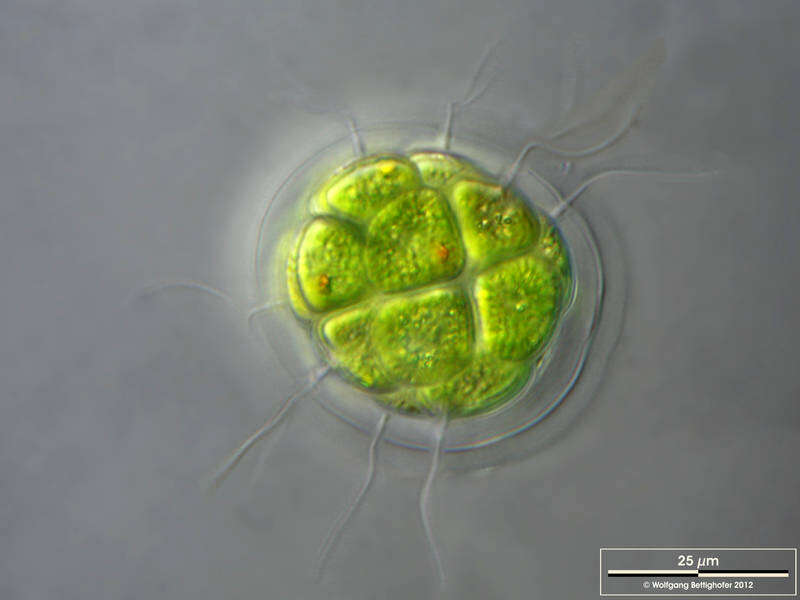
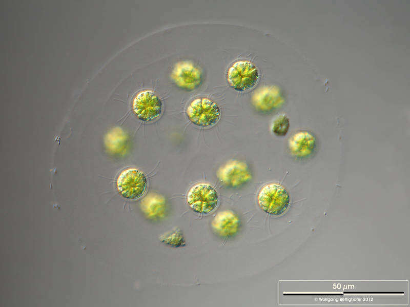
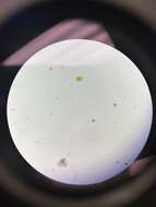
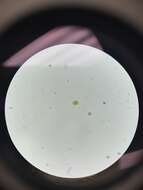
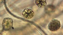
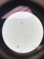
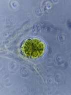
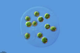
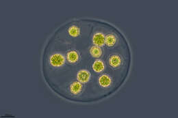
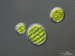
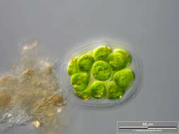
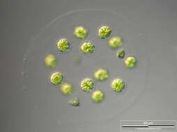
Sampling date 05/2011. Scale bars indicate 50 µm.Two images.First:DOF image showing the cell surface and a cross-section through the mucilaginous envelope. We can see eyespots, contractile vacuoles (black arrow), storage particles and holes in the mucilaginous coat, the endpoints of the channels from the contractile vacuoles to the outside (white arrow).Second:Immotile palmella stages.Please click on < or > on the image edges or on the dots at the bottom edge of the images to browse through the slides!Place name: Creek in Oder valley 100 km north east of Berlin (Germany) Latitude: 53.135032 Longitude: 14.348738Microscope Zeiss Universal, camera Olympus C7070WZ. DOF images.© Wolfgang Bettighofer,images under Creative Commons License V 3.0 (CC BY-NC-SA).For permission to use of (high resolution) images please contact
postmaster@protisten.de.For further information about the image, please click here:
Link to protisten.de page