-
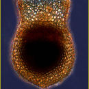
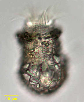
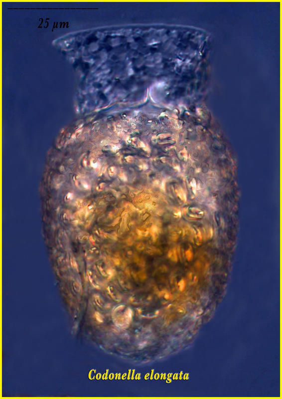
Codonella campanella.
-
-
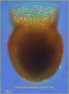
This tintinnid was found in a sample from the Eastern Mediterrean Sea in September
-
Miranda do Douro, Bragana, Portugal
-
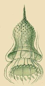
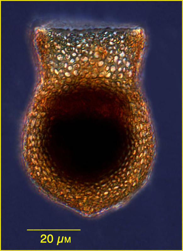
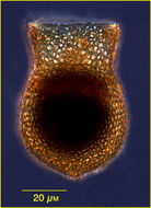
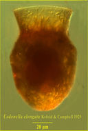
Codonella cratera lorica. Codonella cratera is a large tintinnid ciliate. A distinctive chitinous lorica is coated with xenosomes (foreign particles such as sand particles as in these individuals and diatoms). The posterior lorica is broadly spherical with a cylindrical anterior half. There is a prominent circumferential anterior adoral zone of membranelles. From freshwater pond near Boise, Idaho. Oblique illumination.
-
Miranda do Douro, Bragana, Portugal
-
-
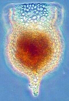
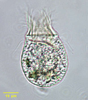
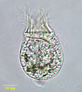
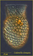
Codonella cratera (Leidy, 1877) Imhof, 1885, which has fled its lorica. The cell body is trumpet shaped when in its lorica, attaching to the lorica base by a drawn-out posterior extension. When free-swimming, the cell body assumes a more globular shape. There is a prominent circumferential anterior adoral zone of membranelles. The buccal cavity is funnel-shaped. Somatic ciliature is uniform and slightly spirals from anterior to posterior. The macronucleus is bipartite. There is a single anterior contractile vacuole. Codonella appears to be omnivorous. Ingested algae are visible in this image. From freshwater pond near Boise, Idaho. Brightfield illumination.
-
Miranda do Douro, Bragana, Portugal
-
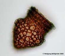
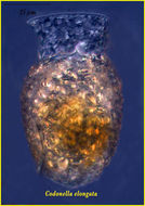
This ciliat belonging to the morphological group of oligotrichids builds marvellous amphora shaped loricae putting together mostly centric diatom frustules. Depth of focus compilation using 11 photos and CombineZ-SW developed by Alan Hadley. For more pictures and 3d-drawing see ZIP archive. Collection from littoral region (stand of Phragmites) of oligotrophic lake near Kiel (Schleswig-Holstein, Germany). Images were taken using Zeiss Universal with Olympus C7070 CCD camera.
-
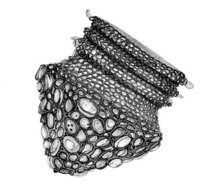
This ciliat belonging to the morphological group of oligotrichids builds marvellous amphora shaped loricae putting together mostly centric diatom frustules. This drawing (it´s an original) was inspired by a SEM picture of a good friend. Collection from littoral region (stand of Phragmites) of oligotrophic lake near Kiel (Schleswig-Holstein, Germany).
-
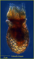
This ciliat belonging to the morphological group of oligotrichids builds marvellous amphora shaped loricae putting together mostly centric diatom frustules. The scale bar indicates 25 µm. The specimen was gathered in the wetlands of Oderbruch (Oder valley 100 km north east of Berlin). The image was built up using several photomicrographic frames with manual stacking technique. Images were taken using Zeiss Universal with Olympus C7070 CCD camera.Image under Creative Commons License V 3.0 (CC BY-NC-SA).
-
-
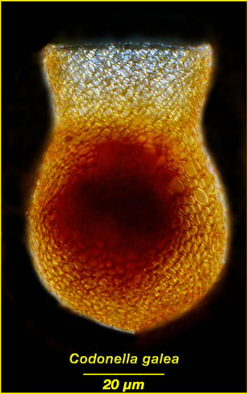
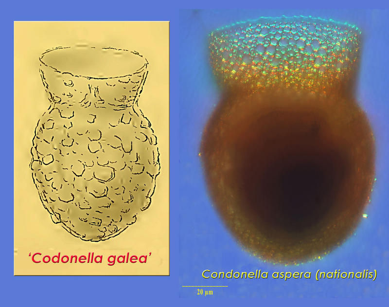
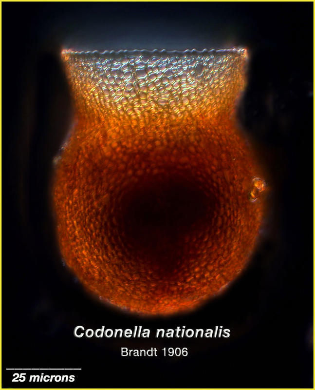
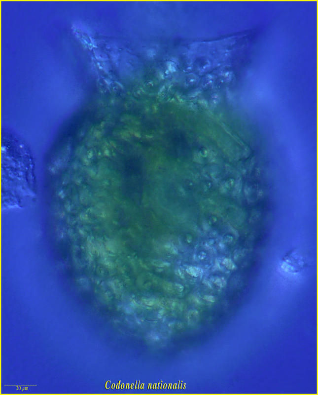
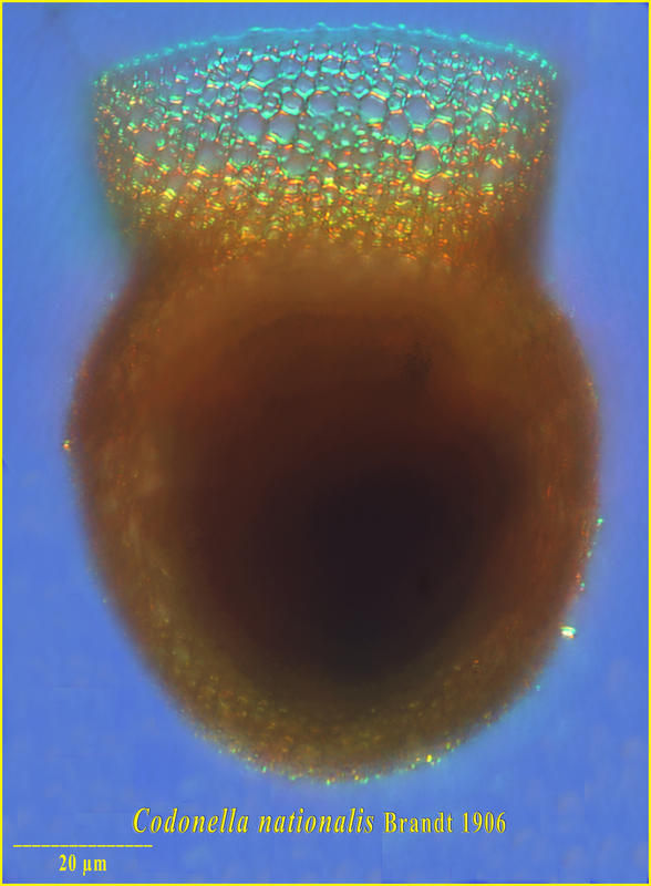
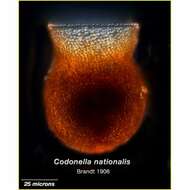
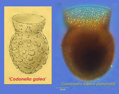
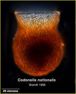
Codonella aspera was first depicted by Fol in 1884 as Codonella galea. The organism described by Fol was later named Codonella aspera. Codonella nationalis described by Brandt in 1906 and now appears to be a morph of C. aspera.
-
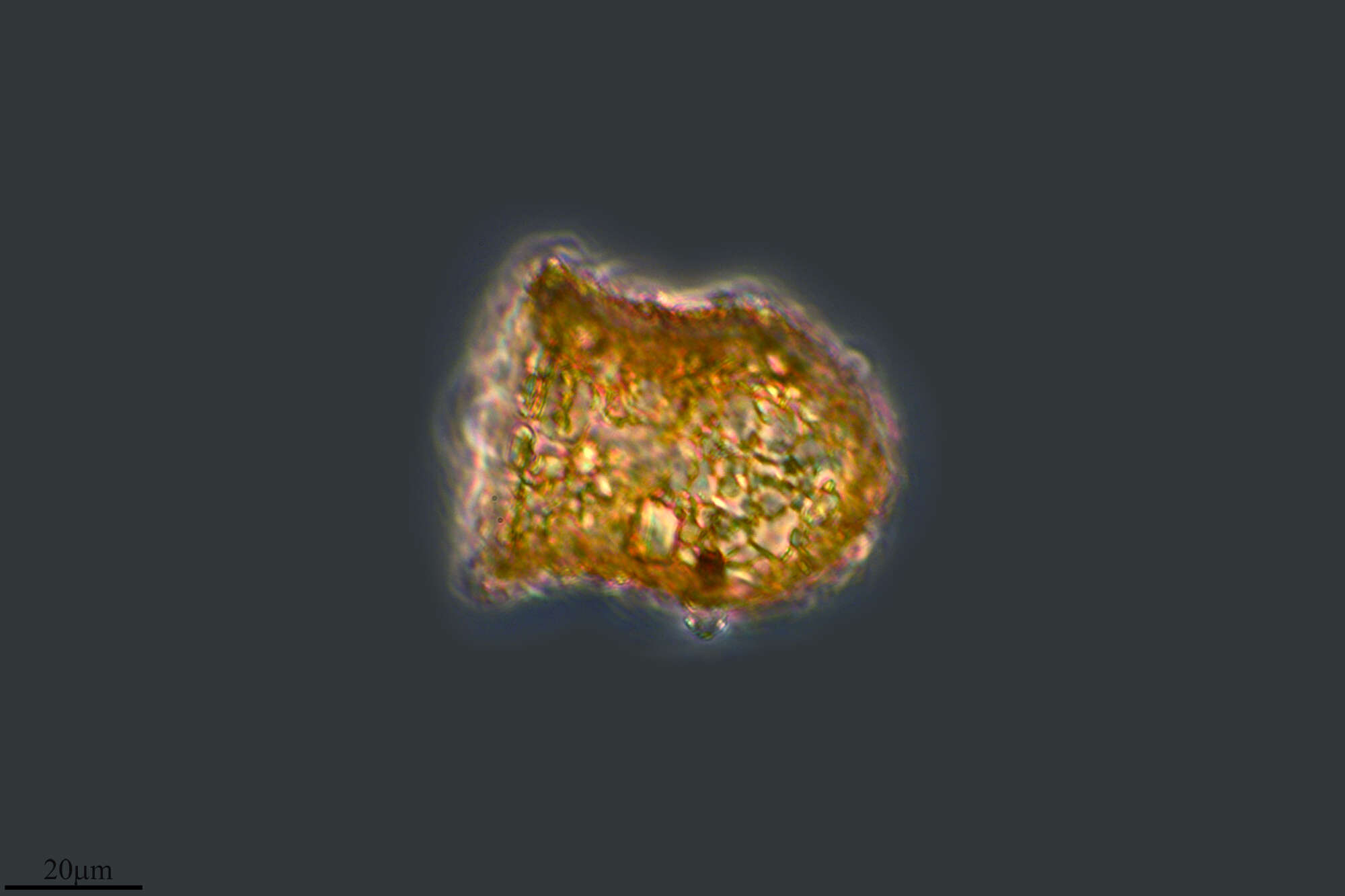
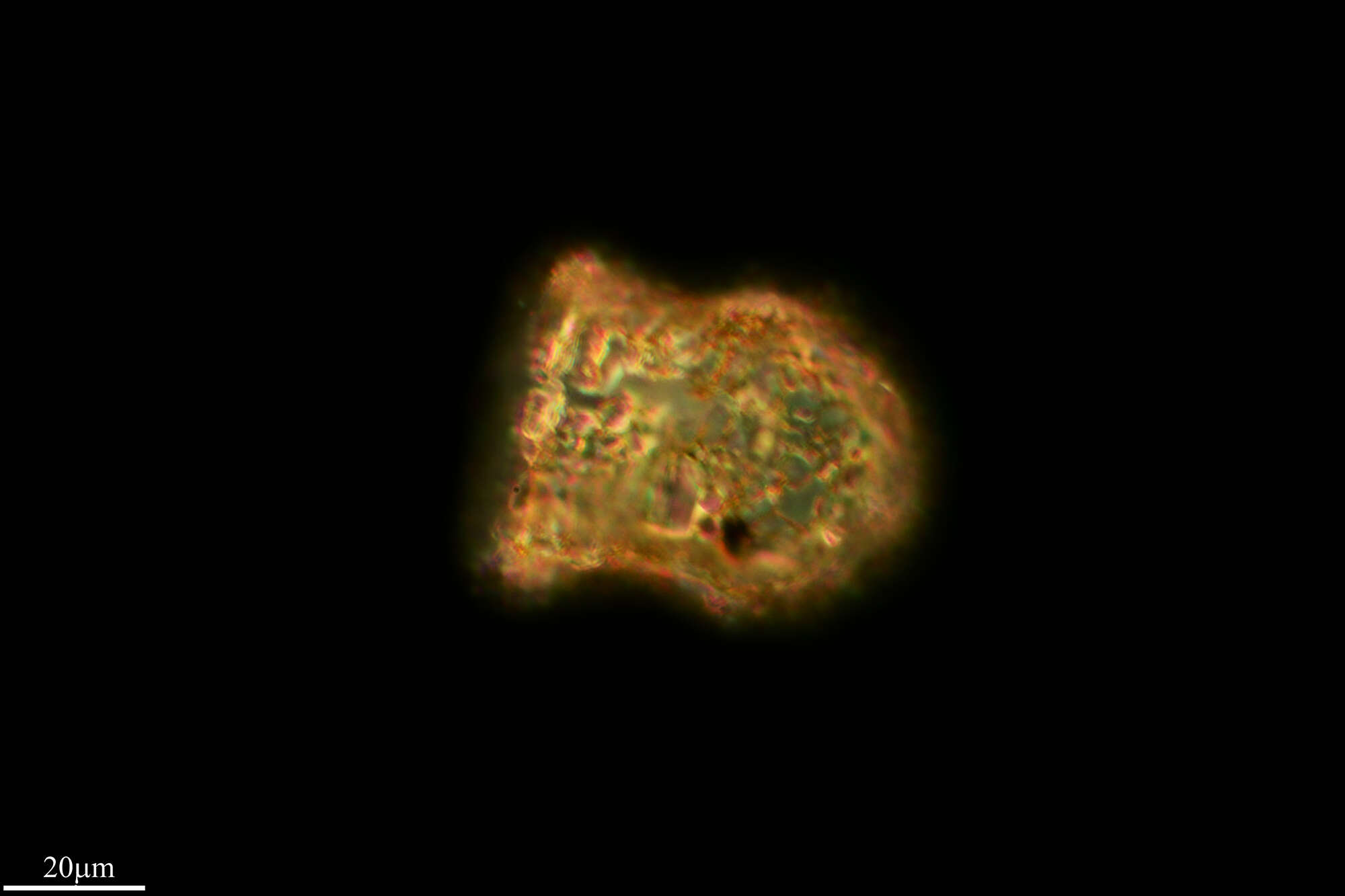
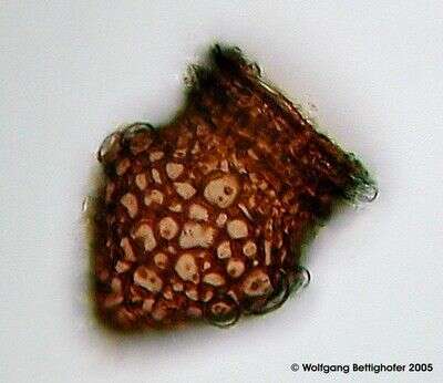
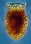
Here only the shell or lorica is visible. The specimen was from the South Pacific
-
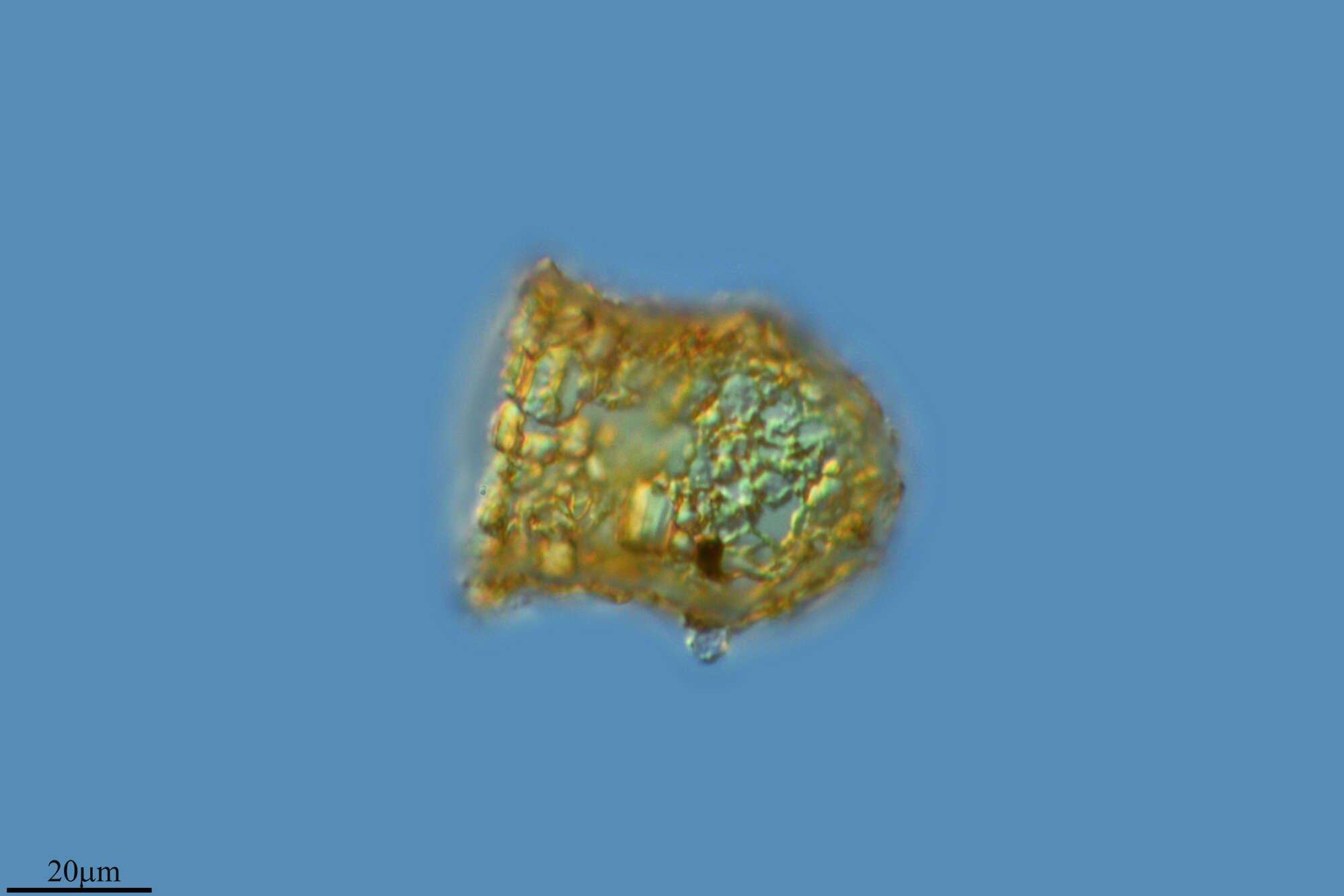
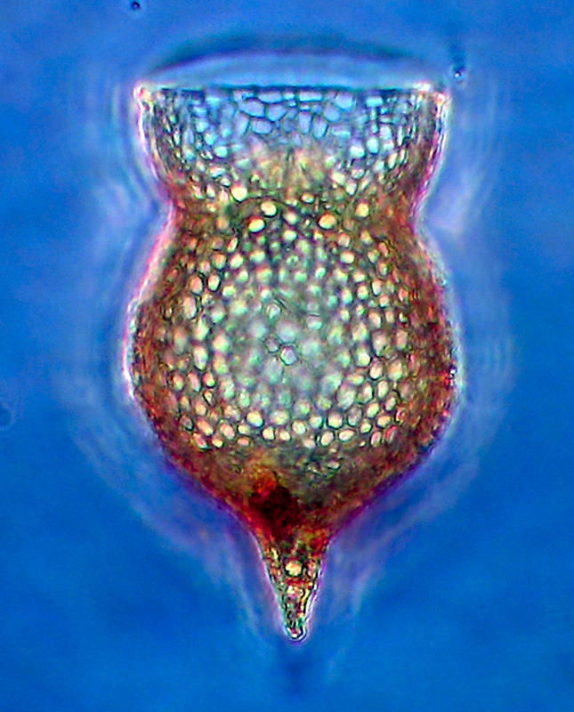
This specimen is from the central Med found in material from the Boum cruise.
-
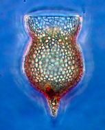
The species is likely a morph of Codonella aspera
-
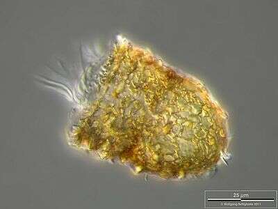
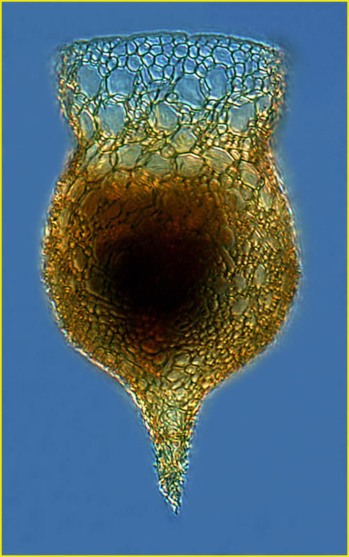
Specimen from the Bay of Villefrtanche in April 2010
-
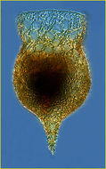
Specimen from the Bay of Villefranche in Jan 2003- lugol's fixed. The species is likely a morph of C. aspera.
-
Specimen from the Tara expedition, Station 68. Lugol's-fixed specimen probably had coccoliths covering lost with fixation.
-
-
From the central Med.
-
Lugol's-fixed specimen from Feb 2003 in the Bay of Villefranche.
-
Live specimen from Bay in Villefranche in Sept 2010. The lorica is covered in coccoliths.