-
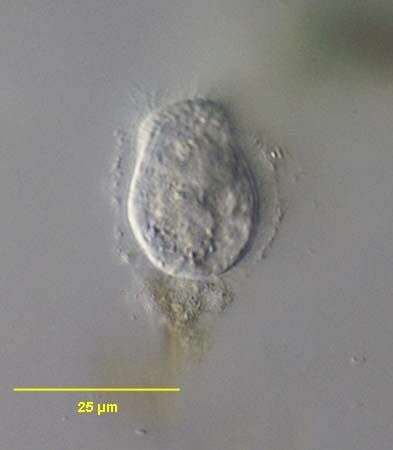
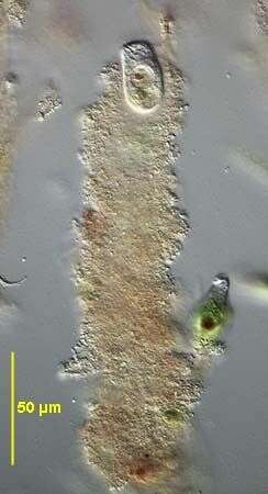
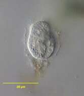
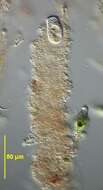
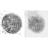
Sagittaria hyalina (FOISSNER, CZAPIK & WIACKOWSKI, 1981) just beginning its mucus case. Unlike the tube-dwelling colpodid, Maryna, Sagittaria usually has its anterior end protruding from the mucus tube.Collected from an ephemeral puddle on the lawn of a public park in Boise, Idaho.September 2006.DIC.
-
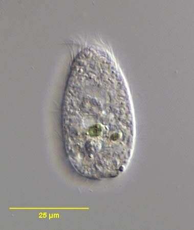
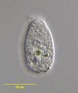
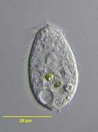
Sagittaria hyalina (FOISSNER, CZAPIK & WIACKOWSKI, 1981) which has fled its mucus case. Collected from an ephemeral puddle on the lawn of a public park in Boise, Idaho.September 2006.DIC.
-
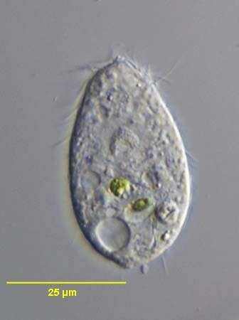
Sagittaria hyalina (FOISSNER, CZAPIK & WIACKOWSKI, 1981) which has fled its mucus case. Collected from an ephemeral puddle on the lawn of a public park in Boise, Idaho.September 2006.DIC.
-
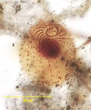
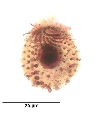
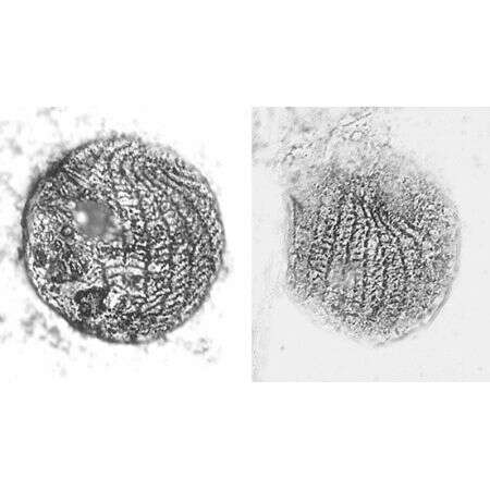
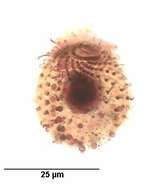
Infraciliature of the cyrtolophosidid ciliate, Sagittaria hyalina (FOISSNER, CZAPIK & WIACKOWSKI, 1981) viewed from the right apicoolateral aspect. Collected from an ephemeral puddle on the lawn of a public park in Boise, Idaho.September 2006.Stained by the silver carbonate technique (see Foissner, W. Europ. J. Protistol., 27:313-330;1991).Brightfield.
-
Infraciliature of the cyrtolophosidid ciliate, Sagittaria hyalina (FOISSNER, CZAPIK & WIACKOWSKI, 1981) viewed from the right apicoolateral aspect. Larger densely stained circular structures are mucocysts which the organism extrudes to form its mucus case.Collected from an ephemeral puddle on the lawn of a public park in Boise, Idaho.September 2006.Stained by the silver carbonate technique (see Foissner, W. Europ. J. Protistol., 27:313-330;1991).Brightfield.
-
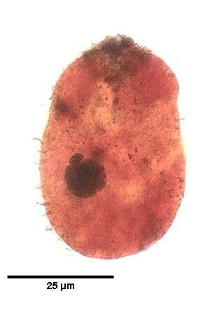
Sagittaria hyalina (FOISSNER, CZAPIK & WIACKOWSKI, 1981). The densely stained macronucleus and ellipsoid micronucleus in its perinuclear space (11 o'clock) are seen here.Collected from an ephemeral puddle on the lawn of a public park in Boise, Idaho.September 2006.Stained by the silver carbonate technique (see Foissner, W. Europ. J. Protistol., 27:313-330;1991).Brightfield.
-
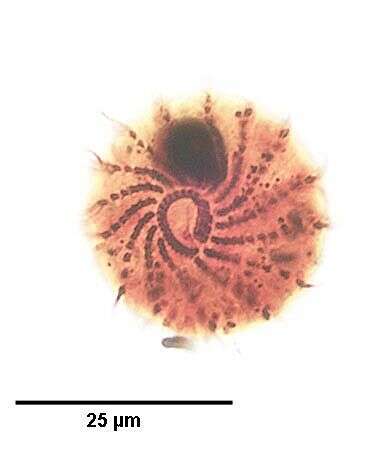
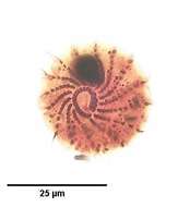
Infraciliature of the cyrtolophosidid ciliate, Sagittaria hyalina (FOISSNER, CZAPIK & WIACKOWSKI, 1981) viewed from the anterior apical aspect.The right paraoral membrane consists of ciliated dikinetids.Four adoral membranelles lie along the left border of the cytostome between the two ends of the C-shaped paraoral membrane. Collected from an ephemeral puddle on the lawn of a public park in Boise, Idaho.September 2006.Stained by the silver carbonate technique (see Foissner, W. Europ. J. Protistol., 27:313-330;1991).Brightfield.
-
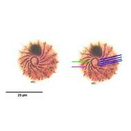
Infraciliature of the cyrtolophosidid ciliate, Sagittaria hyalina (FOISSNER, CZAPIK & WIACKOWSKI, 1981) viewed from the anterior apical aspect.The right paraoral membrane consists of ciliated dikinetids (green line).Four adoral membranelles (blue lines) lie along the left border of the cytostome between the two ends of the C-shaped paraoral membrane. About 15 somatic kineties spiral around the long axis to abut the cytostome anteriorly (pink line). Collected from an ephemeral puddle on the lawn of a public park in Boise, Idaho.September 2006.Stained by the silver carbonate technique (see Foissner, W. Europ. J. Protistol., 27:313-330;1991).Brightfield.
-
Sagittaria hyalina (FOISSNER, CZAPIK & WIACKOWSKI, 1981) seen in its mucus case. Unlike the tube-dwelling colpodid, Maryna, Sagittaria usually has its anterior end protruding from the mucus tube.Collected from an ephemeral puddle on the lawn of a public park in Boise, Idaho.September 2006.Stained by the silver carbonate technique (see Foissner, W. Europ. J. Protistol., 27:313-330;1991).DIC.
-
Stained by the Klein-Foissner dry silver nitrate technique (see Foissner, W. Europ. J. Protistol., 27:313-330;1991).