-
Canencia, Madrid, Spain
-
Galende, Castile and Len, Spain
-
Galende, Castile and Len, Spain
-
Ribadelago de Franco, Castille and Leon, Spain
-
San Martin De Castaneda, Castille and Leon, Spain
-
Ribadelago, Castille and Leon, Spain
-
Galende, Castile and Len, Spain
-
Galende, Castile and Len, Spain
-
Ribadelago de Franco, Castille and Leon, Spain
-
Galende, Castile and Len, Spain
-
Canencia, Madrid, Spain
-
Galende, Castile and Len, Spain
-
Galende, Castille and Leon, Spain
-
Ribadelago, Castille and Leon, Spain
-
Ribadelago de Franco, Castille and Leon, Spain
-
Galende, Castile and Len, Spain
-
Ribadelago de Franco, Castille and Leon, Spain
-
Ribadelago, Castille and Leon, Spain
-
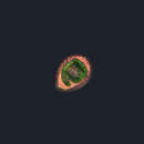
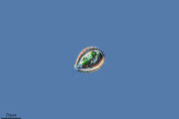
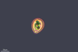
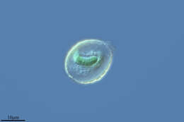
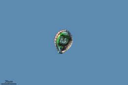
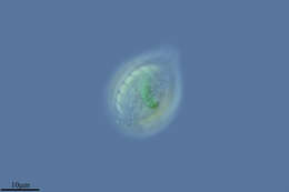
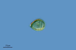
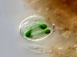
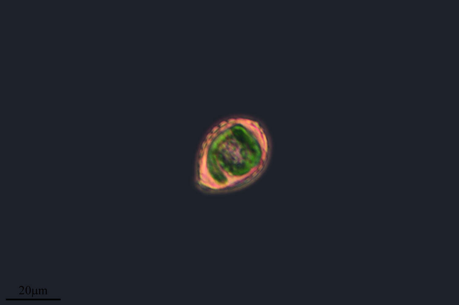
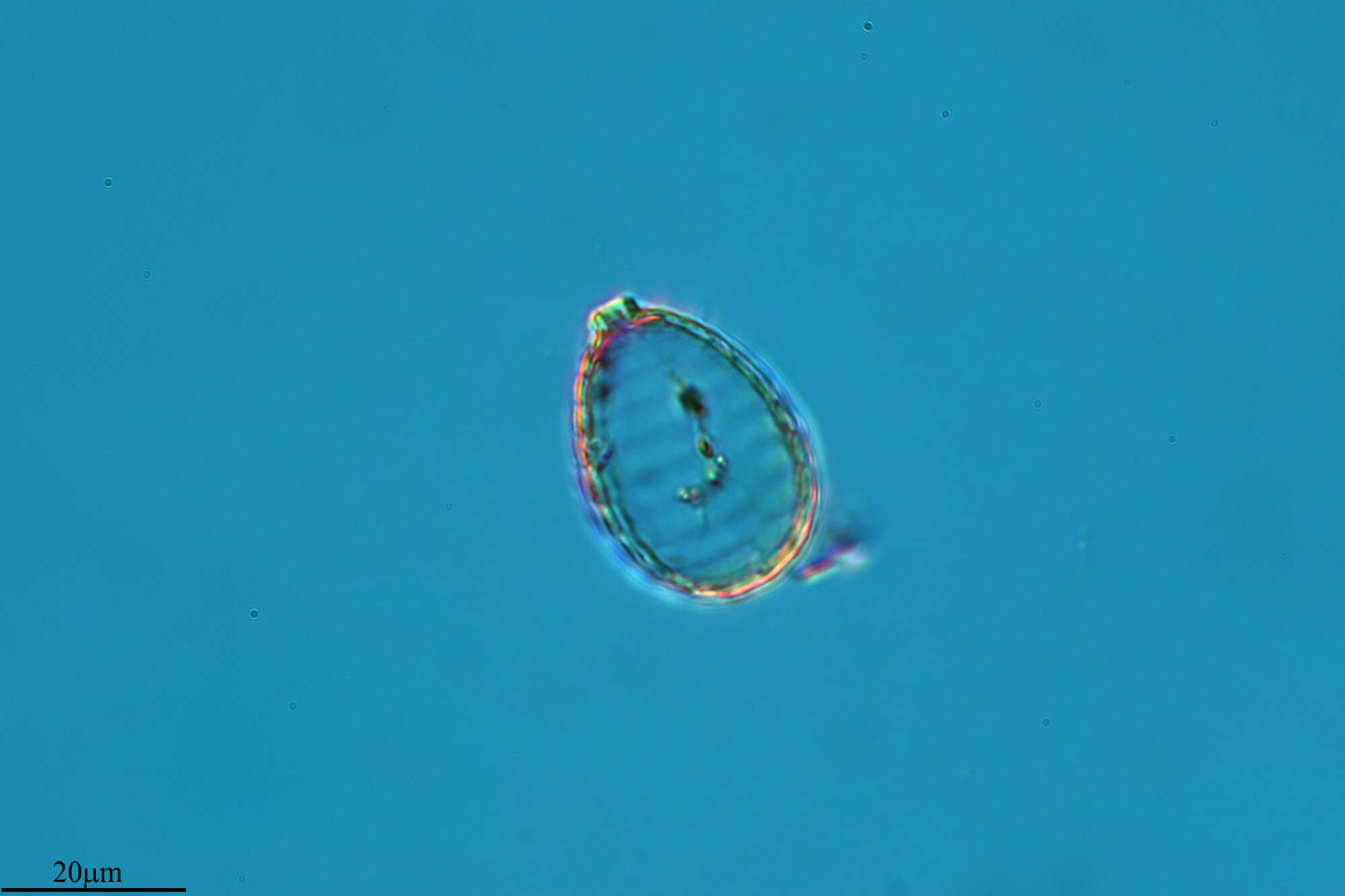
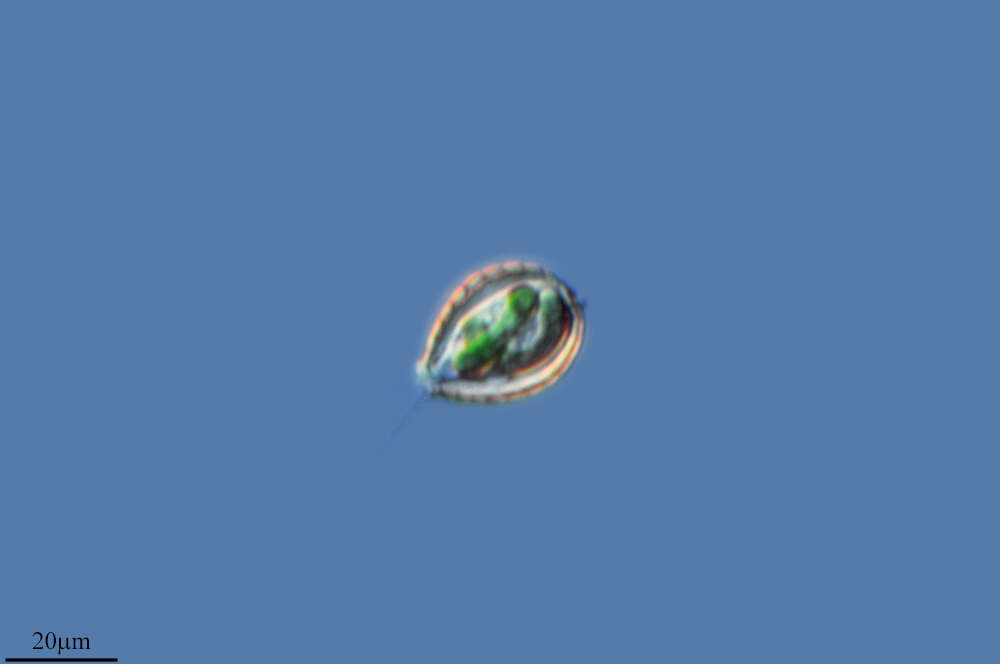
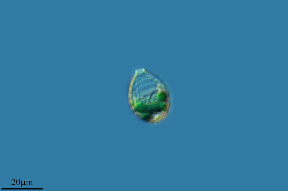
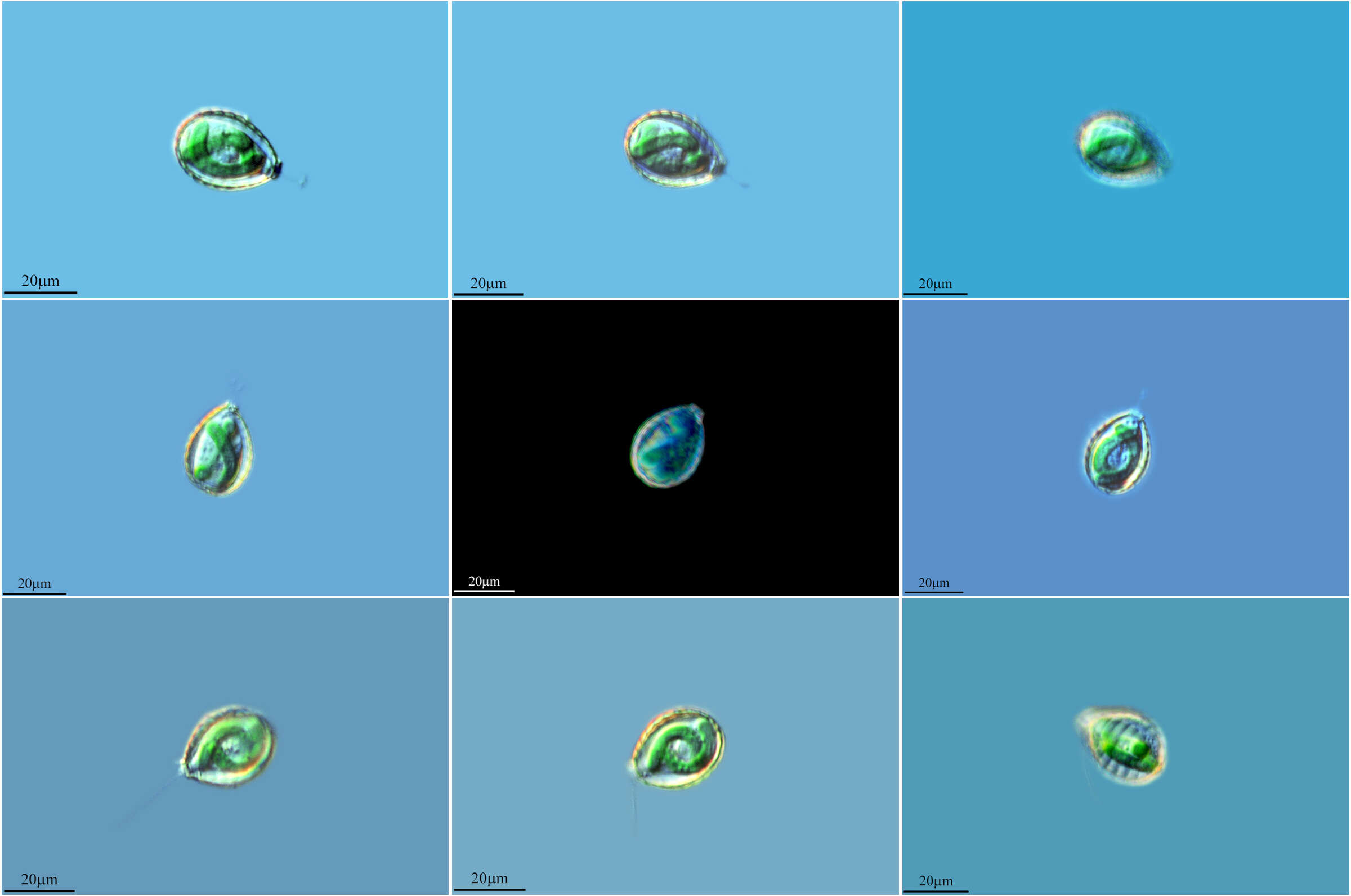
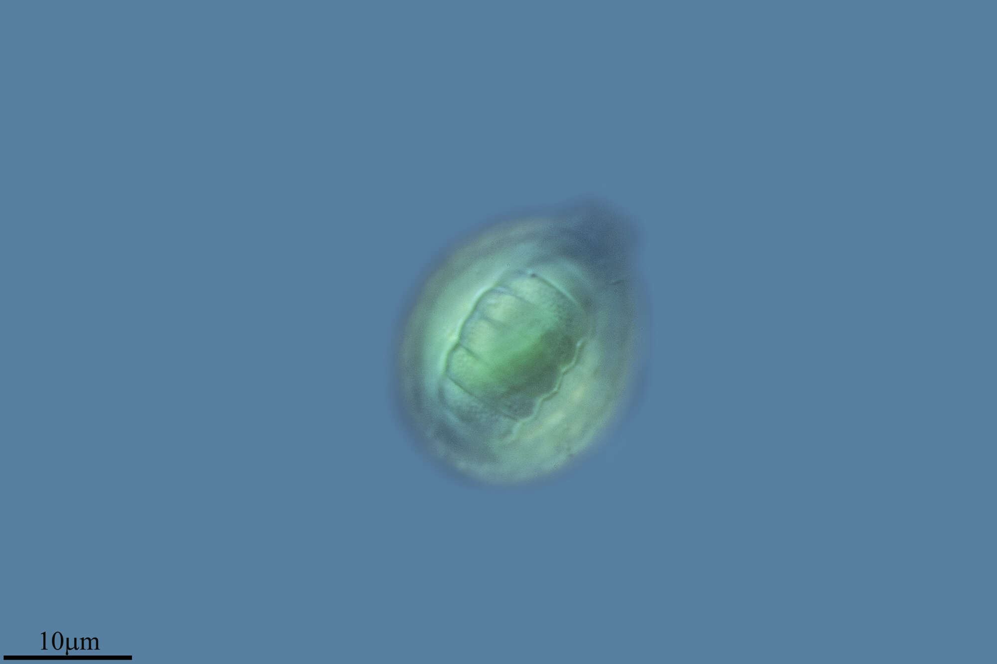
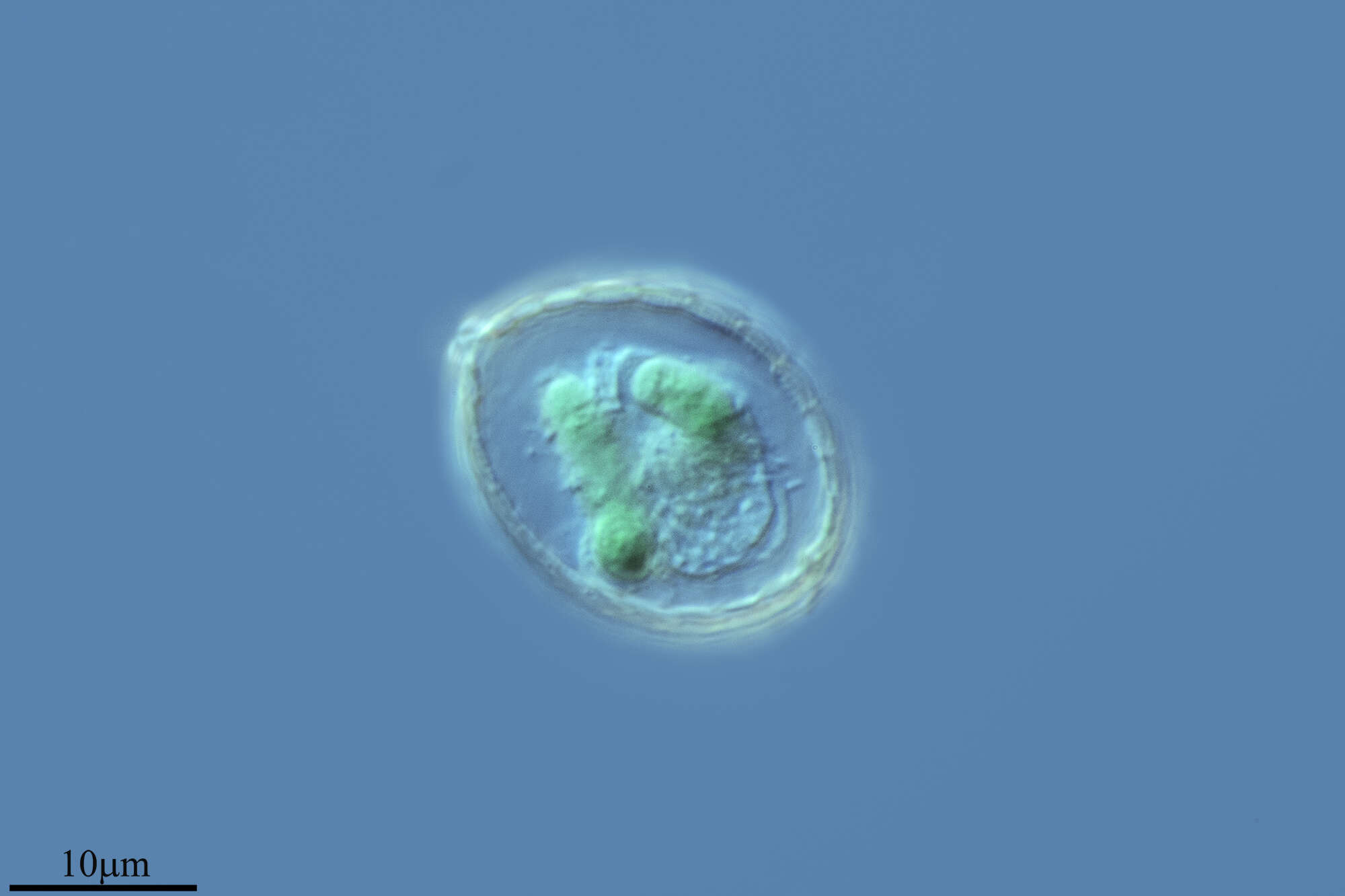
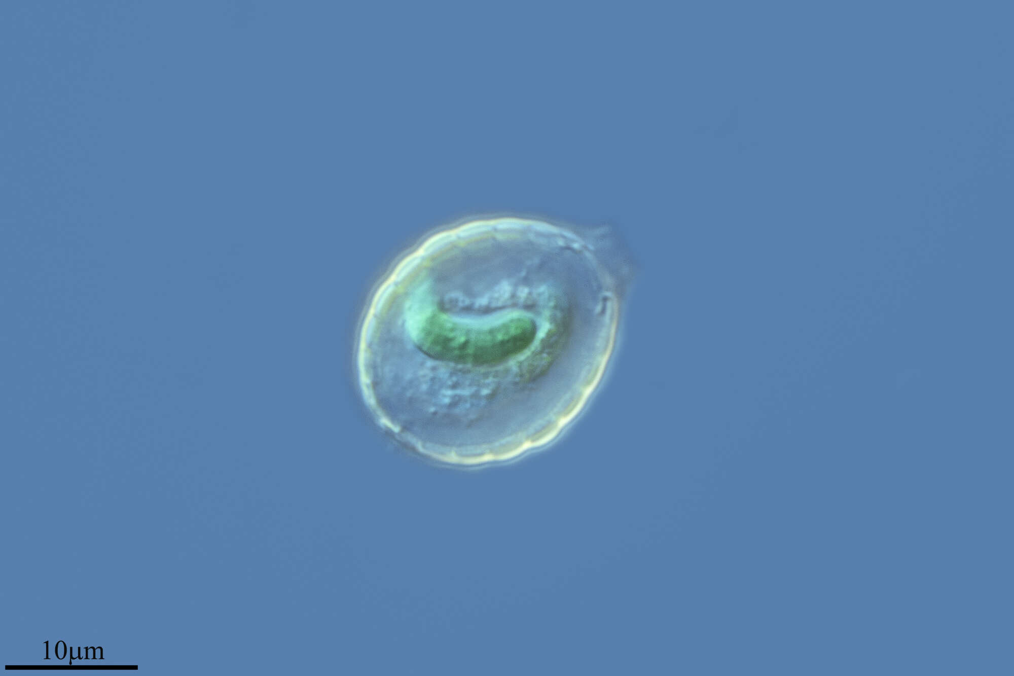
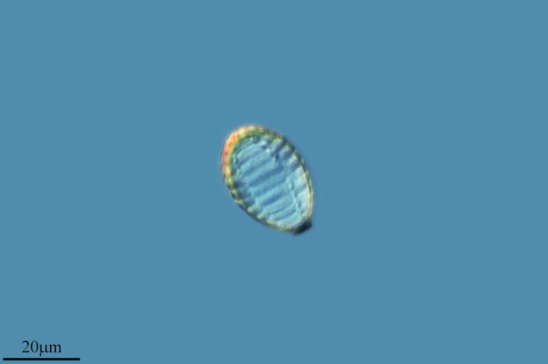
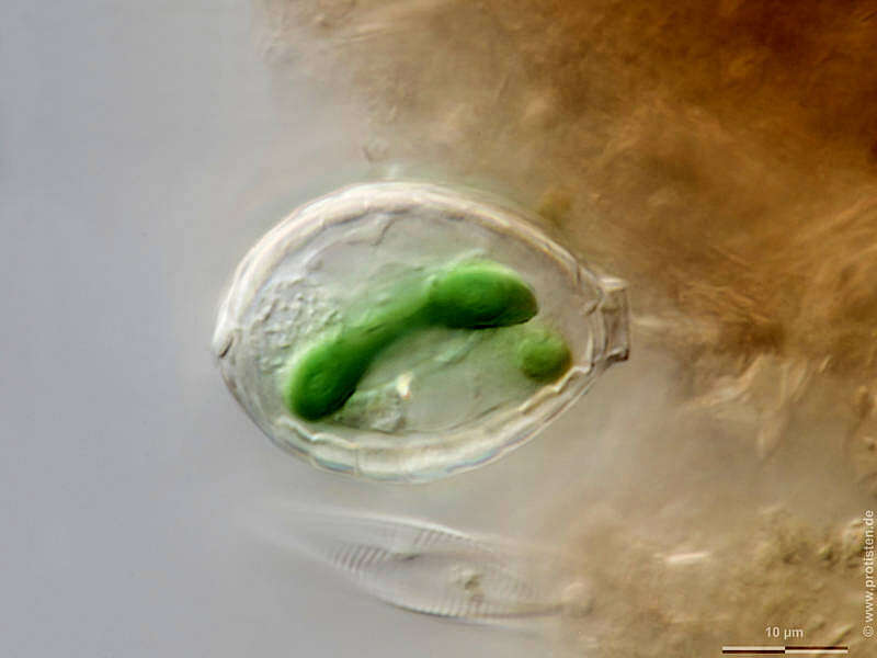
Paulinella (paul-in-ella) is a testate amoeba but this species is distinguished by the presence of (usually two) curved endosymbiotic blue green algae. The small aperture of the lorica is to the top of the image. Differential interference contrast.
-
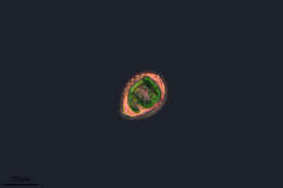
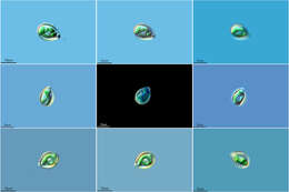
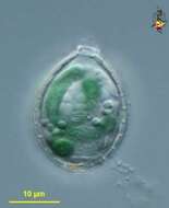
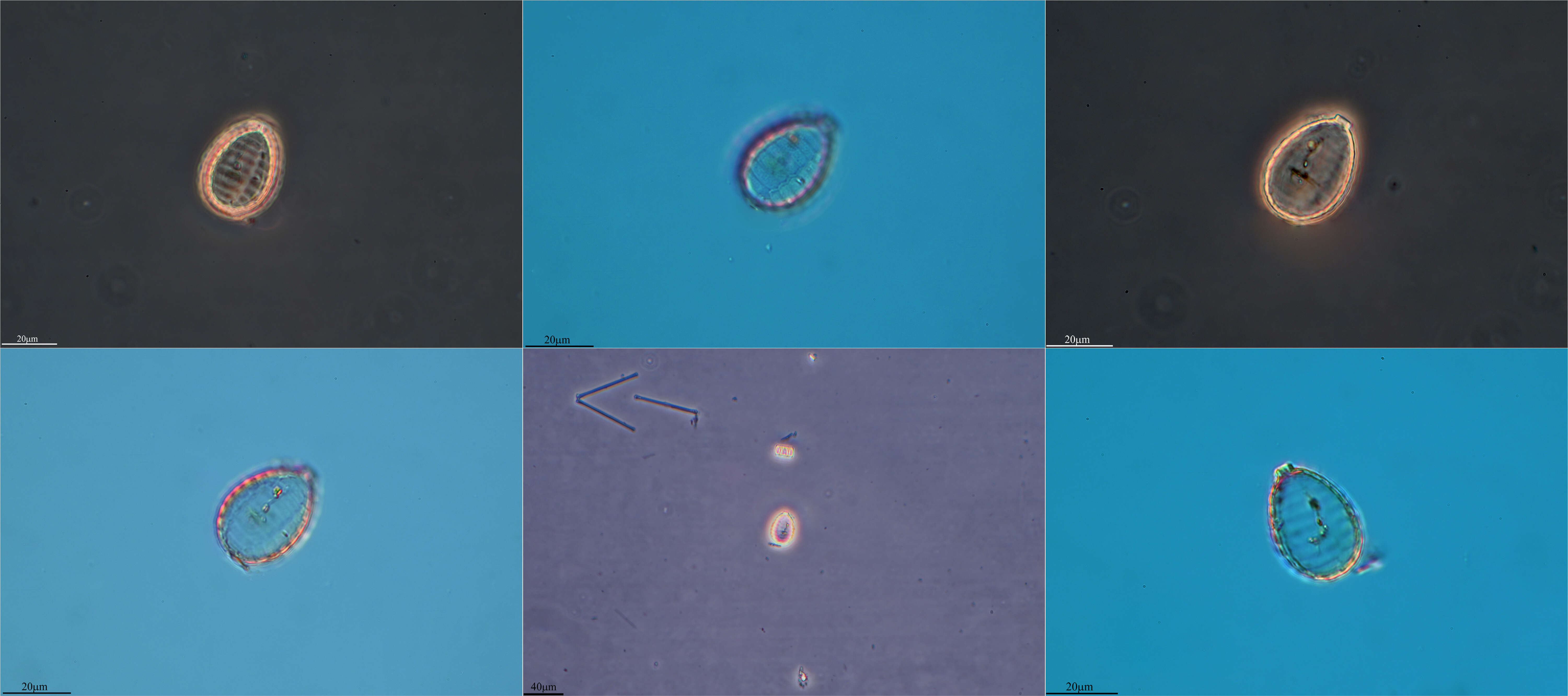
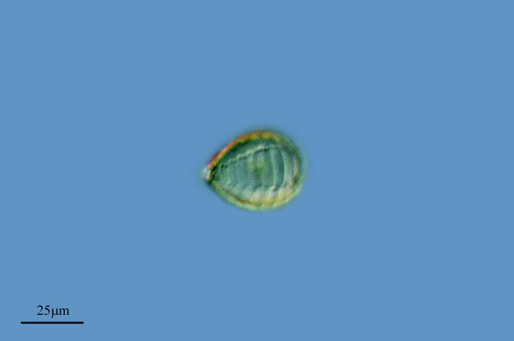
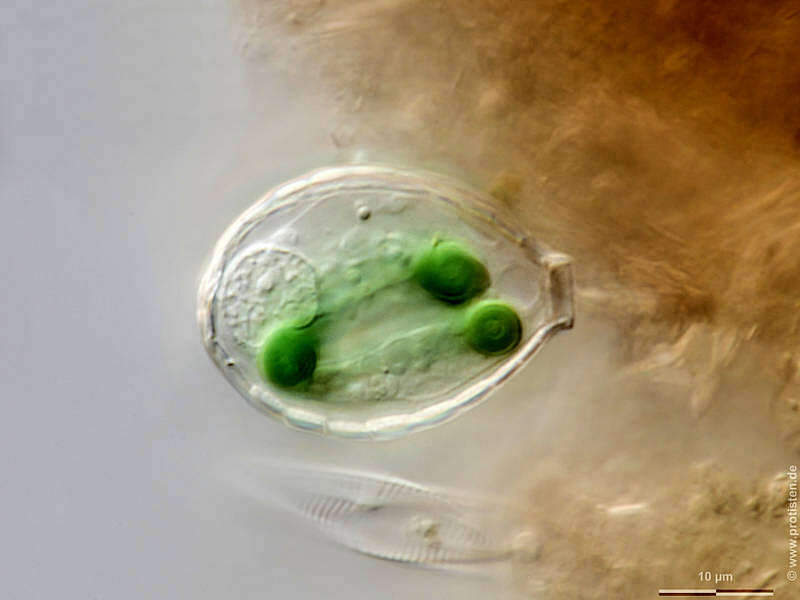
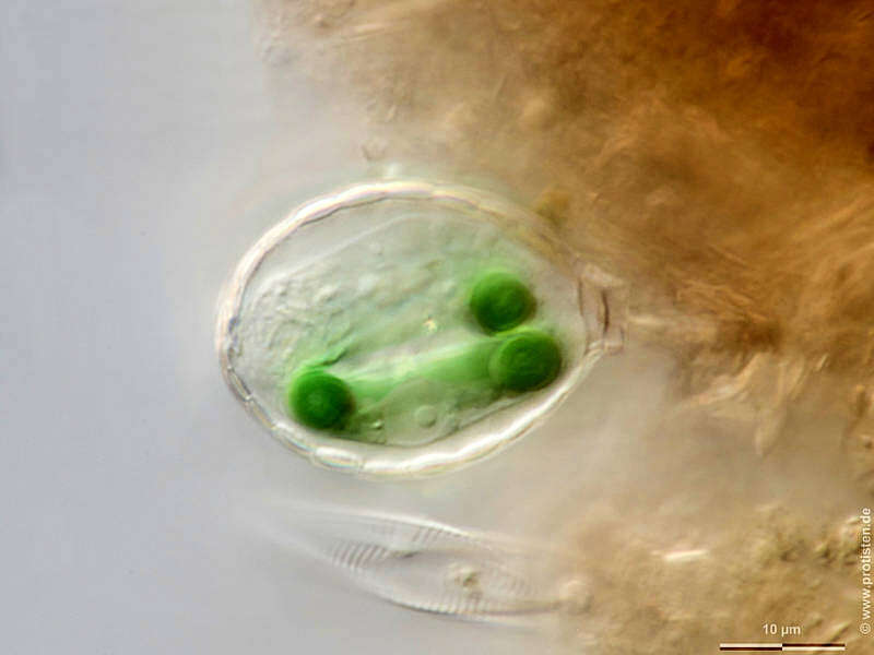
Sampling date 06/2020. Scale bars indicate 10 µm.Eight images.First:Synoptic representation of the test (shell).Second to eighth:Optical cross-sections showing chromatophores. The nucleus is located posteriorly in the cell’s fundus opposite the aperture (images No. 6 and 7).Please click on < or > on the image edges or on the dots at the bottom edge of the images to browse through the slides!Place name: Creek in 63649 Rossbach (Lower Franconia, Germany) Latitude: 49.87747709 Longitude: 9.23824668Microscope Zeiss Axioplan, camera Olympus OM-D M5 MKII. DOF images.© Wolfgang Bettighofer,images under Creative Commons License V 3.0 (CC BY-NC-SA).For permission to use of (high resolution) images please contact
postmaster@protisten.de.For further information about the image, please click here:
Link to protisten.de page
-
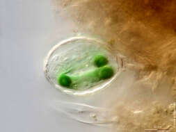
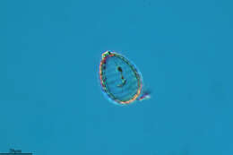
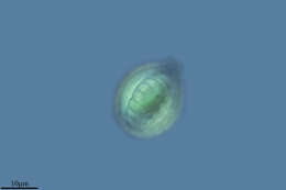
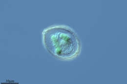
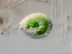
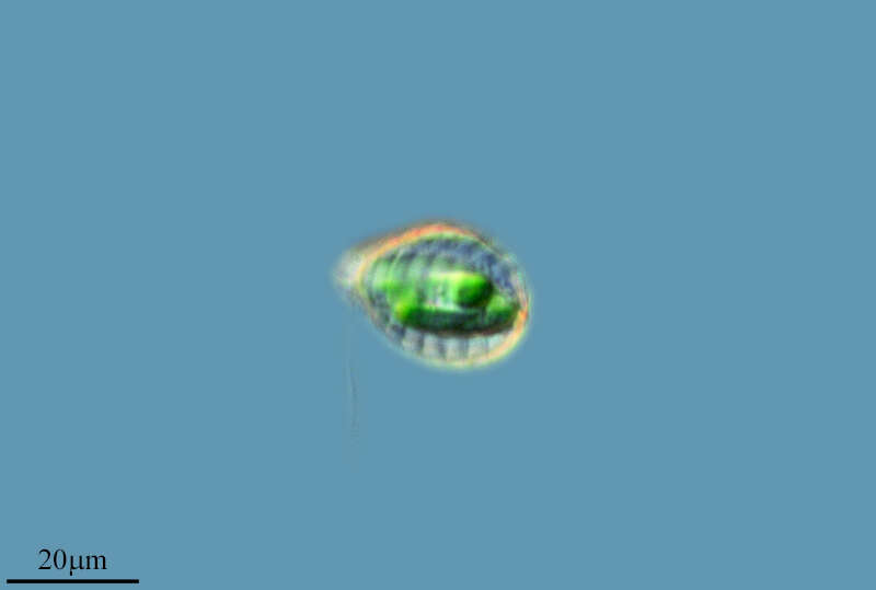
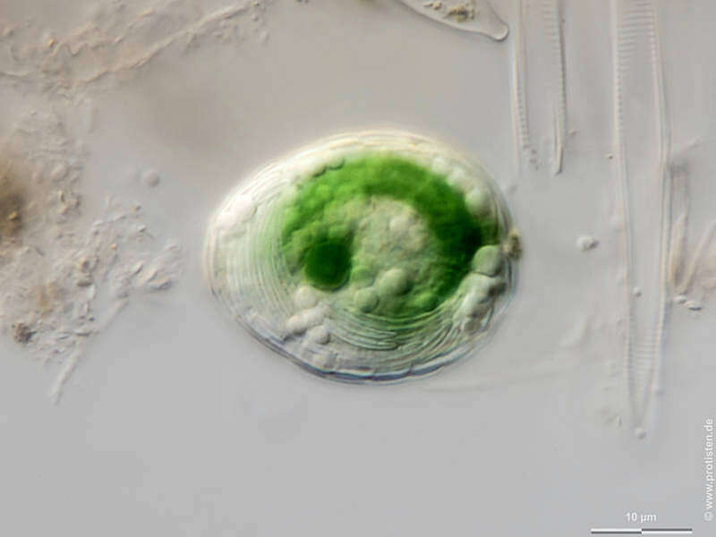
Scale bars indicate 10 µm.Five images. Another specimen.First:Synoptic representation of the test (shell).Second to fifth:Optical cross-sections showing the two chromatophores and numerous storage particles. The last image shows many shell plates pre-produced for the daughter cell.Please click on < or > on the image edges or on the dots at the bottom edge of the images to browse through the slides!Place name: Creek in 63649 Rossbach (Lower Franconia, Germany) Latitude: 49.87747709 Longitude: 9.23824668Microscope Zeiss Axioplan, camera Olympus OM-D M5 MKII. DOF images.© Wolfgang Bettighofer,images under Creative Commons License V 3.0 (CC BY-NC-SA).For permission to use of (high resolution) images please contact
postmaster@protisten.de.For further information about the image, please click here:
Link to protisten.de page
-
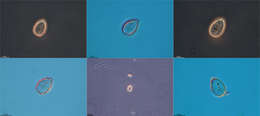
Sampling date 06/2020. Scale bars indicate 10 µm.Eight images.First:Synoptic representation of the test (shell).Second to eighth:Optical cross-sections showing chromatophores. The nucleus is located posteriorly in the cell’s fundus opposite the aperture (images No. 6 and 7).Please click on < or > on the image edges or on the dots at the bottom edge of the images to browse through the slides!Place name: Creek in 63649 Rossbach (Lower Franconia, Germany) Latitude: 49.87747709 Longitude: 9.23824668Microscope Zeiss Axioplan, camera Olympus OM-D M5 MKII. DOF images.© Wolfgang Bettighofer,images under Creative Commons License V 3.0 (CC BY-NC-SA).For permission to use of (high resolution) images please contact
postmaster@protisten.de.For further information about the image, please click here:
Link to protisten.de page
-
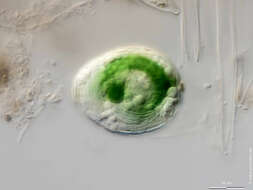
Scale bars indicate 10 µm.Five images. Another specimen.First:Synoptic representation of the test (shell).Second to fifth:Optical cross-sections showing the two chromatophores and numerous storage particles. The last image shows many shell plates pre-produced for the daughter cell.Please click on < or > on the image edges or on the dots at the bottom edge of the images to browse through the slides!Place name: Creek in 63649 Rossbach (Lower Franconia, Germany) Latitude: 49.87747709 Longitude: 9.23824668Microscope Zeiss Axioplan, camera Olympus OM-D M5 MKII. DOF images.© Wolfgang Bettighofer,images under Creative Commons License V 3.0 (CC BY-NC-SA).For permission to use of (high resolution) images please contact
postmaster@protisten.de.For further information about the image, please click here:
Link to protisten.de page