nomes no trilho de navegação
“Marseniopsis pacifica, n. sp. (Pl. I. figs. 7-27).
Habitat.-Southern Ocean (Kerguelen Island).
Only one representative (female) of the species was obtained, by the dredge between 10 and 100 fathoms.
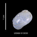
In its preserved state the animal measured 18 mm. in length, 13.5 in breadth, and 12 in height. The foot had a length of 13, and a breadth of mostly about 5.5 mm., increasing to 8 in front. The length of the tail was 6 mm., and that of the tentacles 4 mm. The colour of the upper surface was reddish yellow with several almost purple-red spots, and also apparently with scattered white nodules; the under surface of the animal was
yellowish white.
The back was covered with coarse tubercles, very much as in the Onchidiopsides, which this form on the whole resembled. The respiratory canal measured 4-5 mm., was anteriorly bent slightly upwards, but otherwise exhibited the usual characters. The mantle border was thick, swollen, and not very broad. The ordinary stripes round the base of the foot were absent; the foot was as usual, except that the furrow of the anterior margin was very strongly developed, especially towards the middle line, where a slit-like hole was formed. The head and tentacles exhibited the ordinary form. The shell and the viscera were indistinctly seen as a greyish mass shining through the dorsal surface. The inside of the mantle, adjacent to the shell, was almost white. The shell (fig. 7) measured 15 mm. in length, about 12 in breadth, and 10 in height. The spiral was small, and exhibited two turns; the last turn was very large and much vaulted. Through the thin calcareous shell was seen the dark-grey coating of the superior visceral mass.
After removing the shell and the enveloping coat, the superior viscera exhibited the following arrangement (fig. 8). Far back lay the large, whitish, apparently granular ovary (fig. 8, a); in front of this was situated the large, very faint greenish-grey foliated gland (fig. 8, b), and across the lower surface of the latter the whitish intestine could be seen. Further forward to the right lay the faint yellow mucus- and albumen-gland, with the white rectum along the left side; both rectum and gland extended forwards. To the left was seen the roof of the branchial cavity, apparently of a white colour, owing to the presence of the secreted substance (fig. 8, d) of the kidney or of the foliated gland. Before this white portion, to the left, lay the branchia, with the small kidney at its posterior left end, while in front, more to the left, the olfactory organ was situated, and at the posterior extremity of the latter, the pericardium. Near the hinder end of the mucus and albumen gland, the larger, right, triangular, shell-muscle facette arose (fig. 8, c); the left facette, which was longer, narrower, and on the whole smaller, lay on the left side of the anterior margin of the pericardium and posterior end of the olfactory organ. On the under side of the last turn of the shell, on the right, lay the ovary. Shining through, on the left side of this turn, the stomach could be seen for a short distance, of a grey colour, but elsewhere covered by the whitish liver.
The bulbus pharyngeus, the more distinct radula sheath, and the foliated stomach, were seen shining through the floor of the branchial cavity, and all exhibited the normal relations.
The central nervous system was in essential agreement with that of the typical
Marseniæ. The arrangement of the ganglia was perfectly typical. Of the two cerebral ganglia, which lay anteriorly (fig. 10, a), the left was smaller than the right and less markedly separated from the pleural. The two pleural ganglia, which (fig. 10, b, b) lay behind the cerebral, were almost as large as the latter, and again the left was somewhat smaller than the right. The supra-intestinal ganglion, situated above the left cerebro-pleural mass, had (fig. 10, d) a long oval form, and was united by a very short connective with the left pleural, and by a long connective with the right (fig. 10). The infra-intestinal ganglion (fig. 10, c), was as usual compressed, had a somewhat bent form, and was superiorly connected with the right pleural by a short band, while inwards (to the left) it was united to the left pleural by a long connective. The pedal ganglia (fig. 11) lay beneath the others, and were almost entirely enveloped by a continuation of the white glandular layer covering the foliated stomach. They approached one another in the middle line, and each was short and irregularly pear-shaped, with the tubercle-like process (fig. 11, a, a) at the anterior end. The right pedal was connected as by a short process with the right cerebro-pleural mass, while the other was united to the left mass by two longer, separate, cerebro- and pleuro-pedal connectives (fig. 11, b, b). The buccal ganglia (fig. 10, c) were plano-convex, and were about a third the size of the cerebral. They were united by a commissure which was about twice as long as the diameter of the ganglia. The nerves seemed to originate in the same way as in the typical Marseniæ.
The eyes appeared as in the Marseniæ (fig. 9), and two white calcareous points visible below the pedal ganglia and embedded in the pedal musculature represented the usual otocysts (fig. 11, c, c). They measured about 0.24 mm. in diameter, and the dull calcareous, spherical otolith about 0.1 mm. The auditory vesicles seemed to be connected by a fine nerve with the external inferior portion of the relative pedal ganglion. The feather-like olfactory organ exhibited a structure exactly like that of the Marseniæ proper, and was provided on each side with thirty-five leaflets.
The external mouth (fig. 9) lay further forward on the under side of the head than in the true Marseniæ. The whole rostrum, the mouth-tube, was as usual (fig. 12, a). The whitish bulbus pharyngeus (fig. 12) exhibited the ordinary characters; it was about 3 mm. long by 2.5 broad and 2 thick; the cartilaginous or tendinous patches (fig. 12, b) lay as usual on the hinder end, and the Musculi laterales, superior and inferior (fig. 12, b) had the normal relations. The usual radula sheath (fig. 12, d) projected from the cleft posterior end; when unrolled and extended it measured approximately 5 mm. The sides of the downward-directed, slit-like, internal mouth (fig. 13, a) each bore a firm, dull horny-yellow lip plate, somewhat narrowed anteriorly and posteriorly, and attaining a length of about 0.6 mm. (fig. 13, b, b). The ends of the plates did not meet. The plates were composed of as many as twenty, closely packed, indistinctly separated rows of rods, which attained a height of as much as 0.068 mm., and were slightly bent above (fig. 14). The whole internal surface of the external mouth opening was lined by a colourless, somewhat thick cuticle, shining through which the ends of the epithelial cells were visible, producing a tabulated appearance (fig. 15). The yellow radula (fig. 16) bore 20 rows of tooth plates, 9 of which were covered, while, within the sheath, there were 30 developed and 5 immature rows, making a total of 55. The tooth plates were of a bright yellow colour; the median attained a length of 0.12 mm., the lateral had a maximum height of 0.18, and the first (innermost) and second external plates measured respectively 0.17 and 0.16 mm. The sides of the median plates were usually parallel, four or five denticles lay on each side of the strongly developed point (figs. 16, a, 17, 18, 20, a). The form of the lateral plates (figs. 16, b, b, 19, a, 20, b) was essentially the same as that in the Marseniadæ generally. The basal portion was bevelled, and had a compressed conical shape, the point was turned upwards and inwards, and bent in a somewhat hook-like curve. The under surface of the basal portion (fig. 20, b) was broad and almost square, and in front of this lay the cleft which received the hook of the basal portion of the tooth in front (fig. 20). The basal portion passes into the small hook (fig. 19, a), which is bent almost at right angles and exhibits a strong sharp point; the hook has usually 6 to 12 or sometimes only 4 to 5 small denticles on the posterior upper margin, and almost always 4 of a coarser character on the turned-down anterior lower edge (figs. 16, b, b, 19, a). On the outside of the lateral plates, the first external plate was hook-shaped, upright, and strongly developed, with a broad and somewhat peculiar basal surface (figs. 19, b, 21, a); the second external tooth, outside the former and a little behind it, was less strongly developed, and had a smaller, simpler base (fig. 21, b). Both these external tooth plates were entirely destitute of denticles.
The æsophagus (fig. 12, e, 22, a) was wide, and dilated into a sort of crop posteriorly and inferiorly (fig. 22, b). The second crop which always occurs in the true Marseniæ was here entirely absent. The yellowish-white foliated stomach (fig. 22, c) was as usual hemispherical, was somewhat markedly concave on the anterior side, measured about 2.5 in length, 4.5 in maximum breadth, and as much 2.25 mm. in height, and exhibited the ordinary foliated structure. The folia themselves were as usual; they seemed only 10 in number, and were sometimes united in pairs at their origin on the periphery. The superior surface, part of the anterior end, but especially the under side of this stomach, were enveloped in the ordinary glandular layer of the Marseniadæ. This white coat extended further along the lower side of the bulbus pharyngeus, quite covering and partly enveloping the pedal ganglia. From the pyloric end of the foliated stomach a short canal extended in the usual fashion (fig. 22, d) to the true stomach. The latter was rather spacious, rounded at the right-hand end, and its walls almost without a fold. The stomach was continued on into the intestine almost without a boundary. The latter formed the ordinary bend (fig. 8), and ended at the anal papilla, on the left side of the vulva (fig. 27, b). Close to the pyloric was situated the large round bile-duct opening.
The stomach and the intestine were filled with indeterminable contents, and the rectum contained compressed white excrement balls, having a diameter of as much as 1 mm.
The liver was large, white externally and greyish yellow internally.
The pericardium and heart exhibited the usual structure. Posteriorly, on the internal wall of the former, the round pericardiaco-renal aperture was seen with unusual distinctness (fig. 24, a).
The kidney was formed of the ordinary glandular bands; the pericardiaco-renal aperture above mentioned was more distinct than the opening into the branchial cavity which lay at the base of the posterior wall somewhat to the left.
The large foliated gland (fig. 8, b) exhibited the typical structure, which was, however, specially well developed (fig. 25).
The branchia was provided with about 75 leaflets, which (fig. 23) hung down to a less extent and straighter than is elsewhere the case among the true Marseniæ. On the sides of the leaflets the characteristic transverse folds of the Marseniæ were also observed (fig. 23).
The ovary was very large, and occupied most of the posterior half of the wide, terminal, visceral winding (fig. 8, a). The ordinary structure was exhibited, and large oogenetic cells were seen in the lobules. The short oviduct led, posteriorly and to the right, into the upper end of the mucus- and albumen-gland. Behind this, shining through the thin layer of the foliated gland, the calcareous-white, spherical, seminal receptacles (fig. 8) could be seen. These attain a maximum diameter of 1 mm. Four were present, swollen with semen, and of almost equal size. The ducts were about twice as long as the seminal receptacles, and opened close together on the right side of the mucus- and albumen-gland (fig. 26, b). This gland (fig. 8, c) measured about 6 mm. in length, with a maximum breadth of 4 mm., and a thickness up to 4 mm., was of a yellow white colour, and presented the ordinary finely granular appearance (fig. 26, a). It consisted, as usual, of a right and a left portion, differing slightly in colour. The structure and the flattened cavity exhibited the usual characters. The vagina (fig. 27, a) was short and strongly developed; the thick, muscular diverticulum (fig. 27, c) about 3 mm. long, ascending along the right side of the mucus gland; the cavity was narrow, with longitudinal folds on the wall.”
(Bergh, 1886: 19-22)
Marseniopsis pacifica is een slakkensoort uit de familie van de Velutinidae.[1] De wetenschappelijke naam van de soort is voor het eerst geldig gepubliceerd in 1886 door Bergh.
Bronnen, noten en/of referenties